. 2017 Sep 21;7(10):121.
doi: 10.3390/brainsci7100121.

Integrity of Cerebellar Fastigial Nucleus Intrinsic Neurons Is Critical for the Global Ischemic Preconditioning

Affiliations

Integrity of Cerebellar Fastigial Nucleus Intrinsic Neurons Is Critical for the Global Ischemic Preconditioning

Eugene V Golanov et al. Brain Sci. .

Abstract

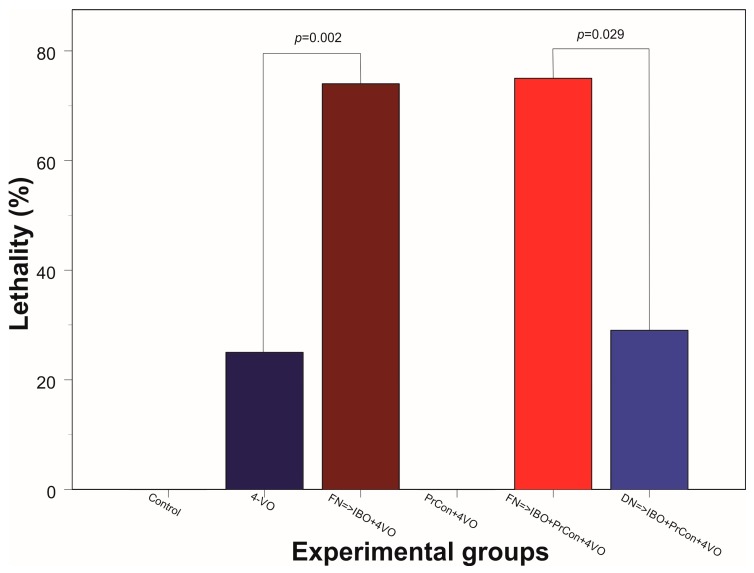
Excitation of intrinsic neurons of cerebellar fastigial nucleus (FN) renders brain tolerant to local and global ischemia. This effect reaches a maximum 72 h after the stimulation and lasts over 10 days. Comparable neuroprotection is observed following sublethal global brain ischemia, a phenomenon known as preconditioning. We hypothesized that FN may participate in the mechanisms of ischemic preconditioning as a part of the intrinsic neuroprotective mechanism. To explore potential significance of FN neurons in brain ischemic tolerance we lesioned intrinsic FN neurons with excitotoxin ibotenic acid five days before exposure to 20 min four-vessel occlusion (4-VO) global ischemia while analyzing neuronal damage in Cornu Ammoni area 1 (CA1) hippocampal area one week later. In FN-lesioned animals, loss of CA1 cells was higher by 22% compared to control (phosphate buffered saline (PBS)-injected) animals. Moreover, lesion of FN neurons increased morbidity following global ischemia by 50%. Ablation of FN neurons also reversed salvaging effects of five-minute ischemic preconditioning on CA1 neurons and morbidity, while ablation of cerebellar dentate nucleus neurons did not change effect of ischemic preconditioning. We conclude that FN is an important part of intrinsic neuroprotective system, which participates in ischemic preconditioning and may participate in naturally occurring neuroprotection, such as "diving response".

Keywords: cerebral ischemia; fastigial nucleus; hypoxic-ischemic brain injury; ischemic preconditioning; neuroprotection; pathophysiology; physiology; stroke.


Conflict of interest statement

The authors declare no conflict of interest.

Figures

Figure 1
Figure 1
Summarized localization of the lesioned areas in the cerebellar fastigial nucleus. (A) Outer borders of overlapped outlines of histological damage 5 days following the injection of IBO depicted on the appropriate level of stereotaxic atlas [23]. (B) Gliosis at the IBO injection site.
Figure 2
Figure 2
Changes of the hippocampal Cornu Ammoni area 1 (CA1) pyramidal layer neurons 7 days after sham or 4-vessels occlusion (4-VO) in different experiments. (A) CA1 neurons in animal, which underwent PBS injection into fastigial nucleus (FN) and sham 4-VO; (B) CA1 neurons following 20-min 4-VO; (C) CA1 neurons in the animal, which received microinjection of ibotenic acid (IBO) into fastigial nucleus five days before 4-VO; (D) CA1 neurons in the animal which underwent 5-min 4-VO three days before the 20-min 4-VO; (E) CA1 neurons in the animal, which received microinjection of IBO into FN 5 days before the 5-min 4-VO followed by 20-min 4-VO three days later; (F) CA1 neurons in the animal which received microinjection of IBO into DN 5 days before the 5-min 4-VO followed by 20-min VO three days later. Bar of panel F equals 50 μm. Other panels have the same magnification.
Figure 3
Figure 3
Changes of the average number of hippocampal CA1 pyramidal layer neurons 7 days after sham or 4-vessels occlusion (4-VO) in different experiments. Dark green box: CA1 neurons in animals, which underwent PBS injection into fastigial nucleus (FN) and sham 4-VO (n = 5); Dark blue boxes. CA1 neurons following 20-min 4-VO (6); Brown box: CA1 neurons in the animals, which received microinjection of ibotenic acid into (IBO) fastigial nucleus five days before 4-VO (n = 5); Light green box: CA1 neurons in the animals which underwent 5-min 4-VO three days before the 20-min 4-VO (n = 5); Red box: CA1 neurons in the animal, which received microinjection of IBO into FN 5 days before the 5-min 4-VO followed by 20-min 4-VO three days later (n = 5); Light blue box: CA1 neurons in the animal which received microinjection of IBO into DN 5 days before the 5-min 4-VO followed by 20-min VO three days later (n = 5). Boxes indicate minimum–maximum spread; middle rhombus indicates mean; whiskers indicate 2 × SD. p indicate significance of the difference (One-way ANOVA F (5, 25) = 42.632, p < 0.001, Sheffe test).
Figure 4
Figure 4
Animal lethality within 7 days following 20-min 4 vessel occlusion in different experiments. Control group did not have lethal cases (0/5); Dark blue bar: lethality within one week following 20-min 4-VO (2/8); Brown bar: lethality in the animals, which received microinjection of ibotenic acid into (IBO) fastigial nucleus five days before 4-VO (14/19); PrCon + 4-VO: lethality in the animals which underwent 5-min 4-VO three days before the 20-min 4-VO (0/5); Red bar: lethality in the animals, which received microinjection of IBO into FN 5 days before the 5-min 4-VO followed by 20-min 4-VO three days later (15/20); Light blue box: lethality in the animals which received microinjection of IBO into DN 5 days before the 5-min 4-VO followed by 20-min VO three days later (2/7). p—significance χ2.

Similar articles

Cited by

References

    1. Wang J., Dong W.W., Zhang W.H., Zheng J., Wang X. Electrical stimulation of cerebellar fastigial nucleus: Mechanism of neuroprotection and prospects for clinical application against cerebral ischemia. CNS Neurosci. Ther. 2014;20:710–716. doi: 10.1111/cns.12288. - DOI - PMC - PubMed
    1. Mandel M., Fonoff E.T., Bor-Seng-Shu E., Teixeira M.J., Chadi G. Neurogenic neuroprotection: Future perspectives. Transl. Neurosci. 2012;3:399–412. doi: 10.2478/s13380-012-0047-4. - DOI - PMC - PubMed
    1. Golanov E.V., Zhou P. Neurogenic neuroprotection. Cell. Mol. Neurobiol. 2003;23:651–663. doi: 10.1023/A:1025088516742. - DOI - PMC - PubMed
    1. Yamamoto S., Golanov E.V., Reis D.J. Reductions in focal ischemic infarctions elicited from cerebellar fastigial nucleus do not result from elevations in cerebral blood flow. J. Cereb. Blood Flow Metab. 1993;13:1020–1024. doi: 10.1038/jcbfm.1993.128. - DOI - PubMed
    1. Reis D.J., Berger S.B., Underwood M.D., Khayata M. Electrical stimulation of cerebellar fastigial nucleus reduces ischemic infarction elicited by middle cerebral artery occlusion in rat. J. Cereb. Blood Flow Metab. 1991;11:810–818. doi: 10.1038/jcbfm.1991.139. - DOI - PubMed

LinkOut - more resources