

Mobile-Based Analysis of Malaria-Infected Thin Blood Smears: Automated Species and Life Cycle Stage Determination
- PMID: 28934170
- PMCID: PMC5677014
- DOI: 10.3390/s17102167
Mobile-Based Analysis of Malaria-Infected Thin Blood Smears: Automated Species and Life Cycle Stage Determination
Abstract
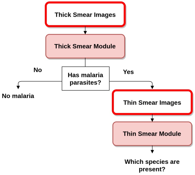
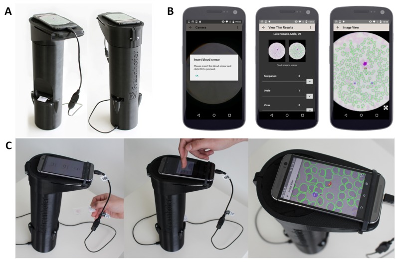
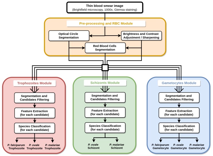
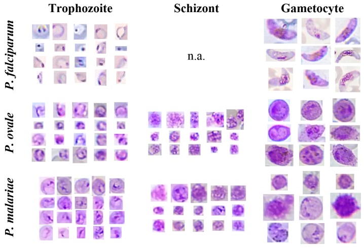
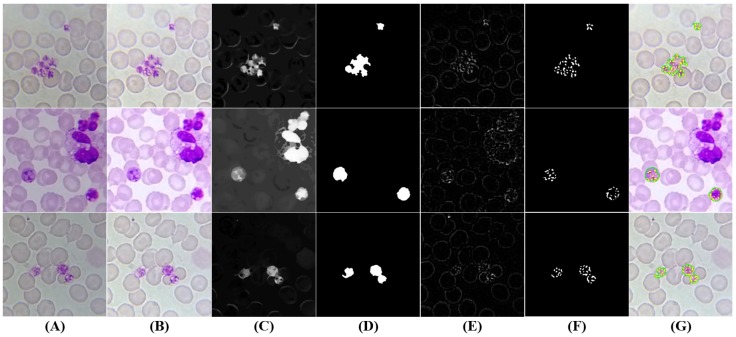
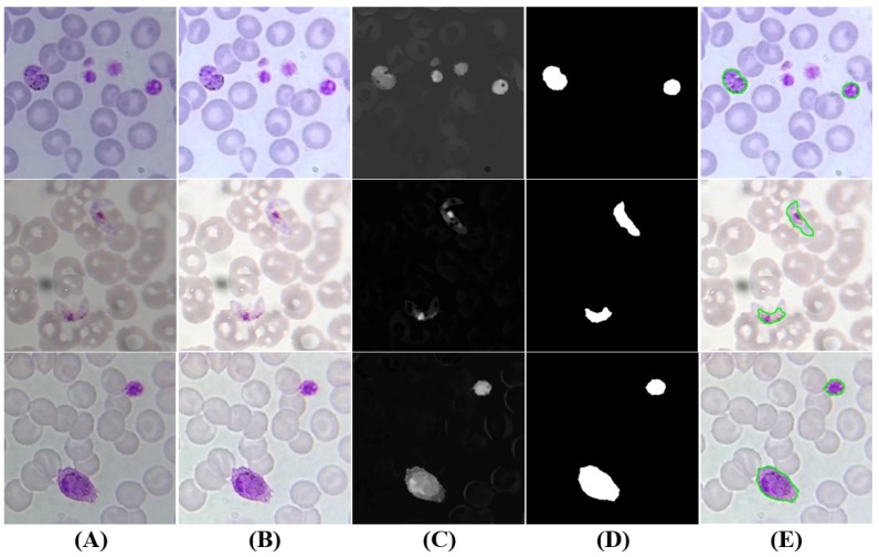
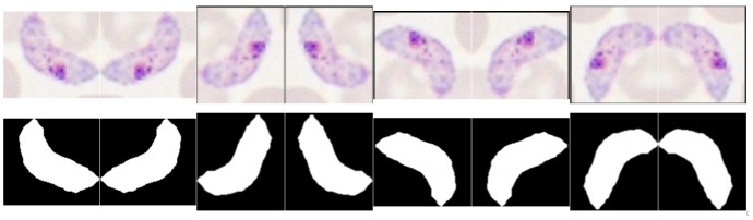
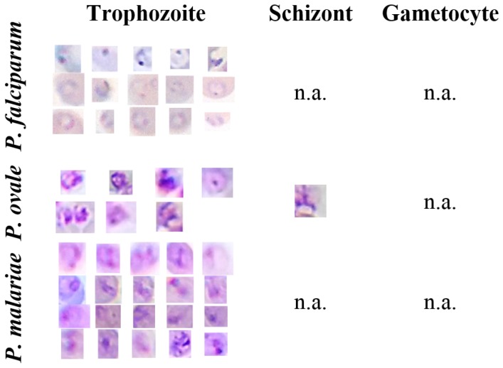
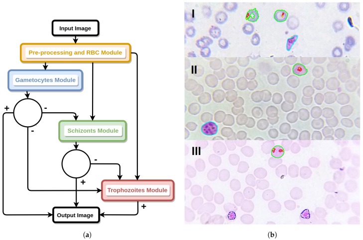
Microscopy examination has been the pillar of malaria diagnosis, being the recommended procedure when its quality can be maintained. However, the need for trained personnel and adequate equipment limits its availability and accessibility in malaria-endemic areas. Rapid, accurate, accessible diagnostic tools are increasingly required, as malaria control programs extend parasite-based diagnosis and the prevalence decreases. This paper presents an image processing and analysis methodology using supervised classification to assess the presence of malaria parasites and determine the species and life cycle stage in Giemsa-stained thin blood smears. The main differentiation factor is the usage of microscopic images exclusively acquired with low cost and accessible tools such as smartphones, a dataset of 566 images manually annotated by an experienced parasilogist being used. Eight different species-stage combinations were considered in this work, with an automatic detection performance ranging from 73.9% to 96.2% in terms of sensitivity and from 92.6% to 99.3% in terms of specificity. These promising results attest to the potential of using this approach as a valid alternative to conventional microscopy examination, with comparable detection performances and acceptable computational times.
Keywords: computer-aided diagnosis; image analysis; malaria; microscopy; mobile devices.
Conflict of interest statement
The authors declare no conflict of interest.
Figures

Similar articles
-
An automatic device for detection and classification of malaria parasite species in thick blood film.BMC Bioinformatics. 2012;13 Suppl 17(Suppl 17):S18. doi: 10.1186/1471-2105-13-S17-S18. Epub 2012 Dec 13. BMC Bioinformatics. 2012. PMID: 23281600 Free PMC article.
-
Plasmodium life cycle stage classification based quantification of malaria parasitaemia in thin blood smears.Microsc Res Tech. 2019 Mar;82(3):283-295. doi: 10.1002/jemt.23170. Epub 2018 Dec 21. Microsc Res Tech. 2019. PMID: 30575213
-
Automated system for characterization and classification of malaria-infected stages using light microscopic images of thin blood smears.J Microsc. 2015 Mar;257(3):238-52. doi: 10.1111/jmi.12206. Epub 2014 Dec 18. J Microsc. 2015. PMID: 25523795
-
[Advances in automatic detection technology for images of thin blood film of malaria parasite].Zhongguo Xue Xi Chong Bing Fang Zhi Za Zhi. 2017 May 5;29(3):388-392. doi: 10.16250/j.32.1374.2017015. Zhongguo Xue Xi Chong Bing Fang Zhi Za Zhi. 2017. PMID: 29469543 Review. Chinese.
-
Recent Advances of Malaria Parasites Detection Systems Based on Mathematical Morphology.Sensors (Basel). 2018 Feb 8;18(2):513. doi: 10.3390/s18020513. Sensors (Basel). 2018. PMID: 29419781 Free PMC article. Review.
Cited by
-
Detection and stage classification of Plasmodium falciparum from images of Giemsa stained thin blood films using random forest classifiers.Diagn Pathol. 2020 Oct 23;15(1):130. doi: 10.1186/s13000-020-01040-9. Diagn Pathol. 2020. PMID: 33097073 Free PMC article.
-
Advances in Portable Optical Microscopy Using Cloud Technologies and Artificial Intelligence for Medical Applications.Sensors (Basel). 2024 Oct 17;24(20):6682. doi: 10.3390/s24206682. Sensors (Basel). 2024. PMID: 39460161 Free PMC article. Review.
-
The Impact of Artificial Intelligence on Microbial Diagnosis.Microorganisms. 2024 May 23;12(6):1051. doi: 10.3390/microorganisms12061051. Microorganisms. 2024. PMID: 38930432 Free PMC article. Review.
-
Detection of Intestinal Protozoa in Trichrome-Stained Stool Specimens by Use of a Deep Convolutional Neural Network.J Clin Microbiol. 2020 May 26;58(6):e02053-19. doi: 10.1128/JCM.02053-19. Print 2020 May 26. J Clin Microbiol. 2020. PMID: 32295888 Free PMC article.
-
Systematic Review and Meta-Analysis of the Diagnostic Accuracy of Mobile-Linked Point-of-Care Diagnostics in Sub-Saharan Africa.Diagnostics (Basel). 2021 Jun 12;11(6):1081. doi: 10.3390/diagnostics11061081. Diagnostics (Basel). 2021. PMID: 34204848 Free PMC article. Review.
References
-
- World Health Organization . World Malaria Report 2016. WHO; Geneva, Switzerland: 2016.
-
- World Health Organization . World Malaria Report 2015. WHO; Geneva, Switzerland: 2015.
-
- Blycroft Limited . Africa & Middle East Mobile Factbook 2Q 2014. Blycroft Publishing; Aylesbury, UK: 2014.
-
- Dolgin E. Portable pathology for Africa. IEEE Spectr. 2015;52:37–39. doi: 10.1109/MSPEC.2015.6995631. - DOI
-
- Rosado L., Correia da Costa J.M., Elias D., Cardoso J.S. A Review of Automatic Malaria Parasites Detection and Segmentation in Microscopic Images. Anti-Infect. Agents. 2016;14:11–22. doi: 10.2174/221135251401160302121107. - DOI
MeSH terms
LinkOut - more resources
Full Text Sources
Other Literature Sources
Medical
