

HLA-DR7 and HLA-DQ2: Transgenic mouse strains tested as a model system for ximelagatran hepatotoxicity
- PMID: 28934241
- PMCID: PMC5608249
- DOI: 10.1371/journal.pone.0184744
HLA-DR7 and HLA-DQ2: Transgenic mouse strains tested as a model system for ximelagatran hepatotoxicity
Abstract
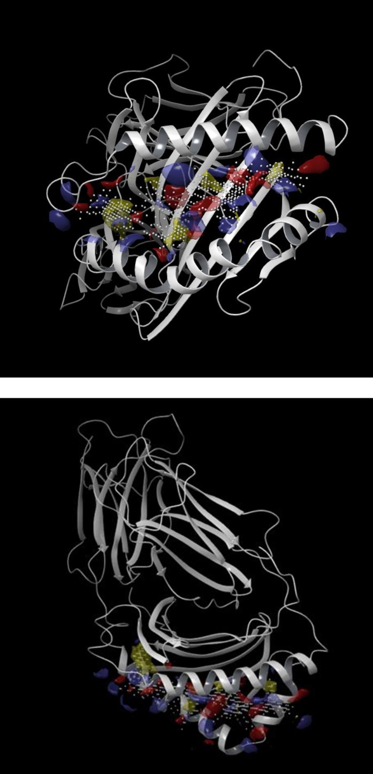
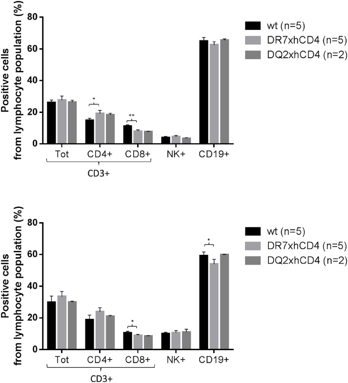
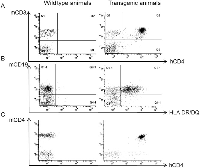
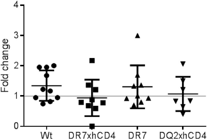
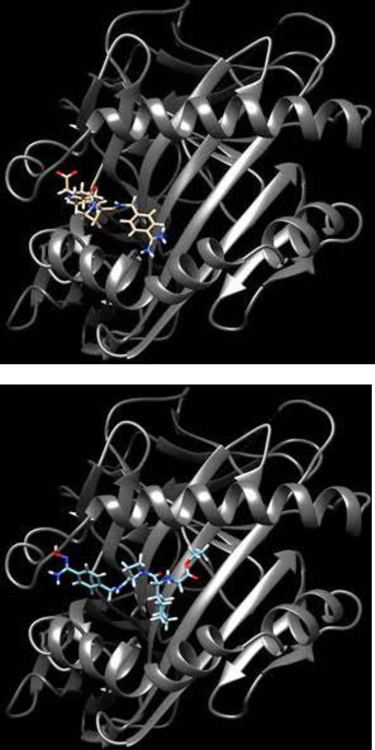
The oral thrombin inhibitor ximelagatran was withdrawn in the late clinical trial phase because it adversely affected the liver. In approximately 8% of treated patients, drug-induced liver injury (DILI) was expressed as transient alanine transaminase (ALT) elevations. No evidence of DILI had been revealed in the pre-clinical in vivo studies. A whole genome scan study performed on the clinical study material identified a strong genetic association between the major histocompatibility complex alleles for human leucocyte antigens (HLA) (HLA-DR7 and HLA-DQ2) and elevated ALT levels in treated patients. An immune-mediated pathogenesis was suggested. Here, we evaluated whether HLA transgenic mice models could be used to investigate whether the expression of relevant HLA molecules was enough to reproduce the DILI effects in humans. In silico modelling performed in this study revealed association of both ximelagatran (pro-drug) and melagatran (active drug) to the antigen-presenting groove of the homology modelled HLA-DR7 molecule suggesting "altered repertoire" as a key initiating event driving development of DILI in humans. Transgenic mouse strains (tgms) expressing HLA of serotype HLA-DR7 (HLA-DRB1*0701, -DRA*0102), and HLA-DQ2 (HLA-DQB1*0202,-DQA1*0201) were created. These two lines were crossed with a human (h)CD4 transgenic line, generating the two tgms DR7xhCD4 and DQ2xhCD4. To investigate whether the DILI effects observed in humans could be reproduced in tgms, the mice were treated for 28 days with ximelagatran. Results revealed no signs of DILI when biomarkers for liver toxicity were measured and histopathology was evaluated. In the ximelagatran case, presence of relevant HLA-expression in a pre-clinical model did not fulfil the prerequisite for reproducing DILI observed in patients. Nonetheless, for the first time an HLA-transgenic mouse model has been investigated for use in HLA-associated DILI induced by a low molecular weight compound. This study shows that mimicking of genetic susceptibility, expressed as DILI-associated HLA-types in mice, is not sufficient for reproducing the complex pathogenesis leading to DILI in man.
Conflict of interest statement
Figures
Similar articles
-
In Silico and In Vitro Analysis of Interaction between Ximelagatran and Human Leukocyte Antigen (HLA)-DRB1*07:01.Int J Mol Sci. 2017 Mar 24;18(4):694. doi: 10.3390/ijms18040694. Int J Mol Sci. 2017. PMID: 28338626 Free PMC article.
-
Prediction of drug-induced liver injury in humans by using in vitro methods: the case of ximelagatran.Toxicol In Vitro. 2008 Apr;22(3):730-46. doi: 10.1016/j.tiv.2007.11.014. Epub 2007 Dec 3. Toxicol In Vitro. 2008. PMID: 18191936
-
Drug-induced liver injury in humans: the case of ximelagatran.Handb Exp Pharmacol. 2010;(196):407-18. doi: 10.1007/978-3-642-00663-0_13. Handb Exp Pharmacol. 2010. PMID: 20020269 Review.
-
Evidence of a strong, positive association between atopy and the HLA class II alleles DR4 and DR7.Clin Exp Allergy. 1996 Jul;26(7):821-8. Clin Exp Allergy. 1996. PMID: 8842557
-
Pharmacogenetic testing in idiosyncratic drug-induced liver injury: current role in clinical practice.Liver Int. 2015 Jul;35(7):1801-8. doi: 10.1111/liv.12836. Epub 2015 Apr 20. Liver Int. 2015. PMID: 25809692 Review.
Cited by
-
Hepatotoxicity of Herbal Supplements Mediated by Modulation of Cytochrome P450.Int J Mol Sci. 2017 Nov 8;18(11):2353. doi: 10.3390/ijms18112353. Int J Mol Sci. 2017. PMID: 29117101 Free PMC article. Review.
-
Idiosyncratic Drug-Induced Liver Injury: Mechanistic and Clinical Challenges.Int J Mol Sci. 2021 Mar 14;22(6):2954. doi: 10.3390/ijms22062954. Int J Mol Sci. 2021. PMID: 33799477 Free PMC article. Review.
-
Preclinical models of idiosyncratic drug-induced liver injury (iDILI): Moving towards prediction.Acta Pharm Sin B. 2021 Dec;11(12):3685-3726. doi: 10.1016/j.apsb.2021.11.013. Epub 2021 Nov 18. Acta Pharm Sin B. 2021. PMID: 35024301 Free PMC article. Review.
-
Application of hepatocyte-like cells to enhance hepatic safety risk assessment in drug discovery.Philos Trans R Soc Lond B Biol Sci. 2018 Jul 5;373(1750):20170228. doi: 10.1098/rstb.2017.0228. Philos Trans R Soc Lond B Biol Sci. 2018. PMID: 29786562 Free PMC article. Review.
-
What have we learned from animal models of idiosyncratic, drug-induced liver injury?Expert Opin Drug Metab Toxicol. 2020 Jun;16(6):475-491. doi: 10.1080/17425255.2020.1760246. Epub 2020 May 4. Expert Opin Drug Metab Toxicol. 2020. PMID: 32324077 Free PMC article. Review.
References
-
- Lee WM (2003) Drug-induced hepatotoxicity. N Engl J Med 349: 474–485. doi: 10.1056/NEJMra021844 - DOI - PubMed
-
- Cook D, Brown D, Alexander R, March R, Morgan P, et al. (2014) Lessons learned from the fate of AstraZeneca's drug pipeline: a five-dimensional framework. Nat Rev Drug Discov 13: 419–431. doi: 10.1038/nrd4309 - DOI - PubMed
-
- Albers GW, Diener HC, Frison L, Grind M, Nevinson M, et al. (2005) Ximelagatran vs warfarin for stroke prevention in patients with nonvalvular atrial fibrillation: a randomized trial. JAMA 293: 690–698. doi: 10.1001/jama.293.6.690 - DOI - PubMed
-
- Petersen P, Grind M, Adler J, Investigators SI (2003) Ximelagatran versus warfarin for stroke prevention in patients with nonvalvular atrial fibrillation. SPORTIF II: a dose-guiding, tolerability, and safety study. J Am Coll Cardiol 41: 1445–1451. - PubMed
-
- Lee WM, Larrey D, Olsson R, Lewis JH, Keisu M, et al. (2005) Hepatic findings in long-term clinical trials of ximelagatran. Drug Saf 28: 351–370. - PubMed
MeSH terms
Substances
LinkOut - more resources
Full Text Sources
Other Literature Sources
Medical
Molecular Biology Databases
Research Materials
