

Quantitative optical coherence tomography angiography of macular vascular structure and foveal avascular zone in glaucoma
- PMID: 28934255
- PMCID: PMC5608222
- DOI: 10.1371/journal.pone.0184948
Quantitative optical coherence tomography angiography of macular vascular structure and foveal avascular zone in glaucoma
Abstract
Objective: The study aimed to evaluate the quantitative characteristics of the macular vessel density (VD) and foveal avascular zone (FAZ) in glaucoma using optical coherence tomography angiography (OCT-A).
Design: Cross-sectional, age- and sex-matched case-control study.
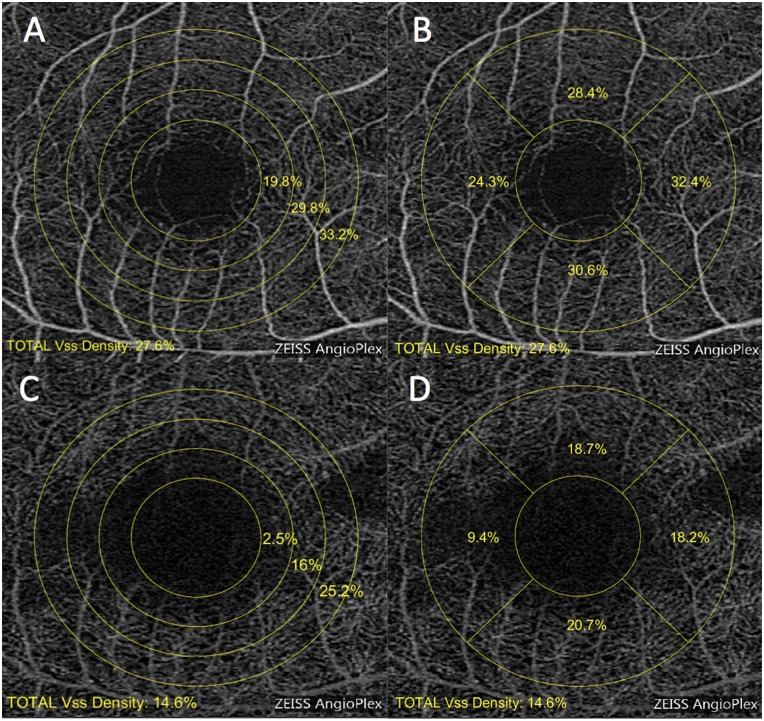
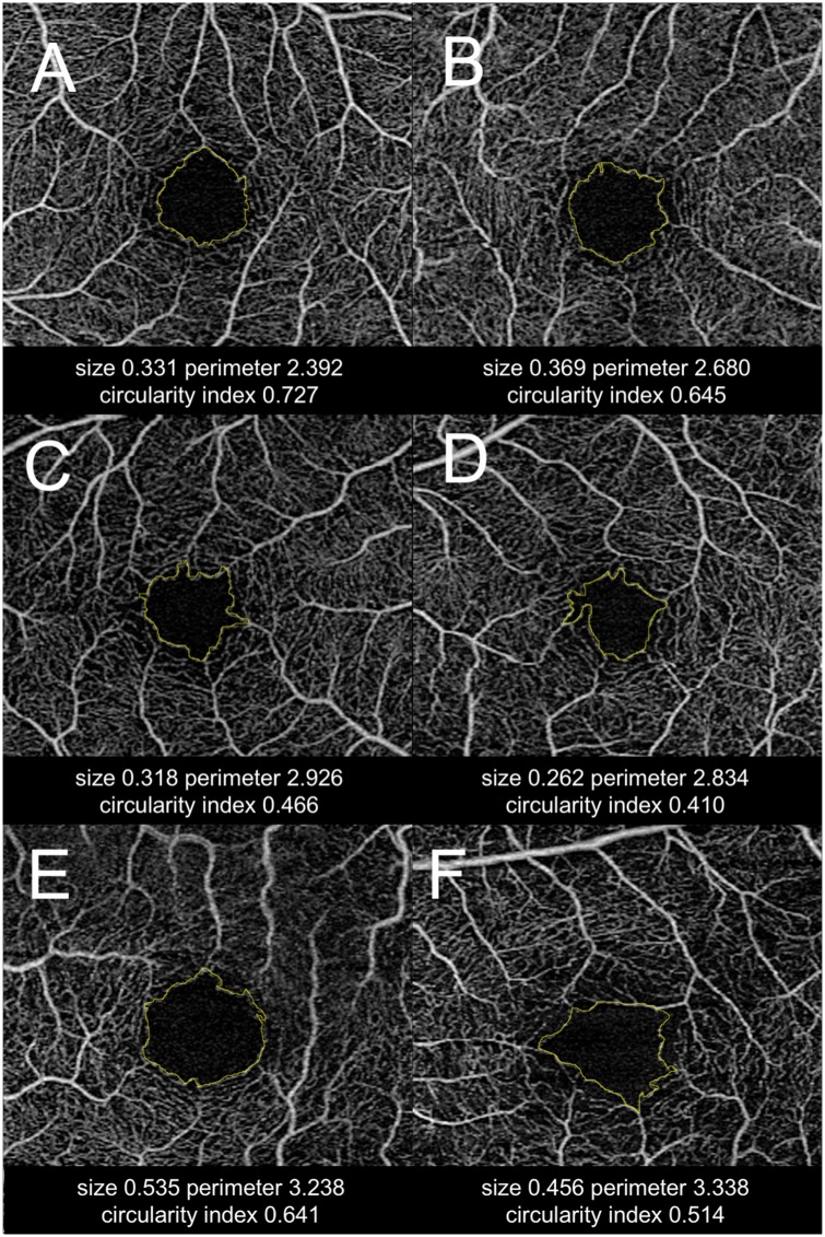
Methods: Fifty-two eyes of 52 patients with primary open angle glaucoma and 52 eyes from 52 healthy participants were recruited retrospectively. OCT-A was performed on a 3 x 3-mm macular region centered on the fovea. OCT-A scans were manually graded to define the FAZ. Parafoveal VD in superficial and deep retina were analyzed in the circular- and quadrant-segmented zone. The FAZ parameters included size, perimeter, and circularity index. The regression analysis among VD and FAZ-related parameters and ocular parameters was performed, and the diagnostic ability was calculated with refractive error adjusted.
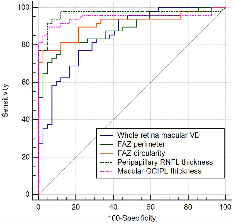
Results: For both groups, the mean age and the sex ratio was not different between groups. With refractive error adjusted, the average macular VD was lower in glaucoma than in the control group for superficial (P = 0.013), deep (P<0.001), and the whole retina (P = 0.002). There were increased FAZ perimeter and decreased FAZ circularity index in glaucoma when compared with controls (P<0.001). In the multivariate regression models, FAZ circularity index were significantly associated with decreased peripapillary RNFL thickness (P = 0.007) and macular GCIPL thickness (P = 0.009) measured by OCT. The refractive-error adjusted area under receiver operating characteristics was highest for FAZ circularity index (0.905; 95% CI, 0.844-0.966), followed by temporal deep retinal VD (0.870; 95% CI, 0.803-0.937) and FAZ perimeter (0.858; 95% CI, 0.784-0.932).
Conclusions: Decreased macular VD, increased FAZ perimeter, and decreased FAZ circularity index were observed in eyes with glaucoma using OCT-A. With refractive error adjusted, these parameters showed considerable diagnostic value for glaucoma. FAZ circularity index may be a novel biomarker representing disruption of the parafoveal capillary network in glaucoma, as supported by its association with structural parameters.
Conflict of interest statement
Figures
Similar articles
-
Evaluating the Quantitative Foveal Avascular Zone and Retino-Choroidal Vessel Density Using Optical Coherence Tomography Angiography in a Healthy Indian Population.Cureus. 2022 Aug 4;14(8):e27669. doi: 10.7759/cureus.27669. eCollection 2022 Aug. Cureus. 2022. PMID: 36072178 Free PMC article.
-
Macular vessel density and foveal avascular zone parameters in patients after acute primary angle closure determined by OCT angiography.Sci Rep. 2020 Oct 30;10(1):18717. doi: 10.1038/s41598-020-73223-9. Sci Rep. 2020. PMID: 33127916 Free PMC article.
-
Optical coherence tomography angiography analysis of foveal microvascular changes and inner retinal layer thinning in patients with diabetes.Br J Ophthalmol. 2018 Sep;102(9):1226-1231. doi: 10.1136/bjophthalmol-2017-311149. Epub 2017 Dec 19. Br J Ophthalmol. 2018. PMID: 29259019
-
Retinal microvasculature features in patients with Behcet's disease: a systematic review and meta-analysis.Sci Rep. 2022 Jan 14;12(1):752. doi: 10.1038/s41598-021-04730-6. Sci Rep. 2022. PMID: 35031636 Free PMC article.
-
Application of Optical Coherence Tomography Angiography Macular Analysis for Systemic Hypertension. A Systematic Review and Meta-analysis.Am J Hypertens. 2022 Apr 2;35(4):356-364. doi: 10.1093/ajh/hpab172. Am J Hypertens. 2022. PMID: 34718393
Cited by
-
Qualitative and quantitative comparison of two semi-manual retinal vascular density analyzing methods on optical coherence tomography angiography images of healthy individuals.Sci Rep. 2023 Oct 9;13(1):16981. doi: 10.1038/s41598-023-44234-z. Sci Rep. 2023. PMID: 37813968 Free PMC article.
-
Macular Thickness and Microvasculature Loss in Glaucoma Suspect Eyes.Ophthalmol Glaucoma. 2022 Mar-Apr;5(2):170-178. doi: 10.1016/j.ogla.2021.07.009. Epub 2021 Jul 30. Ophthalmol Glaucoma. 2022. PMID: 34339877 Free PMC article.
-
Macular vessel density before and after panretinal photocoagulation in patients with proliferative diabetic retinopathy.Int J Retina Vitreous. 2022 Mar 14;8(1):21. doi: 10.1186/s40942-022-00369-1. Int J Retina Vitreous. 2022. PMID: 35287760 Free PMC article.
-
Quantitative analysis of retinal and choroidal microvascular changes in patients with diabetes.Sci Rep. 2018 Aug 14;8(1):12146. doi: 10.1038/s41598-018-30699-w. Sci Rep. 2018. PMID: 30108264 Free PMC article.
-
Optical coherence tomography angiography (OCTA) flow speed mapping technology for retinal diseases.Expert Rev Med Devices. 2018 Dec;15(12):875-882. doi: 10.1080/17434440.2018.1548932. Epub 2018 Nov 22. Expert Rev Med Devices. 2018. PMID: 30460869 Free PMC article. Review.
References
-
- Choi J, Jeong J, Cho HS, Kook MS. Effect of nocturnal blood pressure reduction on circadian fluctuation of mean ocular perfusion pressure: a risk factor for normal tension glaucoma. Investigative ophthalmology & visual science. 2006; 47: 831–836. doi: 10.1167/iovs.05-1053 - DOI - PubMed
-
- Choi J, Kim KH, Jeong J, Cho HS, Lee CH, Kook MS. Circadian fluctuation of mean ocular perfusion pressure is a consistent risk factor for normal-tension glaucoma. Investigative ophthalmology & visual science. 2007; 48: 104–111. doi: 10.1167/iovs.06-0615 - DOI - PubMed
-
- Flammer J, Orgul S. Optic nerve blood-flow abnormalities in glaucoma. Progress in retinal and eye research. 1998; 17: 267–289. - PubMed
-
- Flammer J, Orgul S, Costa VP, Orzalesi N, Krieglstein GK, Serra LM, et al. The impact of ocular blood flow in glaucoma. Progress in retinal and eye research. 2002; 21: 359–393. - PubMed
-
- Fuchsjager-Mayrl G, Wally B, Georgopoulos M, Rainer G, Kircher K, Buehl W, et al. Ocular blood flow and systemic blood pressure in patients with primary open-angle glaucoma and ocular hypertension. Investigative ophthalmology & visual science. 2004; 45: 834–839. - PubMed
MeSH terms
LinkOut - more resources
Full Text Sources
Other Literature Sources
Medical
