

Activation of phosphatidylinositol 3-kinase/AKT/snail signaling pathway contributes to epithelial-mesenchymal transition-induced multi-drug resistance to sorafenib in hepatocellular carcinoma cells
- PMID: 28934275
- PMCID: PMC5608310
- DOI: 10.1371/journal.pone.0185088
Activation of phosphatidylinositol 3-kinase/AKT/snail signaling pathway contributes to epithelial-mesenchymal transition-induced multi-drug resistance to sorafenib in hepatocellular carcinoma cells
Abstract
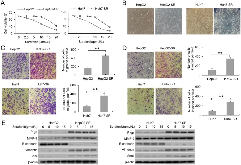
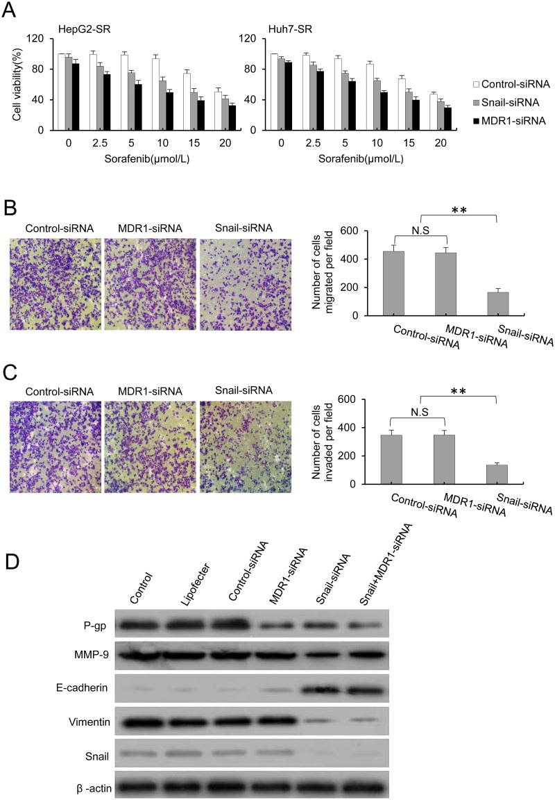
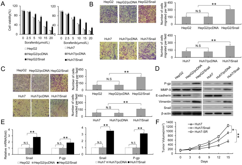
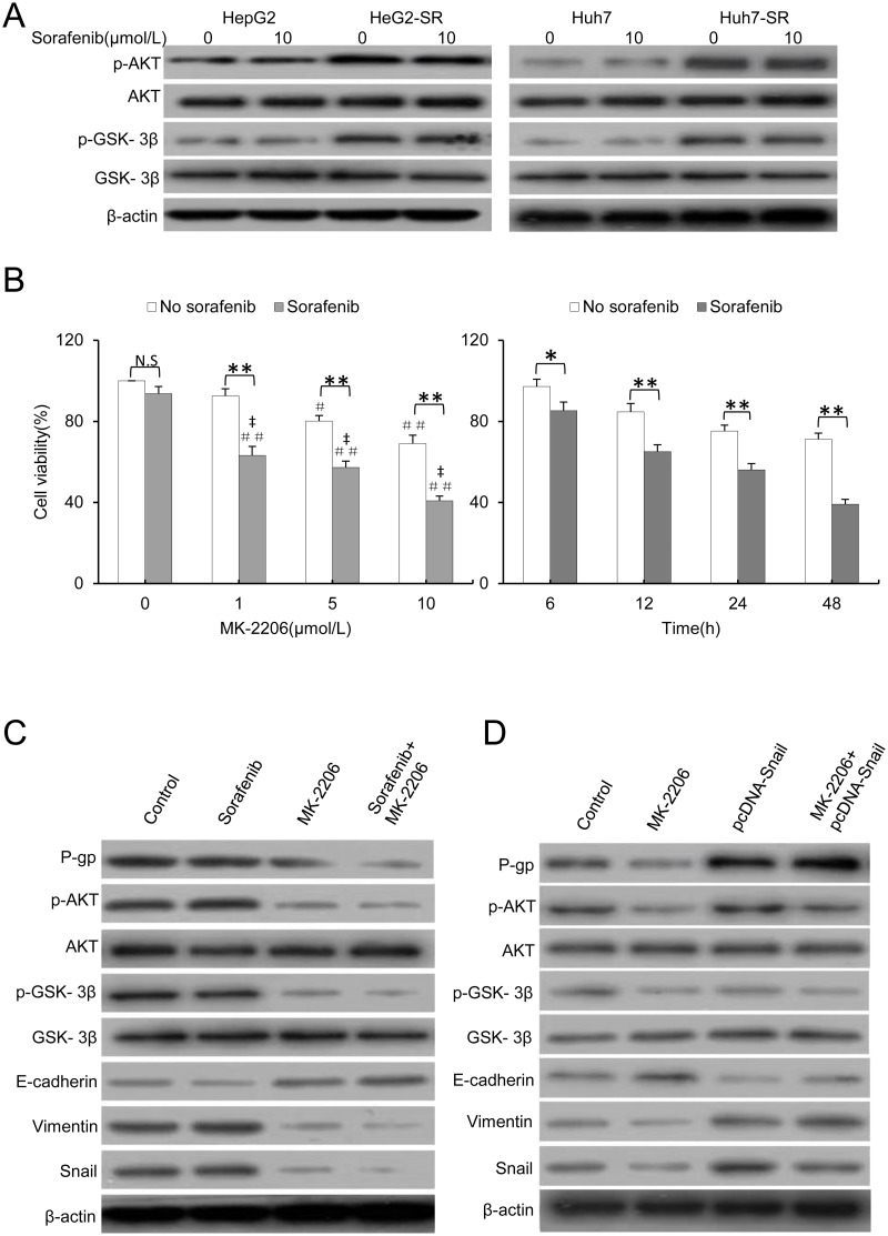
Sorafenib, an orally available kinase inhibitor, is the standard first-line systemic drug for advanced hepatocellular carcinoma (HCC), and it exerts potent inhibitory activity against epithelial-mesenchymal transition (EMT) and multidrug resistance (MDR) by inhibiting mitogen-activated protein kinase (MAPK) signaling in HCC. However, after long-term exposure to sorafenib, HCC cells exhibit EMT and resistance to sorafenib. The activation of AKT by sorafenib is thought to be responsible for the development of these characteristics. The present study aims to examine the underlying mechanism and seek potential strategies to reverse this resistance and the progression to EMT. Sorafenib-resistant cells showed increased metastatic and invasive ability, with a higher expression of P-glycoprotein (P-gp), compared with the parental cells. This phenomenon was at least partially due to EMT and the appearance of MDR in sorafenib-resistant HCC cells. Moreover, MDR was a downstream molecular event of EMT. Silencing Snail with siRNA blocked EMT and partially reversed the MDR, thereby markedly abolishing invasion and metastasis in sorafenib-resistant HCC cells, but silencing of MDR1 had no effect on the EMT phenotype. Additionally, HCC parental cells that were stably transfected with pCDNA3.1-Snail exhibited EMT and MDR. Two sorafenib-resistant HCC cell lines, established from human HCC HepG2 and Huh7 cells, were refractory to sorafenib-induced growth inhibition but were sensitive to MK-2206, a novel allosteric AKT inhibitor. Thus, the combination of sorafenib and MK-2206 led to significant reversion of the EMT phenotype and P-gp-mediated MDR by downregulating phosphorylated AKT. These findings underscore the significance of EMT, MDR and enhanced PI3K/AKT signaling in sorafenib-resistant HCC cells.
Conflict of interest statement
Figures
Similar articles
-
Activation of phosphatidylinositol 3-kinase/Akt signaling mediates sorafenib-induced invasion and metastasis in hepatocellular carcinoma.Oncol Rep. 2014 Oct;32(4):1465-72. doi: 10.3892/or.2014.3352. Epub 2014 Jul 23. Oncol Rep. 2014. PMID: 25070581
-
MicroRNA-216a/217-induced epithelial-mesenchymal transition targets PTEN and SMAD7 to promote drug resistance and recurrence of liver cancer.Hepatology. 2013 Aug;58(2):629-41. doi: 10.1002/hep.26369. Epub 2013 Jun 25. Hepatology. 2013. PMID: 23471579
-
MiR-21 mediates sorafenib resistance of hepatocellular carcinoma cells by inhibiting autophagy via the PTEN/Akt pathway.Oncotarget. 2015 Oct 6;6(30):28867-81. doi: 10.18632/oncotarget.4814. Oncotarget. 2015. PMID: 26311740 Free PMC article.
-
Potential molecular, cellular and microenvironmental mechanism of sorafenib resistance in hepatocellular carcinoma.Cancer Lett. 2015 Oct 10;367(1):1-11. doi: 10.1016/j.canlet.2015.06.019. Epub 2015 Jul 10. Cancer Lett. 2015. PMID: 26170167 Review.
-
Epithelial-to-Mesenchymal Transition: A Mediator of Sorafenib Resistance in Advanced Hepatocellular Carcinoma.Curr Cancer Drug Targets. 2017;17(8):698-706. doi: 10.2174/1568009617666170427104356. Curr Cancer Drug Targets. 2017. PMID: 28460616 Review.
Cited by
-
Combined Inhibition of TGF-β1-Induced EMT and PD-L1 Silencing Re-Sensitizes Hepatocellular Carcinoma to Sorafenib Treatment.J Clin Med. 2021 Apr 27;10(9):1889. doi: 10.3390/jcm10091889. J Clin Med. 2021. PMID: 33925488 Free PMC article.
-
Achillin Increases Chemosensitivity to Paclitaxel, Overcoming Resistance and Enhancing Apoptosis in Human Hepatocellular Carcinoma Cell Line Resistant to Paclitaxel (Hep3B/PTX).Pharmaceutics. 2019 Oct 4;11(10):512. doi: 10.3390/pharmaceutics11100512. Pharmaceutics. 2019. PMID: 31590262 Free PMC article.
-
Feasibility of Serum Galectin-1 as a Diagnostic Biomarker for Metabolic Dysfunction-Associated Steatotic Liver Disease: A Study on a Segment of the Chinese Population Using Convenience Sampling.Biomedicines. 2025 Feb 10;13(2):425. doi: 10.3390/biomedicines13020425. Biomedicines. 2025. PMID: 40002838 Free PMC article.
-
Mitochondrial Plasticity Promotes Resistance to Sorafenib and Vulnerability to STAT3 Inhibition in Human Hepatocellular Carcinoma.Cancers (Basel). 2021 Nov 30;13(23):6029. doi: 10.3390/cancers13236029. Cancers (Basel). 2021. PMID: 34885140 Free PMC article.
-
Inhibitor of Differentiation 1 (ID1) Facilitates the Efficacy of Sorafenib in Non-Small Cell Lung Cancer Cells through Suppressing Epithelial to Mesenchymal Transition.Med Sci Monit. 2020 Apr 10;26:e922148. doi: 10.12659/MSM.922148. Med Sci Monit. 2020. PMID: 32275644 Free PMC article.
References
-
- Torre LA, Bray F, Siegel RL, Ferlay J, Lortet-Tieulent J, Jemai A. Global cancer statistics, 2012. CA Cancer J Clin. 2015;65(2):87–108. doi: 10.3322/caac.21262 - DOI - PubMed
-
- Wang L, Yao M, Dong ZZ, Zhang Y, Yao DF. Circulating specific biomarkers in diagnosis of hepatocellular carcinoma and its metastasis monitoring. Tumour Biol. 2014;35(1):9–20. doi: 10.1007/s13277-013-1141-0 - DOI - PMC - PubMed
-
- Cheng AL, Kang YK, Chen ZD, Tsao CJ, Qin SK. Efficacy and safety of sorafenib in patients in the Asia-Pacific region with advanced hepatocellular carcinoma: a phase III randomised, double-blind, placebo-controlled trial. Lancet Oncol. 2009;10(1):25–34. doi: 10.1016/S1470-2045(08)70285-7 - DOI - PubMed
-
- Llovet JM, Ricci S, Mazzaferro V, Hilgard P, Gane E, Blanc JF, et al. Sorafenib in advanced hepatocellular carcinoma. N Engl J Med. 2008;359(4):378–90. doi: 10.1056/NEJMoa0708857 - DOI - PubMed
-
- Nagai T, Arao T, Furuta K, Sakai K, Kudo K, Kaneda H, et al. Sorafenib inhibits the hepatocyte growth factor-mediated epithelial mesenchymal transition in hepatocellular carcinoma. Mol Cancer Ther. 2011;10(1):169–177. doi: 10.1158/1535-7163.MCT-10-0544 - DOI - PubMed
MeSH terms
Substances
LinkOut - more resources
Full Text Sources
Other Literature Sources
Medical
Research Materials
Miscellaneous
