. 2017 Sep 21;12(9):e0185159.
doi: 10.1371/journal.pone.0185159. eCollection 2017.

An analysis of beak shape variation in two ages of domestic turkeys (Meleagris gallopavo) using landmark-based geometric morphometrics

Affiliations

An analysis of beak shape variation in two ages of domestic turkeys (Meleagris gallopavo) using landmark-based geometric morphometrics

Hillary A Dalton et al. PLoS One. .

Abstract

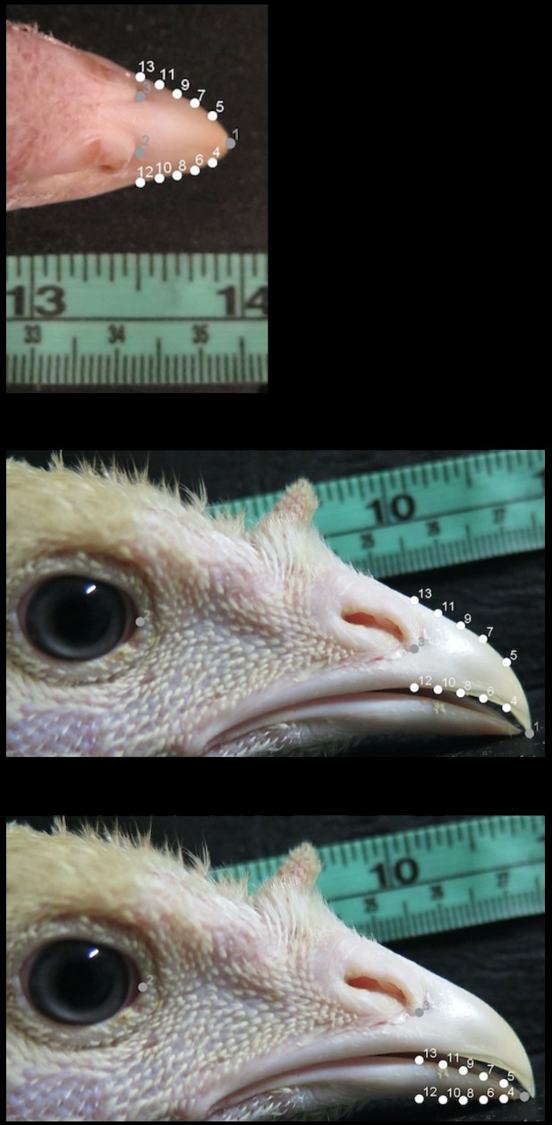
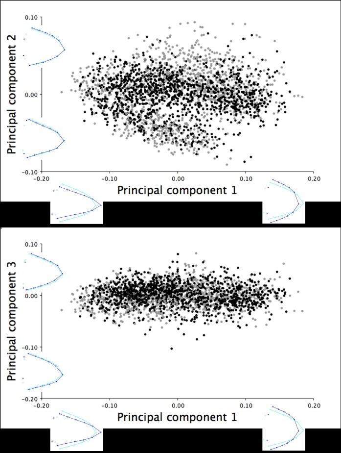
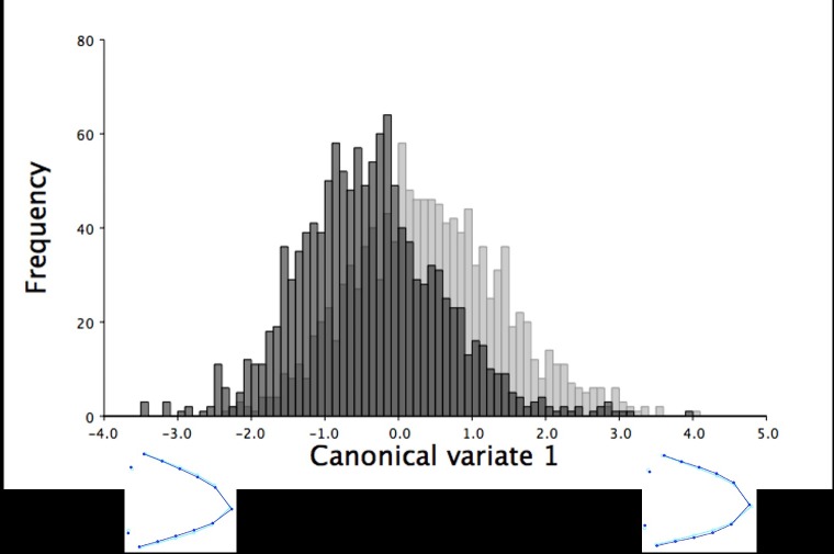
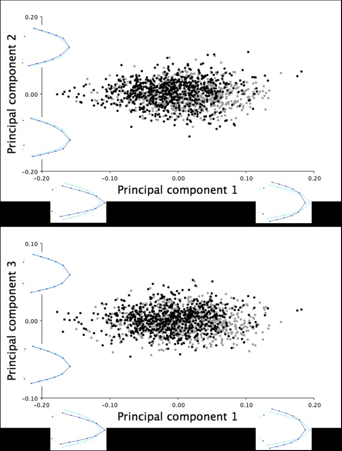
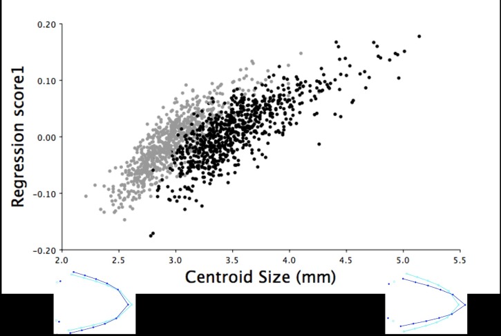
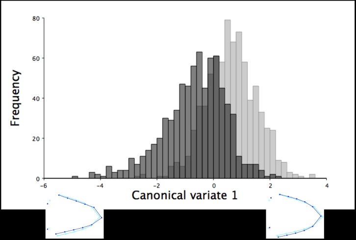
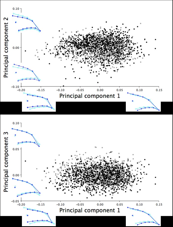
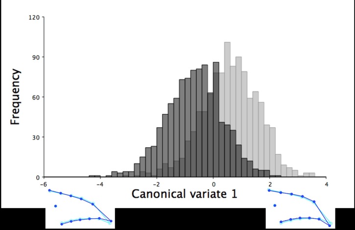
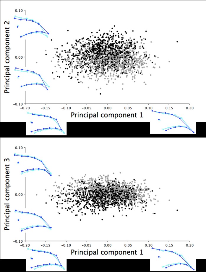
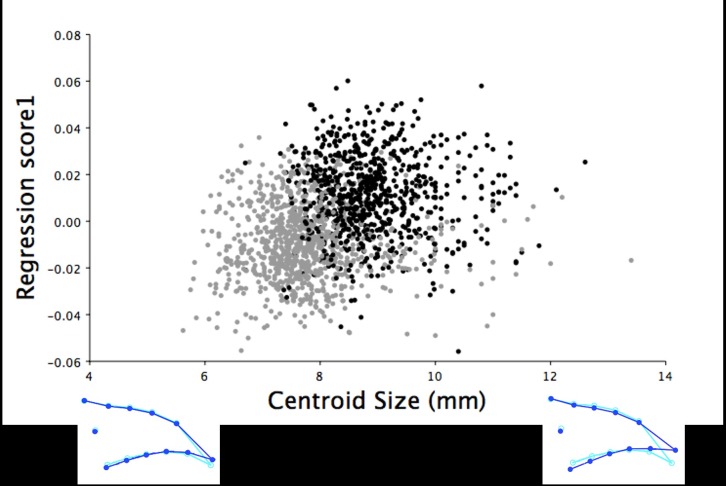
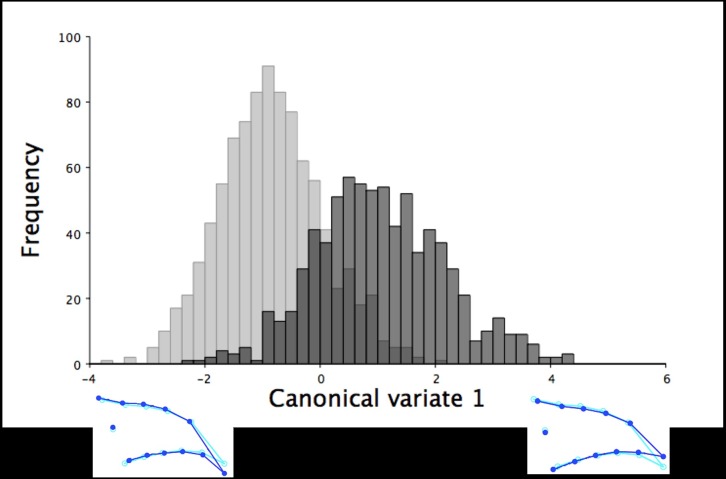
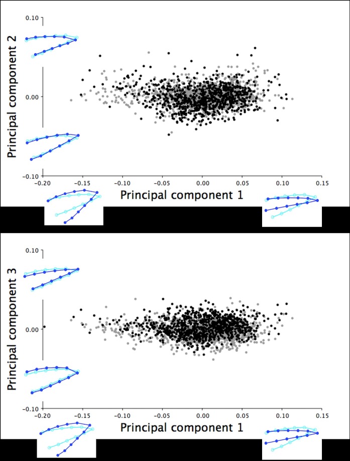
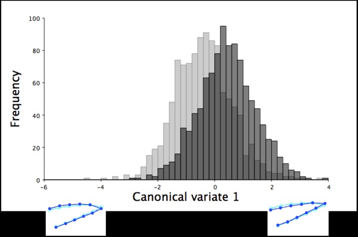
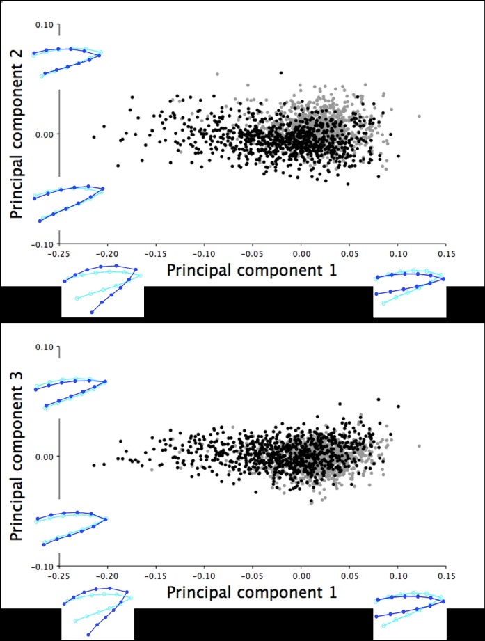
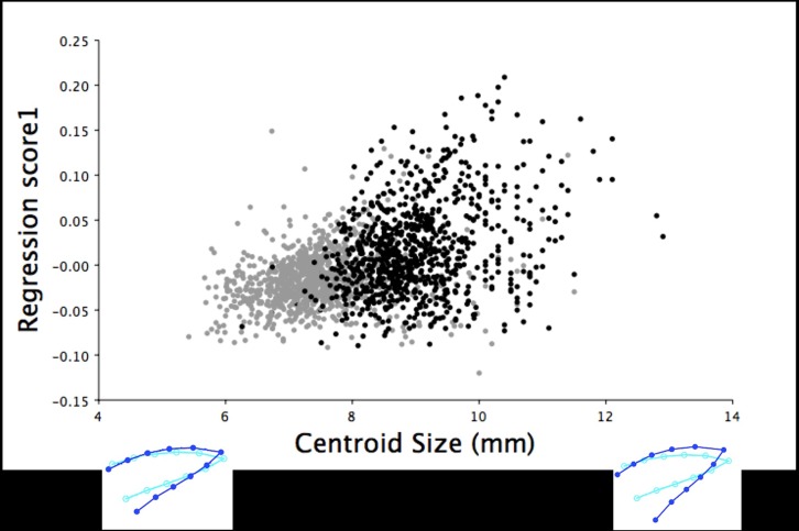
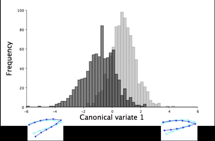
The objective of this study was to assess beak shape variation in domestic turkeys (Meleagris gallopavo) and determine the effects of age, sex, and beak size on beak shape variation using geometric morphometrics. Dorsal and right lateral images were taken of 2442 turkeys at 6 and 18.5 weeks of age. Landmarks were digitized in tpsDig in three analyses of the dorsal upper mandible, lateral upper mandible, and lateral lower mandible shape of each turkey at both ages. The coordinate data were then subjected to a principal components analysis (PCA), multivariate regression, and a canonical variates analysis (CVA) with a Procrustes ANOVA in MorphoJ. For the dorsal images, three principal components (PCs) showed beak shape variation ranged from long, narrow, and pointed to short, wide, and blunt upper mandibles at both ages (6 weeks: 95.36%, 18.5 weeks: 92.21%). Three PCs showed the lateral upper mandible shape variation ranged from long, wide beaks with long, curved beak tips to short, narrow beaks with short, pointed beak tips at both ages (6 weeks: 94.91%, 18.5 weeks: 94.33%). Three PCs also explained 97.80% (6 weeks) and 97.11% (18.5 weeks) of the lateral lower mandible shape variation ranging from wide and round to narrow and thin lower mandibles with superior/inferior beak tip shifts. Beak size accounted for varying proportions of the beak shape variation (0.96-54.76%; P < 0.0001) in the three analyses of each age group. For all the analyses, the CVA showed sexual dimorphism in beak shape (P < 0.0001) with female upper mandibles appearing wider and blunter dorsally with long, curved beak tips laterally. Whereas male turkey upper mandibles had a narrow, pointed dorsal appearance and short, pointed beak tips laterally. Future applications of beak shape variability could have a genetic and welfare value by incorporating beak shape variation to select for specific turkey beak phenotypes as an alternative to beak treatment.


Conflict of interest statement

Competing Interests: BJW is an employee of Hybrid Turkeys. Their employment does not alter our adherence to PLOS ONE policies on sharing data and materials.

Figures

Fig 1
Fig 1. The landmarks and semilandmarks used for the analyses of the dorsal and lateral images.
The landmarks (grey) and semilandmarks (white) used for the geometric morphometric analyses of: (A) the dorsal images of the upper mandible (LM 1, beak tip of the upper mandible; LM 2, rostral-most point of the right nostril; LM 3, rostral-most point of the left nostril; LM 4–13, semilandmarks), (B) the right lateral images of the upper mandible (LM 1, beak tip of the upper mandible; LM 2, rostral-most corner of the right eye; LM 3, rostral-most point along the major axis of the right nostril; LM 4–13, semilandmarks), and (C) the right lateral images of the lower mandible (LM 1, beak tip of the lower mandible; LM 2, rostral-most corner of the right eye; LM 3, rostral-most point along the major axis of the right nostril; LM 4–13, semilandmarks) for the domestic turkeys photographed at 6 and 18.5 weeks of age. The semilandmarks included in the three types of analyses were positioned where the beak outline intersected a standardized grid that divided the length of the beak equidistantly.
Fig 2
Fig 2. Dorsal shape variation in the upper mandible of the six-week old turkeys.
The dorsal shape variation in the upper mandible explained by (A) PC1 and PC2, and (B) PC1 and PC3 for the 2429 male (black) and female (grey) turkeys photographed at six weeks of age. The light blue beak outlines represent the mean dorsal shape of the upper mandible for these six-week old turkeys. The dark blue outlines are visual representations of the dorsal upper mandible shape at the minimum and maximum scores along the axis of each principal component.
Fig 3
Fig 3. The frequency of six-week old turkeys along the first canonical variate for dorsal shape variation.
The first canonical variate accounted for all dorsal shape variation in the upper mandible between male (black) and female (grey) six-week old turkeys. The light blue beak outlines represent the mean dorsal shape of the upper mandible for these six-week old turkeys. The dark blue outlines are visual representations of the dorsal upper mandible shape at the minimum and maximum scores along the axis of the first canonical variate.
Fig 4
Fig 4. Dorsal shape variation in the upper mandible of the 18.5-week old turkeys.
The dorsal shape variation in the upper mandible explained by (A) PC1 and PC2, and (B) PC1 and PC3 for the 1501 male (black) and female (grey) turkeys photographed at 18.5 weeks of age. The light blue beak outlines represent the mean dorsal shape of the upper mandible for these 18.5-week old turkeys. The dark blue outlines are visual representations of the dorsal upper mandible shape at the minimum and maximum scores for this group along the axis of each principal component.
Fig 5
Fig 5. Multivariate regression scores of dorsal shape variation by centroid size for the 18.5-week old turkeys.
The multivariate regression scores of the dorsal upper mandible Procrustes shape coordinates by centroid size for male (black) and female (grey) 18.5-week old turkeys (r = 34.54%, P < 0.0001). The light blue beak outlines represent the mean dorsal shape of the upper mandible for these 18.5-week old turkeys. The dark blue outlines are visual representations of the dorsal upper mandible shape at the minimum and maximum centroid sizes for this group.
Fig 6
Fig 6. The frequency of 18.5-week old turkeys along the first canonical variate for dorsal shape variation.
The first canonical variate accounted for all dorsal shape variation in the upper mandible between male (black) and female (grey) turkeys at 18.5 weeks of age. The light blue beak outlines represent the mean dorsal shape of the upper mandible for these 18.5-week old turkeys. The dark blue outlines are visual representations of the dorsal upper mandible shape at the minimum and maximum scores along the axis of the first canonical variate.
Fig 7
Fig 7. Lateral shape variation in the upper mandible of the six-week old turkeys.
The right lateral shape variation in the upper mandible explained by (A) PC1 and PC2, and (B) PC1 and PC3 for the 2099 six-week old male (black) and female (grey) turkeys. The light blue beak outlines represent the mean lateral shape of the upper mandible for these six-week old turkeys. The dark blue outlines are visual representations of the lateral upper mandible shape at the minimum and maximum scores for this group along the axis of each principal component.
Fig 8
Fig 8. The frequency of six-week old turkeys along the first canonical variate for lateral upper mandible shape variation.
The frequency of male (black) and female (grey) turkeys along the axis of the first canonical variate. The first canonical variate accounted for all right lateral shape variation in the upper mandible between male and female turkeys at six weeks of age. The light blue beak outlines represent the mean right lateral shape of the upper mandible for these six-week old turkeys. The dark blue outlines are visual representations of the lateral upper mandible shape at the minimum and maximum scores along the axis of this canonical variate.
Fig 9
Fig 9. Lateral shape variation in the upper mandible of the 18.5-week old turkeys.
The right lateral shape variation in the upper mandible explained by (A) PC1 and PC2, and (B) PC1 and PC3 for 1689 male (black) and female (grey) turkeys photographed at 18.5 weeks of age. The light blue beak outlines represent the mean lateral shape of the upper mandible for these 18.5-week old turkeys. The dark blue outlines are visual representations of the lateral upper mandible shape at the minimum and maximum scores for this group along the axis of each principal component.
Fig 10
Fig 10. Multivariate regression of lateral upper mandible shape variation by centroid size for the 18.5-week old turkeys.
The multivariate regression scores of the right lateral upper mandible Procrustes shape coordinates by centroid size for male (black) and female (grey) 18.5-week old turkeys (r = 0.96%, P < 0.0001). The light blue beak outlines show the mean lateral shape of the upper mandible for both male and female turkeys at 18.5 weeks of age. The dark blue outlines are visual representations of the right lateral upper mandible shape at the minimum and maximum centroid sizes for these 18.5-week old turkeys.
Fig 11
Fig 11. The frequency of 18.5-week old turkeys along the canonical variate for lateral upper mandible shape variation.
The frequency of male (black) and female (grey) turkeys along the axis of the first canonical variate. The canonical variate accounted for all right lateral shape variation in the upper mandible between the sexes for the 18.5-week old turkeys. The light blue beak outlines represent the mean right lateral shape of the upper mandible for these 18.5-week old turkeys. The dark blue outlines are visual representations of the lateral upper mandible shape at the minimum and maximum scores along the axis of this canonical variate.
Fig 12
Fig 12. Lateral shape variation in the lower mandible of the six-week old turkeys.
The right lateral shape variation in the lower mandible explained by (A) PC1 and PC2, and (B) PC1 and PC3 for 2081 six-week old male (black) and female (grey) turkeys. The light blue beak outlines represent the mean lateral shape of the lower mandible for these six-week old turkeys. The dark blue outlines are visual representations of the lateral lower mandible shape at the minimum and maximum scores for this group along the axis of each principal component.
Fig 13
Fig 13. The frequency of six-week old turkeys along the first canonical variate for lateral lower mandible shape variation.
The frequency of male (black) and female (grey) turkeys along the axis of the first canonical variate. The first canonical variate accounted for all right lateral shape variation in the upper mandible between male and female turkeys at six weeks of age. The light blue beak outlines represent the mean right lateral shape of the lower mandible for these six-week old turkeys. The dark blue outlines are visual representations of the lateral lower mandible shape at the minimum and maximum scores for this group along the first canonical variate.
Fig 14
Fig 14. Lateral shape variation in the lower mandible of the 18.5-week old turkeys.
The right lateral shape variation in the lower mandible explained by (A) PC1 and PC2, and (B) PC1 and PC3 for 1800 male (black) and female (grey) turkeys at 18.5 weeks of age. The light blue beak outlines represent the mean lateral shape of the lower mandible for these 18.5-week old turkeys. The dark blue outlines are visual representations of the lateral lower mandible shape at the minimum and maximum scores for this group along the axis of each principal component.
Fig 15
Fig 15. Multivariate regression of lateral lower mandible shape variation by centroid size for the 18.5-week old turkeys.
The multivariate regression scores of the right lateral lower mandible Procrustes shape coordinates by centroid size for 838 male (black) and 962 female (grey) 18.5-week old turkeys (r = 15.95%, P < 0.0001). The light blue beak outlines show the mean right lateral shape of the lower mandible for both male and female turkeys at 18.5 weeks of age. The dark blue outlines are visual representations of the right lateral lower mandible shape at the minimum and maximum centroid sizes for these 18.5-week old turkeys.
Fig 16
Fig 16. The frequency of 18.5-week old turkeys along the first canonical variate for lateral lower mandible shape variation.
The frequency of male (black) and female (grey) turkeys along the axis of the first canonical variate. The first canonical variate accounted for all right lateral shape variation in the lower mandible for male and female turkeys at 18.5 weeks of age. The light blue beak outlines represent the mean lateral shape of the lower mandible for these 18.5-week old turkeys. The dark blue outlines are visual representations of the lateral lower mandible shape at the minimum and maximum scores for these 18.5-week old turkeys along this canonical variate.

Similar articles

Cited by

References

    1. Hocking P. Welfare of turkeys. In: Savory CJ, Hughes BO, editors. Proceedings of the Fourth European Symposium on Poultry Welfare. UK: Universities Federation for Animal Welfare; 1993. p. 125–38.
    1. Sherwin CM, Lewis PD, Perry GC. Effects of environmental enrichment fluorescent and intermittent lighting on injurious pecking amongst male turkey poults. Br Poult Sci. 1999;40: 592–8. doi: 10.1080/00071669986954 - DOI - PubMed
    1. Duggan G, Widowski TM, Quinton M, Torrey S. The development of injurious pecking in a commercial turkey facility. J Appl Poult Res. 2014;23: 280–90.
    1. Dalton HA, Wood BJ, Torrey S. Injurious pecking in domestic turkeys: development, causes, and potential solutions. World Poult Sci J. 2013;69: 865–76.
    1. Lewis PD, Perry GC, Sherwin CM. Effect of photoperiod and light intensity on the performance of intact male turkeys. Anim Sci. 1998;66: 759–67.

LinkOut - more resources