. 2017 Sep 21;12(9):e0185170.
doi: 10.1371/journal.pone.0185170. eCollection 2017.

Study of myelin structure changes during the nerve fibers demyelination

Affiliations

Study of myelin structure changes during the nerve fibers demyelination

Natalia N Rodionova et al. PLoS One. .

Abstract

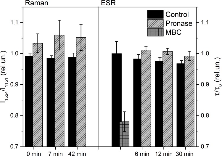
Raman, NMR and EPR spectroscopy and electrophysiology methods were used to investigate the excitability and the packaging of myelin lipid layers and its viscosity during nerve exposure to pronase E. It was established that during exposure of nerve to pronase E the action potential (AP) conduction velocity and the Schwann cell (SC) (or myelin) water ordering increases, but the nerve myelin refractive index and internode incisions numbers decrease. This effect included two periods-short- and long-time period, probably, because the first one depends on SC protein changes and the second one-on the nerve fiber internode demyelination. It was concluded that high electrical resistance of myelin, which is important for a series of AP conduction velocity, not only depends on nerve fiber diameter and the myelin lipid composition, but also on the regularity of myelin lipid fatty acids and myelin lipid layer packing during the axoglial interaction.


Conflict of interest statement

Competing Interests: The authors have declared that no competing interests exist.

Figures

Fig 1
Fig 1
The amplitude (A) and the velocity of AP propagation (B) changes during nerve exposure to pronase E (0,2%). Significant difference (p <0.05).
Fig 2
Fig 2. The phase image parameters of myelin nerve fiber (1–12).
(1–2)—the paranodal areas with Ranvier’s node length; (3–4)—Ranvier’s node diameter; (5–6)–diameter of nerve fiber internodal region (in incisures area); (7–8)—diameter of axon in incisures area; (9–10)—diameter of nerve fiber in internodal region; (1–12)—diameter of axon in internodal region; I—the OPD in Ranvier’s node; II—the OPD in the nerve fiber center; III- OPD in the nerve fiber boundary.
Fig 3
Fig 3. The dynamics of OPD profile changes along the scan-line (μM) through NR.
(A); in internode region of the myelin fiber (B); the perpendicular to the normal nerve fiber and along the nerve fiber (C), during exposure of the nerve to pronase E.
Fig 4
Fig 4
The temperature dependence of nerve fibers: on water T2 (1) and on the intensity ratio I1524/I1151 of carotenoids Raman spectra (2) changes. (Insert) The spin-spin relaxation time of protons (T2) during 40 min nerve exposure to pronase E. (control (nerve exposure to D2O)—white column; nerve exposure—shaded column). The mean value of T2 in the control corresponding to 849 ms is taken as 100%. Significant difference (p <0.05).
Fig 5
Fig 5. Raman spectra of myelin sheath of sciatic nerve fiber.
Raman scattering was excited by 532-nm (A) and 780 nm (B) laser with 0.8 mW power, integration time was 50 s.
Fig 6
Fig 6
Changes in the ratio of I1524/I1151 RS bands of carotenoids during nerve exposure to pronase E (A), and the rotational correlation times (τ) of DS-16 changes during nerve exposure to pronase E and methyl-β-cyclodextrin (10−2 M) (B). Significant difference (p <0.05).
Fig 7
Fig 7. The dynamics of the nerve fatty acids RS peak ratio changes during nerve exposure to pronase E.
Raman scattering was excited by 780 nm laser with power of 0.8 mW, registration time was 50 s. Significant difference (p <0.05).

Similar articles

Cited by

References

    1. Waxman SG, Kocsis JD, Stys PK. The Axon. Structure, function and pathophysiology Oxford Univ. Press; NY-Oxford: 1995; p.325
    1. Chiu SY, Kriegler S. Neurotransmitter-mediated signaling between axons and glial cells. Glia. 1994; 11:191–200 doi: 10.1002/glia.440110213 - DOI - PubMed
    1. Armstrong CM, Bezanilla F, Rojas E. Destruction of sodium conductance inactivation in squid axons perfused with pronase. The Journal of General Physiology. 1973; 62 (4):375–391 - PMC - PubMed
    1. Röper J, Schwarz JR. Heterogeneous distribution of fast and slow potassium channels in myelinated rat nerve fibers. The Journal of Physiology.1989; 416:93–98 - PMC - PubMed
    1. Rojas E, Rudy B. Destruction of the sodium conductance inactivation by a specific protease in perfused nerve fibres from Loligo.The Journal of Physiology.1976; 262 (2):501–531 - PMC - PubMed

LinkOut - more resources