. 2017 Sep 15;216(suppl_4):S499-S507.
doi: 10.1093/infdis/jiw620.

Assessment of Molecular, Antigenic, and Pathological Features of Canine Influenza A(H3N2) Viruses That Emerged in the United States

Affiliations

Assessment of Molecular, Antigenic, and Pathological Features of Canine Influenza A(H3N2) Viruses That Emerged in the United States

Joanna A Pulit-Penaloza et al. J Infect Dis. .

Abstract

Background: A single subtype of canine influenza virus (CIV), A(H3N8), was circulating in the United States until a new subtype, A(H3N2), was detected in Illinois in spring 2015. Since then, this CIV has caused thousands of infections in dogs in multiple states.

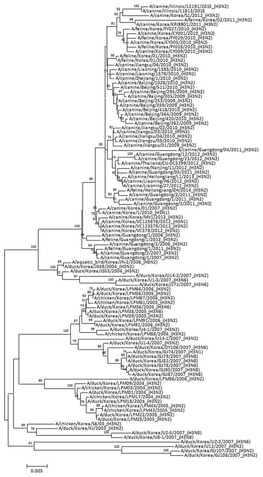
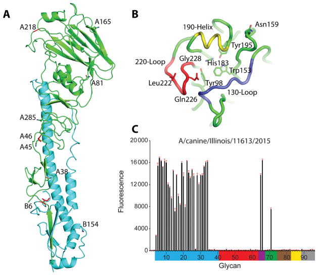
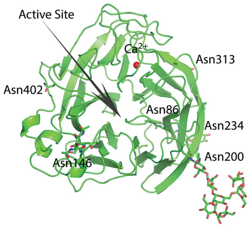
Methods: In this study, genetic and antigenic properties of the new CIV were evaluated. In addition, structural and glycan array binding features of the recombinant hemagglutinin were determined. Replication kinetics in human airway cells and pathogenesis and transmissibility in animal models were also assessed.

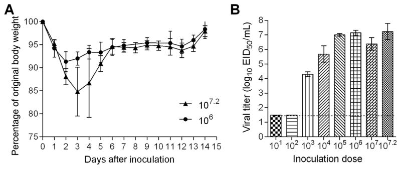
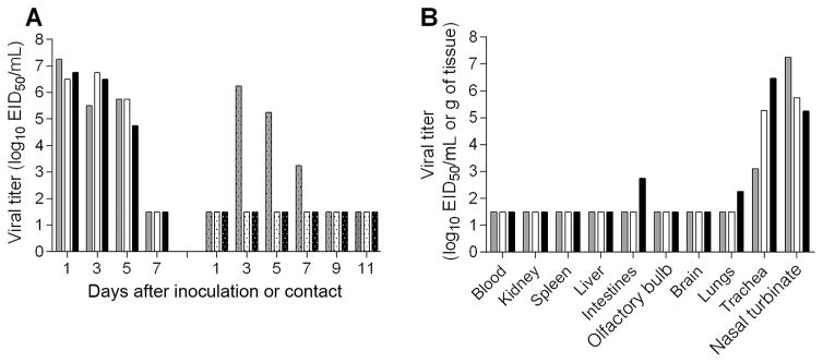
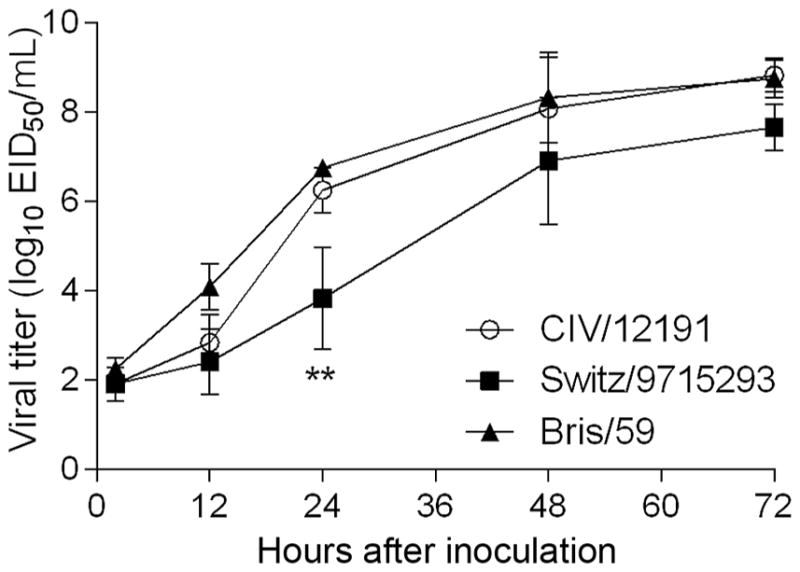
Results: A(H3N2) CIVs maintained molecular and antigenic features related to low pathogenicity avian influenza A(H3N2) viruses and were distinct from A(H3N8) CIVs. The structural and glycan array binding profile confirmed these findings and revealed avian-like receptor-binding specificity. While replication kinetics in human airway epithelial cells was on par with that of seasonal influenza viruses, mild-to-moderate disease was observed in infected mice and ferrets, and the virus was inefficiently transmitted among cohoused ferrets.

Conclusions: Further adaptation is needed for A(H3N2) CIVs to present a likely threat to humans. However, the potential for coinfection of dogs and possible reassortment of human and other animal influenza A viruses presents an ongoing risk to public health.

Keywords: CIV; H3N2; canine influenza; ferrets; transmission.


Conflict of interest statement

Potential conflicts of interest. All authors: No reported conflicts. All authors have submitted the ICMJE Form for Disclosure of Potential Conflicts of Interest. Conflicts that the editors consider relevant to the content of the manuscript have been disclosed.

Figures

Figure 1
Figure 1
Phylogenetic tree of the canine influenza virus hemagglutinin genes. The phylogenetic tree was generated using a general time reversible model and maximum likelihood method with 1000 bootstrap replicates. Bootstrap values of ≥60 are shown at branch nodes. The scale bar represents nucleotide substitutions per site.
Figure 2
Figure 2
Structure and glycan array binding of canine influenza virus hemagglutinin (HA). A, Cartoon representation of the HA monomer. HA1 is green, and HA2 is cyan. The predicted glycosylation sites on HA1 (A38, A45, A81, A165, and A285) and HA2 (B6 and B154) are labeled and shown as sticks. The amino acid differences between A/canine/ IL/12191/2015 and A/canine/IL/11613/2015 viruses are shown in red sticks. B, Binding site elements. The 130-loop is purple, the 190-helix is yellow, and the 220-loop is red. Conserved residues are shown in green sticks. The amino acids discussed in the text are labeled and shown in sticks. C, Glycans on the microarray are grouped according to sialic acid linkage, as follows: α2–3 SA, blue; α2–6 SA, red; α2–6/ α2–3 mixed SA, purple; N-glycolyl SA, green; α2–8 SA, brown; β2–6 and 9-O-acetyl SA, yellow; and asialoglycans, gray (Supplementary Table 2). Error bars reflect the standard error in the signal for 6 independent replicates on the array.
Figure 3
Figure 3
Overall structure of canine influenza virus neuraminidase. The active site is shown with an arrow. Predicted glycosylation sites are shown in sticks.
Figure 4
Figure 4
Pathogenicity of A(H3N2) canine influenza virus in mice. A, Groups of 5 mice were intranasally inoculated with 107.2 or 106.0 50% egg infective doses (EID50) of A/canine/IL/12191/2015 (H3N2) virus and observed for signs of morbidity and mortality for 14 days. The percentage weight loss (±SD) is shown. B, Additional groups of 3 mice were inoculated with 107.2 EID50 or serial 10-fold dilutions ranging from 107.0 to 101.0 EID50 of virus and were euthanized 3 days after inoculation, when lung tissues were collected for viral titer determination. Viral titers are presented as log10 EID50/mL (±SD). The limit of detection is 1.5 log10 EID50/mL.
Figure 5
Figure 5
Pathogenicity and transmission of A(H3N2) canine influenza virus in ferrets. Six ferrets were intranasally inoculated with 107.1 50% egg infective doses (EID50) of A/canine/IL/12191/2015 virus. A, The following day, a serologically naive ferret was placed in the same cage with an inoculated ferret for the assessment of virus transmission between 3 ferret pairs in direct contact. Nasal wash titers from individual ferrets are presented. Viral titers are presented as log10 EID50/mL. B, Three inoculated ferrets were euthanized 3 days after inoculation, and tissues were collected for assessment of viral titers. Blood and nasal turbinate viral titers are presented as log10 EID50/mL, and kidney, spleen, liver, intestines (pooled duodenum, jejunoileal loop, and descending colon), olfactory bulb, brain (pooled anterior and posterior brain), lung, and trachea are presented as log10 EID50/g of tissue. The limit of detection is 1.5 log10 EID50 per g or mL.
Figure 6
Figure 6
Replication kinetics of canine A(H3N2) virus in human airway epithelial (Calu-3) cells. Calu-3 cells were grown on transwell inserts and were inoculated apically in triplicate with a multiplicity of infection of 0.01 of A/canine/ IL/12191/2015 virus (CIV/12191), A/Brisbane/59/2007 (H1N1) virus (Bris/59), or A/ Switzerland/9715293/2013 (H3N2) virus (Switz/9715293). The cells were incubated at 37°C, and culture supernatants were collected 2, 12, 24, 48, and 72 hours after inoculation for viral titer determination in eggs. Statistical significance of the difference between the titers of the canine and each seasonal virus at each time point was determined by 2-way analysis of variance with the Bonferroni post hoc test. No statistically significant difference was observed between CIV/12191 and Bris/59 viruses. **P < .01 for comparison between CIV/12191 and Switz/9715293 viruses.

References

    1. Nikitin A, Cohen D, Todd JD, Lief FS. Epidemiological studies of A-Hong Kong-68 virus infection in dogs. Bull World Health Organ. 1972;47:471–9. - PMC - PubMed
    1. Kilbourne ED, Kehoe JM. Demonstration of antibodies to both hemagglutinin and neuraminidase antigens of H3N2 influenza A virus in domestic dogs. Intervirology. 1975;6:315–8. - PubMed
    1. Crawford PC, Dubovi EJ, Castleman WL, et al. Transmission of equine influenza virus to dogs. Science. 2005;310:482–5. - PubMed
    1. Pecoraro HL, Bennett S, Huyvaert KP, Spindel ME, Landolt GA. Epidemiology and ecology of H3N8 canine influenza viruses in US shelter dogs. J Vet Intern Med. 2014;28:311–8. - PMC - PubMed
    1. Song D, Kang B, Lee C, et al. Transmission of avian influenza virus (H3N2) to dogs. Emerg Infect Dis. 2008;14:741–6. - PMC - PubMed

Publication types

MeSH terms