. 2017 Sep 19;45(16):9388-9397.
doi: 10.1093/nar/gkx563.

Cohesin acetyltransferase Esco2 regulates SAC and kinetochore functions via maintaining H4K16 acetylation during mouse oocyte meiosis

Affiliations

Cohesin acetyltransferase Esco2 regulates SAC and kinetochore functions via maintaining H4K16 acetylation during mouse oocyte meiosis

Yajuan Lu et al. Nucleic Acids Res. .

Abstract

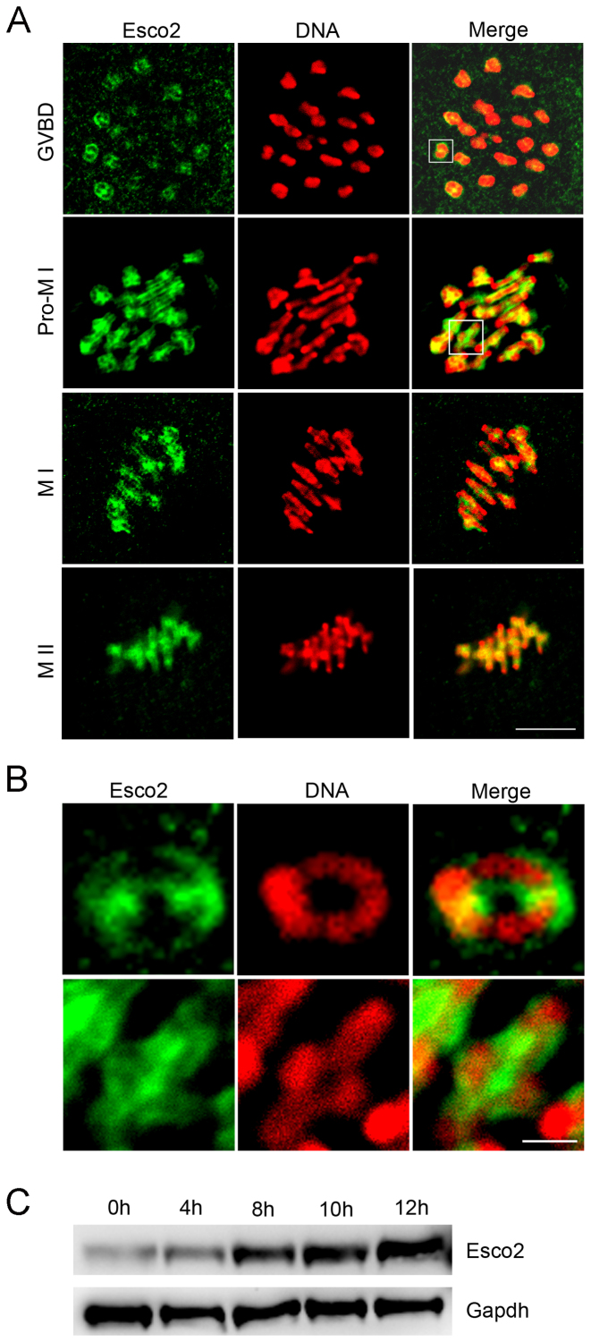
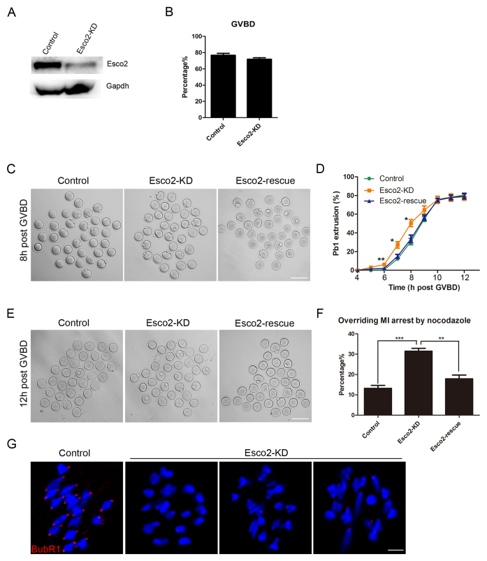
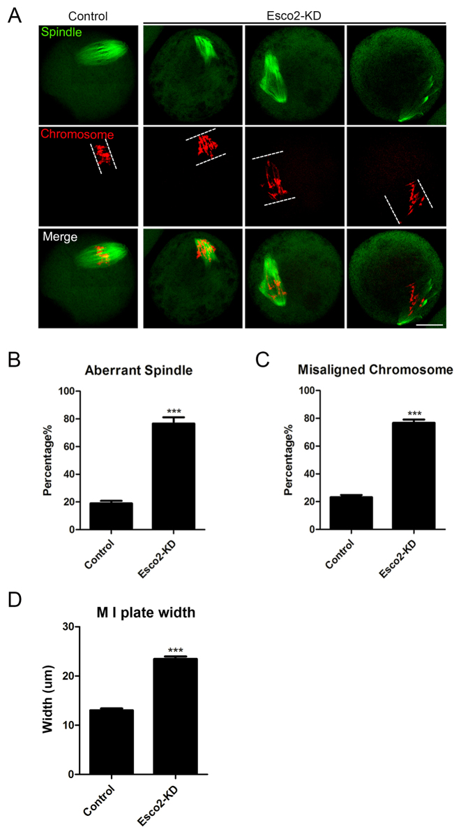
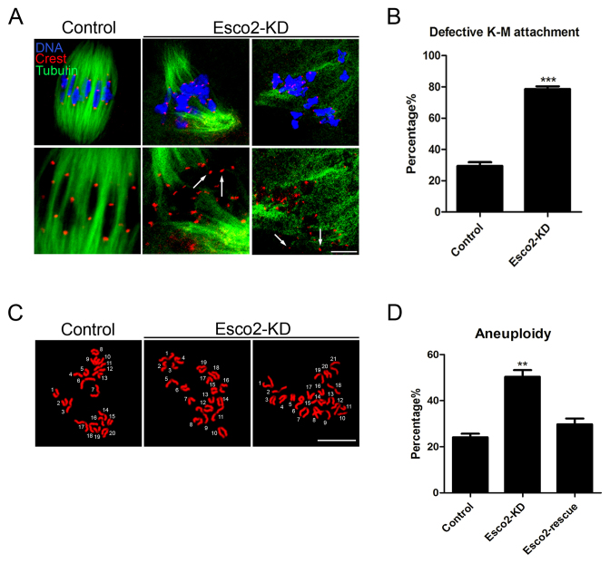
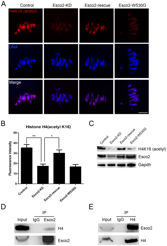
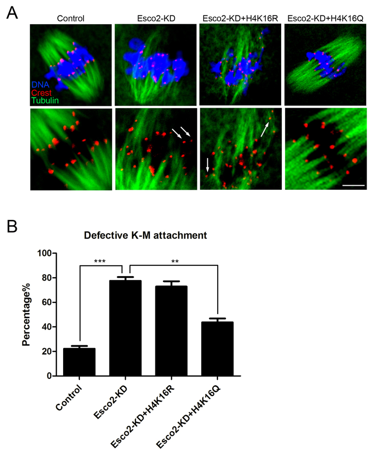
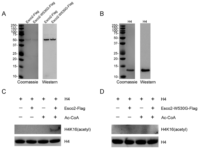
Sister chromatid cohesion, mediated by cohesin complex and established by the acetyltransferases Esco1 and Esco2, is essential for faithful chromosome segregation. Mutations in Esco2 cause Roberts syndrome, a developmental disease characterized by severe prenatal retardation as well as limb and facial abnormalities. However, its exact roles during oocyte meiosis have not clearly defined. Here, we report that Esco2 localizes to the chromosomes during oocyte meiotic maturation. Depletion of Esco2 by morpholino microinjection leads to the precocious polar body extrusion, the escape of metaphase I arrest induced by nocodazole treatment and the loss of BubR1 from kinetochores, indicative of inactivated SAC. Furthermore, depletion of Esco2 causes a severely impaired spindle assembly and chromosome alignment, accompanied by the remarkably elevated incidence of defective kinetochore-microtubule attachments which consequently lead to the generation of aneuploid eggs. Notably, we find that the involvement of Esco2 in SAC and kinetochore functions is mediated by its binding to histone H4 and acetylation of H4K16 both in vivo and in vitro. Thus, our data assign a novel meiotic function to Esco2 beyond its role in the cohesion establishment during mouse oocyte meiosis.


Figures

Figure 1.
Figure 1.
Subcellular localization and expression of Esco2 during mouse meiotic maturation. (A) Mouse oocytes at GVBD, prometaphase I, metaphase I and metaphase II stages were immunolabeled with anti-Esco2 antibody and counterstained with PI. Scale bar, 2.5 μm. (B) The magnified images from panel A. Scale bar, 0.5 μm. (C) Protein levels of Esco2 in oocytes corresponding to GV (0 h), GVBD (4 h), MI (8 h), ATI (10 h) and MII (12 h) stages were examined by western blot.
Figure 2.
Figure 2.
Effects of Esco2 depletion on the meiotic progression and SAC activity in mouse oocytes. (A) Protein levels of Esco2 in control and Esco2-KD oocytes at M I stage were examined by western blot. (B) Quantitative analysis of GVBD rate was shown in control and Esco2-KD oocytes. (C) Representative images of first polar body extrusion(PBE) in control, Esco2-KD and Esco2-rescue oocytes at the time point of 8 h post-GVBD. Scale bar, 200 μm. (D) Quantitative analysis of PBE rate was shown in control, Esco2-KD and Esco2-rescue oocytes at consecutive time points of post-GVBD. (E) Representative images of first PBE in control, Esco2-KD and Esco2-rescue oocytes treated with low dose of nocodazole. Scale bar, 200 μm. (F) The proportion of overriding M I arrest was recorded in control, Esco2-KD and Esco2-rescue oocytes. (G) Localization of BubR1 at prometaphase I stage in control and Esco2-KD oocytes. Scale bar, 2.5 μm. Data of (B), (D) and (F) were presented as mean percentage (mean ± SEM) of at least three independent experiments. Asterisk denotes statistical difference at a P < 0.05 level of significance.
Figure 3.
Figure 3.
Depletion of Esco2 causes spindle/chromosome abnormalities in mouse oocytes. (A) Representative images of spindle morphologies and chromosome alignment in control and Esco2-KD oocytes. Scale bar, 20 μm. (B) The proportion of abnormal spindles was recorded in control and Esco2-KD oocytes. (C) The proportion of misaligned chromosomes was recorded in control and Esco2-KD oocytes. (D) The width of M I plate was measured in control and Esco2-KD oocytes. Data of (B–D) were presented as mean percentage (mean ± SEM) of at least three independent experiments. Asterisk denotes statistical difference at a P < 0.05 level of significance.
Figure 4.
Figure 4.
Depletion of Esco2 compromises K-M attachments and generates aneuploidy in mouse oocytes. (A) Representative images of kinetochore-microtubule attachments in control and Esco2-KD oocytes. Oocytes were immunostained with anti-α-tubulin-FITC antibody to visualize spindles, with CREST to visualize kinetochores and counterstained with Hoechst to visualize chromosomes. Scale bar, 5 μm. (B) The rate of defective kinetochore-microtubule attachments was recorded in control and Esco2-KD oocytes. (C) Representative images of euploid and aneuploid MII eggs. Chromosome spread was performed to count the number of chromosomes in control and Esco2-KD oocytes. Chromosomes were counterstained with Hoechst. Scale bar, 2.5 μm. (D) The rate of aneuploid eggs was recorded in control and Esco2-KD oocytes. Data of (B and D) were presented as mean percentage (mean ± SEM) of at least three independent experiments. Asterisk denotes statistical difference at a P < 0.05 level of significance.
Figure 5.
Figure 5.
Esco2 binds to histone H4 to regulate the acetylation level of H4K16. (A) Representative images of acetylated H4K16 in control, Esco2-KD, Esco2-rescue and Esco2-W530G oocytes. Scale bar, 5 μm. (B) The immunofluorescence intensity of acetylated H4K16 was recorded in control, Esco2-KD, Esco2-rescue and Esco2-W530G oocytes. Data were presented as mean percentage (mean ± SEM) of at least three independent experiments. Asterisk denotes statistical difference at a P < 0.05 level of significance. (C) Acetylation levels of H4K16 in control, Esco2-KD, Esco2-rescue and Esco2-W530G oocytes were examined by western blotting. (D) IP was performed with the Esco2 antibody, and the blots of IP eluate were probed with anti-Esco2 and anti-histone H4 antibodies, respectively. (E) IP was performed with the histone H4 antibody, and the blots of IP eluate were probed with anti-Esco2 and anti-histone H4 antibodies, respectively.
Figure 6.
Figure 6.
H4K16Q rescues the defect of K-M attachments in Esco2-depleted oocytes. (A) Representative images of kinetochore-microtubule attachments in control, Esco2-KD, Esco2-KD+H4K16R and Esco2-KD+H4K16Q oocytes. Scale bar, 5 μm. (B) The rate of defective kinetochore-microtubule attachments was recorded in control, Esco2-KD, Esco2-KD+H4K16R and Esco2-KD+H4K16Q oocytes. Data were presented as mean percentage (mean ± SEM) of at least three independent experiments. Asterisk denotes statistical difference at a P < 0.05 level of significance.
Figure 7.
Figure 7.
Esco2 acetylates histone H4 at Lys16 in vitro. (A) Flag purification of Esco2. Esco2 and enzymatically mutant Esco2-W530G were expressed in HEK293 cells and then purified according to the Flag purification procedure. Purified Esco2-Flag and Esco2-W530G-Flag were detected with sodium dodecyl sulphate-polyacrylamide gel electrophoresis followed by both coomassie staining and western blotting with anti-Esco2 antibody. (B) Commercially obtained recombinant histone H4 was identified by both coomassie staining and western blotting with anti-H4 antibody. (C) In vitro acetylation assay with Esco2-Flag. Recombinant histone H4 was incubated with or without purified Esco2-Flag and Ac-CoA in the acetyltransferase assay buffer at 30°C for 1 h. The reactions were analyzed by western blotting with anti-histone H4 (acetyl K16) antibody for acetylation levels of H4K16 and anti-histone H4 antibody as a loading control. (D) In vitro acetylation assay with Esco2-W530G-Flag. Recombinant histone H4 was incubated with or without purified Esco2-W530G-Flag and Ac-CoA in the acetyltransferase assay buffer at 30°C for 1 h. The reactions were analyzed by western blotting with anti-histone H4 (acetyl K16) antibody for acetylation levels of H4K16 and anti-histone H4 antibody as a loading control.

References

    1. Xiong B., Gerton J.L.. Regulators of the cohesin network. Annu. Rev. Biochem. 2010; 79:131–153. - PubMed
    1. Onn I., Heidinger-Pauli J.M., Guacci V., Unal E., Koshland D.E.. Sister chromatid cohesion: a simple concept with a complex reality. Annu. Rev. Cell Dev. Biol. 2008; 24:105–129. - PubMed
    1. Nasmyth K., Haering C.H.. Cohesin: its roles and mechanisms. Annu. Rev. Genet. 2009; 43:525–558. - PubMed
    1. Saito Y., Zhou H., Kobayashi J.. Chromatin modification and NBS1: their relationship in DNA double-strand break repair. Genes Genet. Syst. 2016; 90:195–208. - PubMed
    1. Rolef Ben-Shahar T., Heeger S., Lehane C., East P., Flynn H., Skehel M., Uhlmann F.. Eco1-dependent cohesin acetylation during establishment of sister chromatid cohesion. Science. 2008; 321:563–566. - PubMed

LinkOut - more resources