. 2017 Sep 19;45(16):9625-9639.
doi: 10.1093/nar/gkx570.

The important conformational plasticity of DsrA sRNA for adapting multiple target regulation

Affiliations

The important conformational plasticity of DsrA sRNA for adapting multiple target regulation

Pengzhi Wu et al. Nucleic Acids Res. .

Abstract

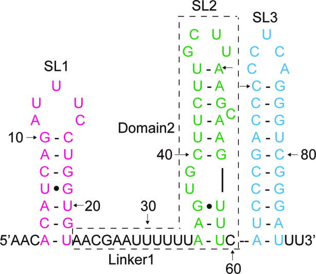
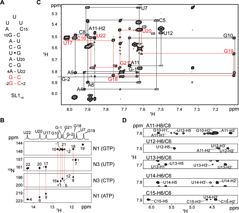
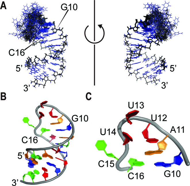
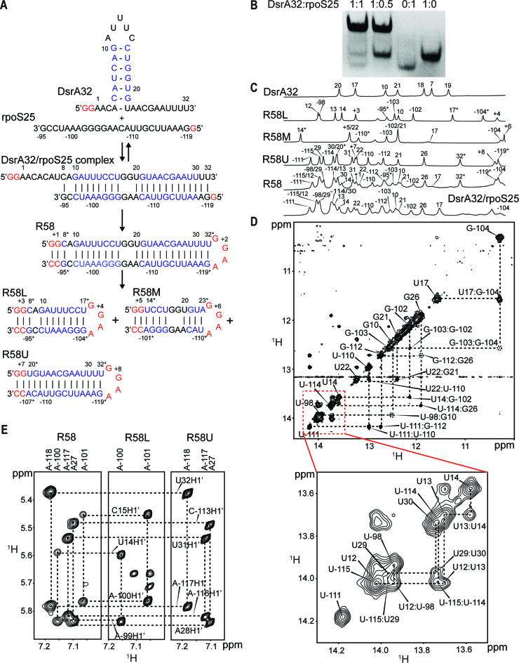
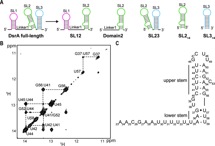
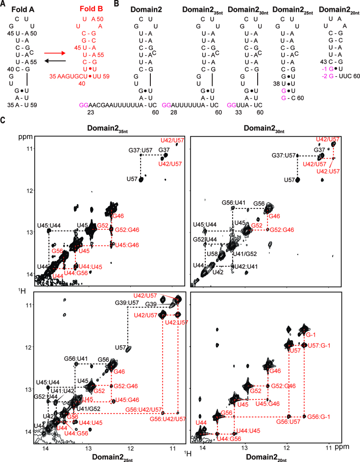
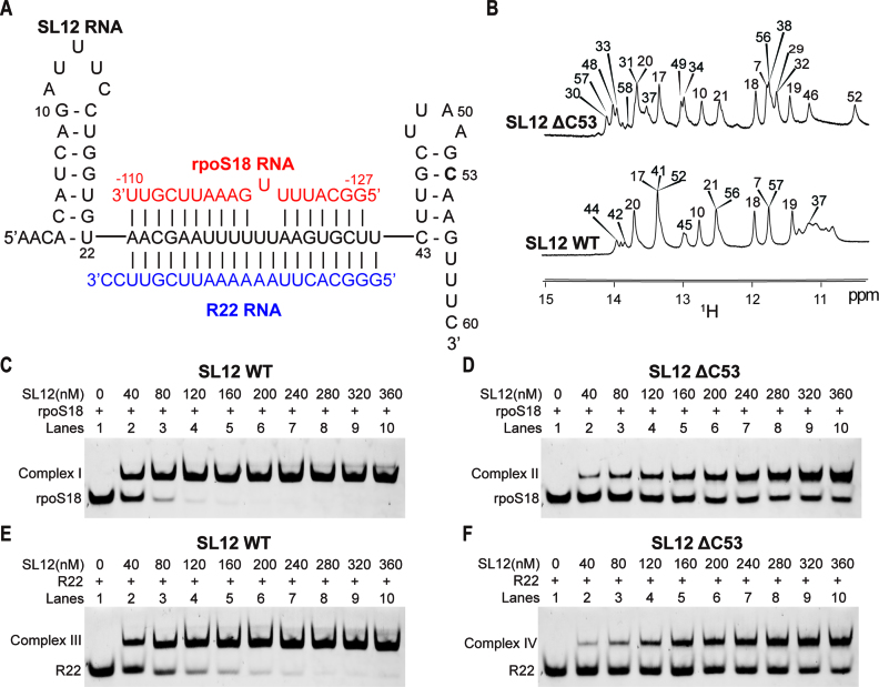
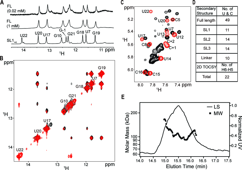
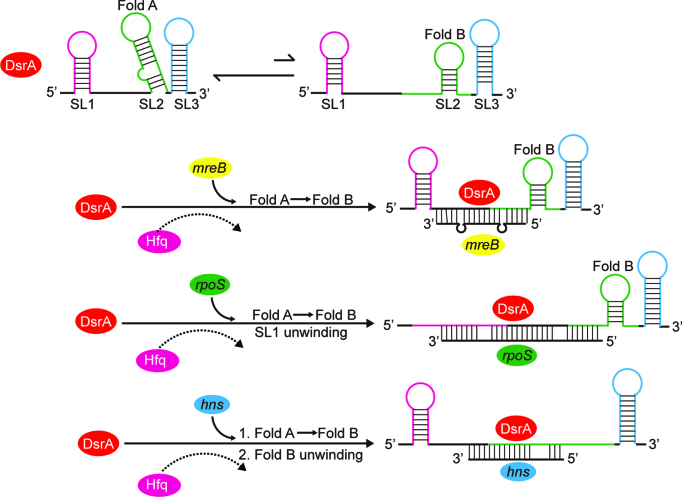
In bacteria, small non-coding RNAs (sRNAs) could function in gene regulations under variable stress responses. DsrA is an ∼90-nucleotide Hfq-dependent sRNA found in Escherichia coli. It regulates the translation and degradation of multiple mRNAs, such as rpoS, hns, mreB and rbsD mRNAs. However, its functional structure and particularly how it regulates multiple mRNAs remain obscure. Using NMR, we investigated the solution structures of the full-length and isolated stem-loops of DsrA. We first solved the NMR structure of the first stem-loop (SL1), and further studied the melting process of the SL1 induced by the base-pairing with the rpoS mRNA and the A-form duplex formation of the DsrA/rpoS complex. The secondary structure of the second stem-loop (SL2) was also determined, which contains a lower stem and an upper stem with distinctive stability. Interestingly, two conformational states of SL2 in dynamic equilibrium were observed in our NMR spectra, suggesting that the conformational selection may occur during the base-pairing between DsrA and mRNAs. In summary, our study suggests that the conformational plasticity of DsrA may represent a special mechanism sRNA employed to deal with its multiple regulatory targets of mRNA.


Figures

Figure 1.
Figure 1.
Schematic of the secondary structure of DsrA. Three stem–loops are colored magenta, green and cyan. The DsrA-Domain2 consists of nucleotides in the dashed box. The single strand linker between SL1 and SL2 is labeled as Linker1. Watson–Crick hydrogen bonds are indicated by continuous lines and G•U wobble base pairs are denoted by a circle.
Figure 2.
Figure 2.
Representative NMR spectra collected for SL1+4. (A) Secondary structure of SL1. SL1+4 is the construct derived from the wild-type (SL1 WT, residues 4–22) and used for structural determination by NMR. Two nonnative G–C base pairs (colored in red) are added to the SL1 WT to increase the yield of RNA transcription. (B) Imino proton region of 1D NMR spectrum and 2D 1H–15N HNN-COSY reveals the base pairs of SL1+4. Imino protons and their hydrogen-bonded nitrogen atoms in the canonical base pairs are shown in red bash lines. In HNN-COSY, the cross-peaks are labeled by the residue number. (C) Portion of 2D NOESY spectrum obtained for unlabeled SL1+4 showing cross-peaks between aromatic H6/H8 protons and ribose H1’ protons. Sequential NOE connectivities are indicated with lines. The base-H1’ sequential walk is traced in black on the 5′-half (G-2 to U12), red on the 3′-half (C16 to U22) and a black dashed line on A11-H2. G-2 falls outside the spectral region shown. The cross-peaks are labeled with the one-letter nucleotide code and the residue number. (D) Expansion of planes corresponding to C6/C8 regions of the loop residues A11 to C15 taken from 3D 1H–13C NOESY-HSQC spectrum collected from uniformly 13C,15N-labeled SL1+4. Unlabeled peaks are due to partially overlapping resonances from other spin systems.
Figure 3.
Figure 3.
NMR solution structure of SL1+4. (A) Superposition of the 20 lowest energy structures. The base and ribose are colored blue and black, respectively. (B) The lowest-energy structure of SL1+4 represented as a ribbon-and-stick model. Nucleotides are colored blue (guanosine), green (cytosine), orange (adenine) and red (uracil). (C) Close-up views of the AUUUC pentaloop.
Figure 4.
Figure 4.
Translation regulation of rpoS by DsrA. (A) RNA constructs used in the present studies. DsrA32 includes nucleotides from 1 to 32 of DsrA, and rpoS25 includes nucleotides from –119 to –95 of rpoS. R58L, R58M and R58U RNAs are derived from the lower stem, middle stem and upper stem of the R58 RNA, respectively. The red letters indicate non-native nucleotides; the blue letters indicate base pairs of which imino proton resonances were observed and assigned in 2D 1H–1H NOESY spectra. The nucleotides with different adjacent nucleotides from DsrA32/rpoS25 complex are labeled by stars. (B) Native PAGE shows that DsrA32 and rpoS25 form a stable heterodimer at 1:1 DsrA32:rpoS25 molar ratios. (C) 1D imino proton spectra of the RNA constructs. From top to bottom are free DsrA32, R58L, R58M, R58U, R58 and DsrA32/rpoS25 complex, respectively. The assignments of all RNA constructs are labeled by the sequence number. (D) Imino proton region of the NOESY spectrum (in 90% H2O/10% D2O) of DsrA32/rpoS25 complex recorded at 900 MHz spectrometer. (E) Portions of the 2D NOESY spectra (in 100% D2O) showing the cross-peaks of adenosine-H2 and ribose-H1’ observed for (from left to right) R58, R58L and R58U RNAs. Adenosine-H2 and ribose-H1’ assignments are labeled vertically and horizontally, respectively.
Figure 5.
Figure 5.
NMR studies of DsrA-Domain2. (A) The constructs of DsrA used for this study. SL1, SL2 and SL3 are colored magenta, green and cyan, respectively. From the left, DsrA full-length, SL12, Domain2, SL23, SL2+4 and SL3+4. Two nonnative G–C base pairs are added to the SL2 and SL3. (B) Portion of 2D 1H–1H NOESY spectrum of Domain2. (C) Secondary structure of Domain2 derived from NMR spectra.
Figure 6.
Figure 6.
Bistable structure of SL2. (A) Schematic of bistable secondary structures of SL2. Nucleotides of Fold A are colored black, and Fold B are colored red. (B) RNA constructs used in the present studies. Non-native nucleotides are shown in magenta. (C) Imino-to-imino NOEs cross-peaks of Domain235nt (top left), Domain230nt (top right), Domain225nt (bottom left), Domain220nt (bottom right) with color-coded signal assignments.
Figure 7.
Figure 7.
The effects of DsrA structure on mRNAs binding affinities. (A) Schematic of the RNA constructs used for this study. SL12 RNA contains the first two stem–loops of DsrA (nucleotides 1–60). C53 is labeled in bold letter and the residue number. SL12, rpoS18 and R22 RNAs are colored black, red and blue, respectively. (B) 1D imino proton spectra of SL12 ΔC53 mutant (top) and SL12 wild type (WT) (bottom). Imino proton resonances are labeled by the residue number. Using gel mobility shift assays, in vitro binding of rpoS18 RNA and SL12 WT (C) or ΔC53 mutant (D) was performed as described in Materials and Methods. 200 nM of 5′-FAM-labeled rpoS18 RNA was incubated with increasing concentrations of unlabeled SL12 WT or ΔC53 mutant (final concentrations above the lanes). Following 20 min incubation on ice, samples were run on a native 10% gel. The same experimental procedure as above but with 5′-FAM-labeled R22 RNA and increasing concentrations of unlabeled SL12 WT (E) or ΔC53 mutant (F).
Figure 8.
Figure 8.
NMR structural studies of full-length DsrA. (A) Comparison of the imino regions of 1D spectra of full-length DsrA (FL) and SL1+4. The imino proton of U22 was not observed in full-length DsrA due to rapid exchange with solvent, while it is protected by two G–C base pairs in SL1+4. (B) Overlap of imino-to-imino regions of 2D 1H–1H NOESY of SL1+4 (red) and full-length DsrA (1 mM, black). The resonances are labeled by the one-letter nucleotide code and the residue number. (C) Overlap of H6-to-H5 regions of 2D 1H-1H TOCSY spectrum of SL1+4 (red) and full-length DsrA (1 mM, black). (D) Table of the number of uracils and cytosines in the secondary structure and number of H6-H5 cross-peaks in 2D 1H–1H TOCSY of full-length DsrA. (E) MALS measurement from SEC of 20 μM full-length DsrA at a flow rate of 0.75 ml/min. Light scattering is shown as a function of elution time (solid line, right axis). Calculated molar mass is shown for the peak (square, left axis).
Figure 9.
Figure 9.
Model of structural selection of DsrA sRNA for adapting multiple target regulation. Three stem–loops of DsrA are colored magenta, green and blue, respectively. The single strand linkers between stem–loops are colored black. DsrA rearranges alternative conformations to adapt to different mRNAs, such as mreB, rpoS and hns mRNAs. The recognition of mRNAs is stimulated by binding of Hfq to free DsrA, followed by the release of Hfq from the sRNA/mRNA complex.

Similar articles

Cited by

References

    1. Waters L.S., Storz G.. Regulatory RNAs in bacteria. Cell. 2009; 136:615–628. - PMC - PubMed
    1. Gottesman S., Storz G.. Bacterial small RNA regulators: versatile roles and rapidly evolving variations. Cold Spring Harbor Perspecti. Biol. 2011; 3:a003798. - PMC - PubMed
    1. Storz G., Vogel J., Wassarman K.M.. Regulation by small RNAs in bacteria: expanding frontiers. Mol. Cell. 2011; 43:880–891. - PMC - PubMed
    1. Gottesman S. The small RNA regulators of Escherichia coli: roles and mechanisms*. Annu. Rev. Microbiol. 2004; 58:303–328. - PubMed
    1. Jousselin A., Metzinger L., Felden B.. On the facultative requirement of the bacterial RNA chaperone, Hfq. Trends Microbiol. 2009; 17:399–405. - PubMed

MeSH terms

LinkOut - more resources