. 2017 Sep 19;45(16):9654-9678.
doi: 10.1093/nar/gkx606.

hnRNP L controls HPV16 RNA polyadenylation and splicing in an Akt kinase-dependent manner

Affiliations

hnRNP L controls HPV16 RNA polyadenylation and splicing in an Akt kinase-dependent manner

Naoko Kajitani et al. Nucleic Acids Res. .

Abstract

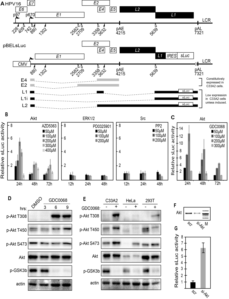
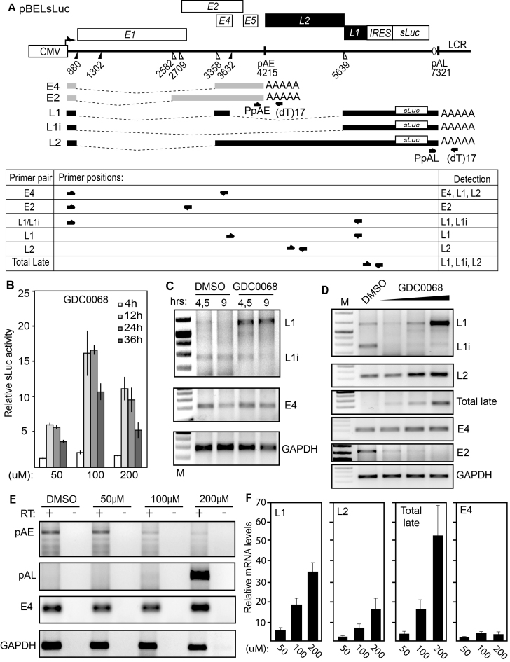
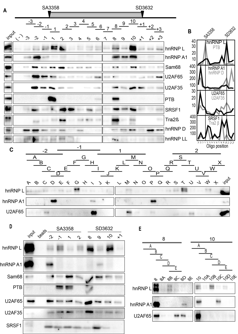
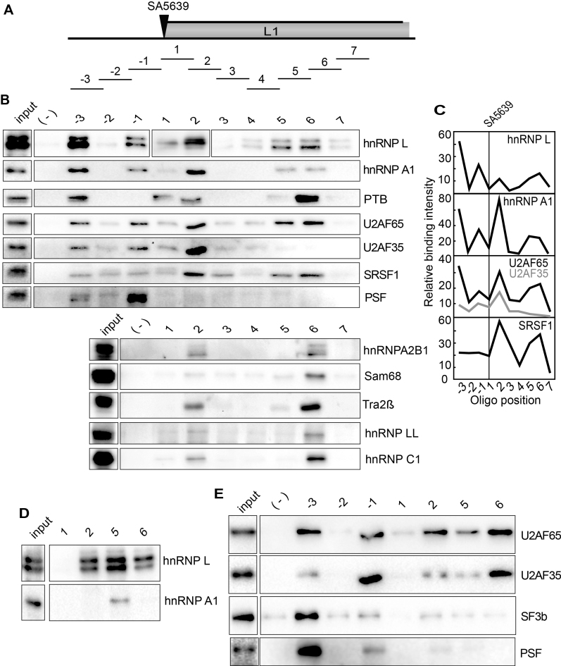
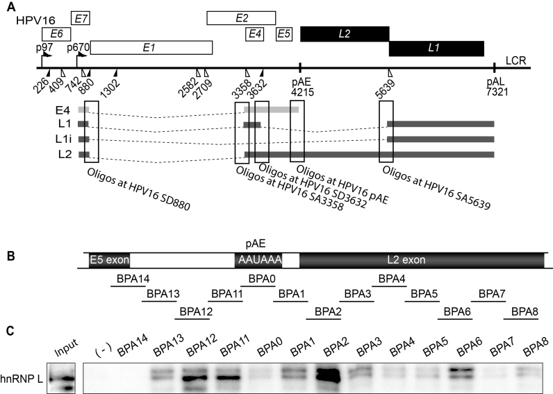
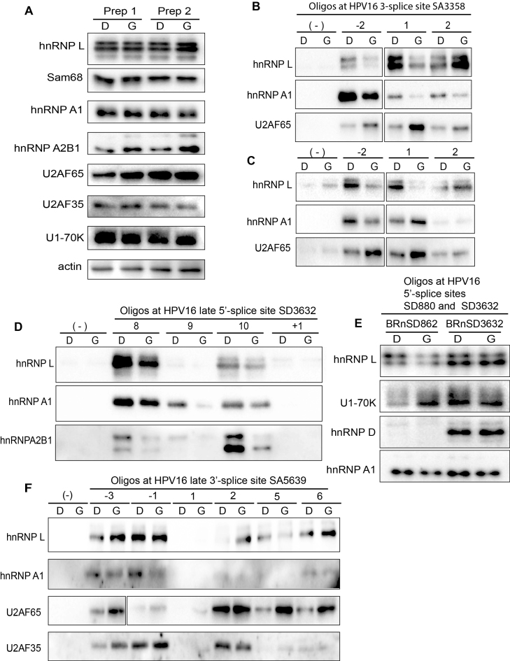
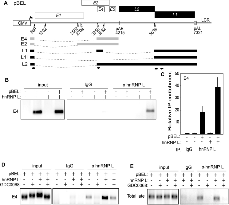
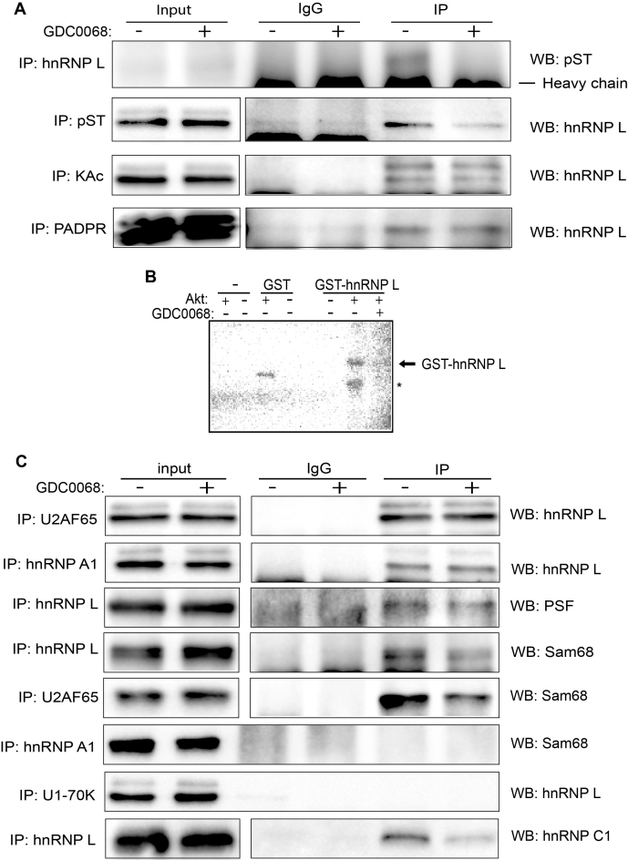
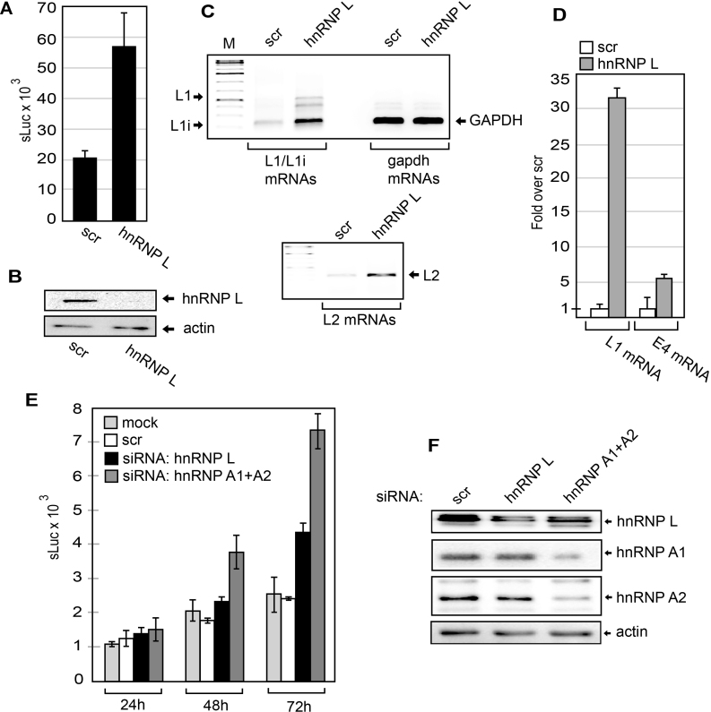
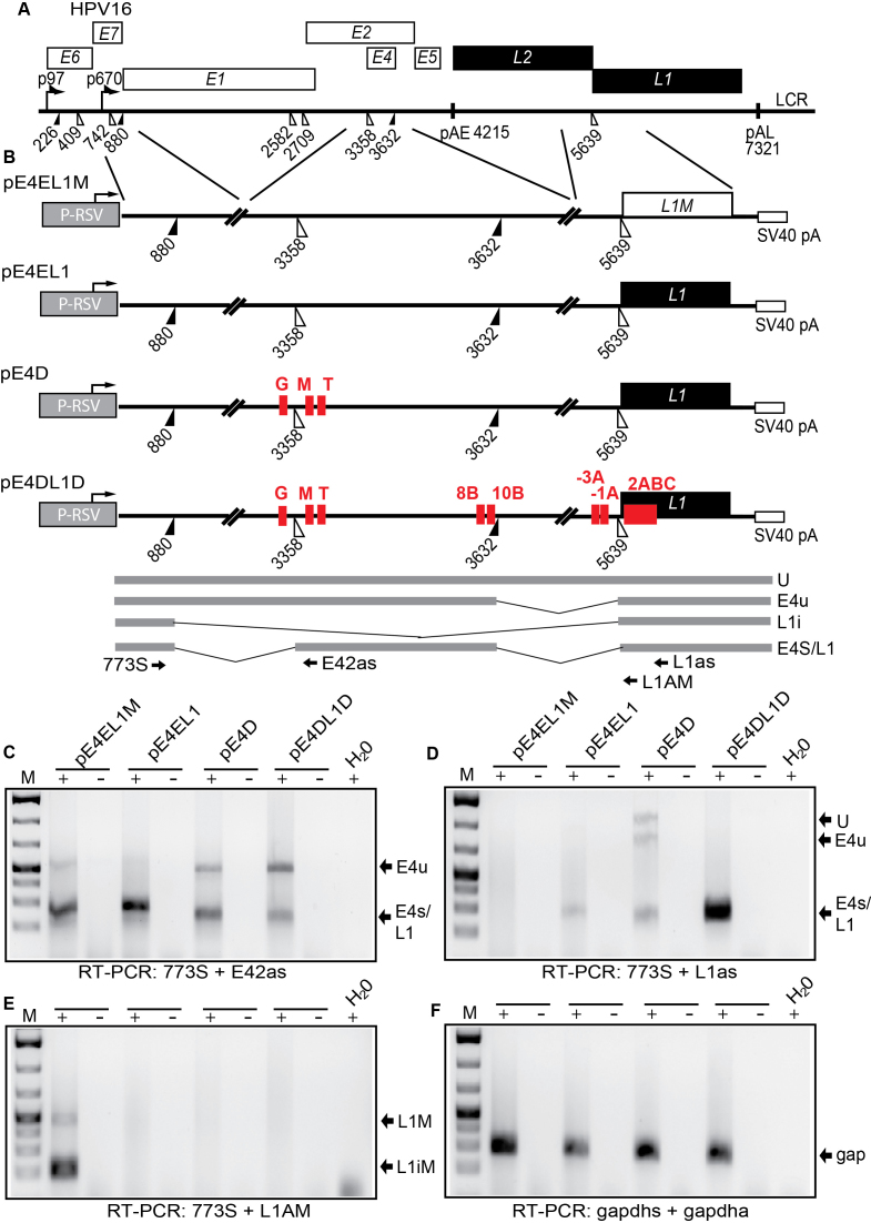
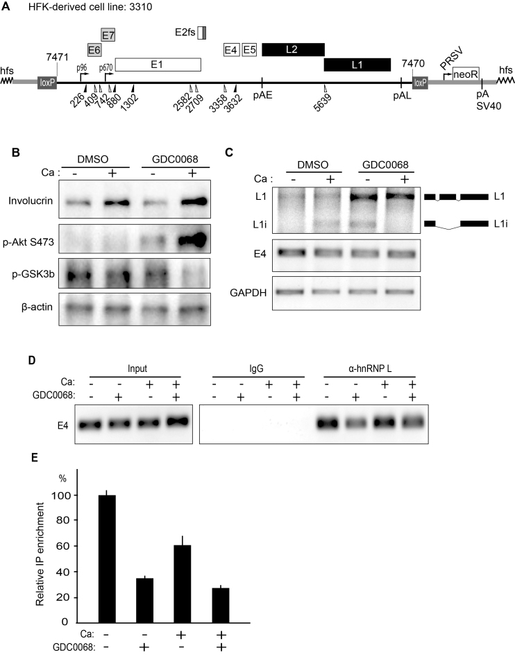
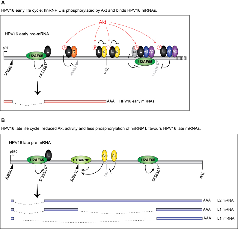
Inhibition of the Akt kinase activates HPV16 late gene expression by reducing HPV16 early polyadenylation and by activating HPV16 late L1 mRNA splicing. We identified 'hot spots' for RNA binding proteins at the early polyA signal and at splice sites on HPV16 late mRNAs. We observed that hnRNP L was associated with sequences at all HPV16 late splice sites and at the early polyA signal. Akt kinase inhibition resulted in hnRNP L dephosphorylation and reduced association of hnRNP L with HPV16 mRNAs. This was accompanied by an increased binding of U2AF65 and Sam68 to HPV16 mRNAs. Furthermore, siRNA knock-down of hnRNP L or Akt induced HPV16 gene expression. Treatment of HPV16 immortalized keratinocytes with Akt kinase inhibitor reduced hnRNP L binding to HPV16 mRNAs and induced HPV16 L1 mRNA production. Finally, deletion of the hnRNP L binding sites in HPV16 subgenomic expression plasmids resulted in activation of HPV16 late gene expression. In conclusion, the Akt kinase inhibits HPV16 late gene expression at the level of RNA processing by controlling the RNA-binding protein hnRNP L. We speculate that Akt kinase activity upholds an intracellular milieu that favours HPV16 early gene expression and suppresses HPV16 late gene expression.


Figures

Figure 1.
Figure 1.
(A) Schematic representation of the HPV16 genome. Rectangles represent open reading frames, promoters p97 and p670 are indicated as arrows, filled and open triangles represent 5′- and 3′-splices sites respectively, HPV16 early and late polyA signals pAE and pAL are indicated. Below the HPV16 genome, a schematic representation of the pBELsLuc reporter plasmid stably integrated in the genome of the C33A2 cells. Transcription of the HPV16 sequences in the pBELsLuc plasmid is driven by the human cytomegalovirus promoter (CMV). The sLuc gene inserted into the L1 region is indicated and is preceded by the poliovirus 2A internal ribosome entry site (IRES). HPV16 E2 and E4 mRNAs mRNAs produced by C33A2 cells are indicated in light grey and HPV16 late mRNAs encoding sLuc that can be induced in this reporter cell line are indicated in black. (B and C) Fold induction of secreted luciferase enzyme activity (sLuc) in the cell culture medium of reporter cell line C33A2 treated with various concentrations of the indicated inhibitors over DMSO treated cells. sLuc activity was monitored at the indicated time points. sLuc activity is displayed as fold over DMSO-treated C33A2 cells at the various time points. (B) Results show the effect on C33A2 cells of inhibitors to the cellular kinases Akt (AZD5363), ERK1/2 (PDO325901) and Src (PP2). (C) Induction of sLuc activity by the Akt kinase inhibitor GDC0068. (D). Western blot of the Akt kinase, various phosphorylated versions of the Akt kinase and phosphorylated GSK3b in C33A2 cells in the absence or presence of Akt kinase inhibitor GDC0068 (100 uM) at different time points. (E) Western blot of the same kinases as in (D) in various cell lines treated with Akt kinase inhibitor GDC0068. Note that Akt kinase inhibitor GDC0068 locks the Akt kinase in an inactive, but phosphorylated state appearing as a band detected with antibodies to phosphorylated forms of Akt. (F) RT-PCR on total RNA extracted from C33A2 cells of the Akt1 mRNA in C33A2 cells transfected with siRNAs to the Akt1 kinase (si-Akt), compared to cells transfected with scrambled siRNAs (scr). (G) Relative sLuc activity induced by transfection of C33A2 cells with siRNAs to the Akt1 kinase (si-Akt), compared to cells transfected with scrambled siRNAs (scr).
Figure 2.
Figure 2.
(A) Schematic representation of the pBELsLuc reporter plasmid stably integrated in the genome of the C33A2 reporter cell line. Transcription of the HPV16 sequences in the pBELsLuc plasmid is driven by the human cytomegalovirus promoter (CMV). The sLuc gene inserted into the L1 region is indicated and is preceded by the poliovirus 2A internal ribosome entry site (IRES). HPV16 E2 and E4 mRNAs mRNAs produced by C33A2 cells are indicated in light grey (E2 and E4 mRNAs) and HPV16 late mRNAs encoding sLuc that can be induced in this reporter cell line are indicated in black. The location of RT-PCR primers is indicated below. (B) Secreted luciferase enzyme activity (sLuc) in the cell culture medium of reporter cell line C33A2 treated with various concentrations of the Akt kinase inhibitor GDC0068. sLuc activity was monitored at the indicated time points. sLuc activity is displayed as fold over DMSO-treated C33A2 cells at the various time points. (C) RT-PCR on total RNA extracted from the C33A2 reporter cell line treated with DMSO alone or Akti kinase inhibitor GDC0068 for 4.5hrs or 9hrs. The RT-PCR primers detected HPV16 L1/L1i-, E4- or GAPDH-mRNAs. (D) RT-PCR on total RNA extracted from the C33A2 reporter cell line treated with DMSO alone or various concentrations of Akt kinase inhibitor GDC0068 (50-, 100- or 200uM) for 9hrs. The RT-PCR primers detected HPV16 L1/L1i-, L2-, total late (L2 + L1 +L1i), E4-, E2- or GAPDH-mRNAs. (E) A 3′-RACE assay on total RNA extracted from C33A2 cells. Primers were F-Set2 and (dT)17-P3 for pAE and sLuc-S-inner and (dT)17-P3 for pAL (for primers see Supplementary Table S1). Spliced HPV16 E4 mRNAs and cellular GAPDH mRNAs are also shown. (F) RT-qPCR on the cDNA samples used for RT-PCR in Figure 2D.
Figure 3.
Figure 3.
(A) Upper panel: Schematic drawing of HPV16 exon 4 and the 35-nucleotide, biotinylated ssDNA oligos (overlapping by 5-nucleotides) used in pull down assays. Location of HPV16 3′-splice site SA3358 and 5′-splice site SD3632 are indicated. Lower panel: Pull downs of cellular factors from nuclear extracts using the indicated ssDNA oligos covering the E4 exon of HPV16 followed by Western blot analysis using antibodies to proteins indicated to the right. (-); mock pull downs using streptavidin beads in the absence of oligo. (B) Quantitation of some of the Western blots of the pull downs in (A). (C) Upper panel: Schematic drawing of shorter oligos (A-X) designed to better map binding sites for hnRNP L, hnRNP A1 and U2AF65. Lower panel: Western blots for hnRNP L, hnRNP A1 and U2AF65 of on proteins pulled down by the shorter biotinylated ssDNA oligos. (D) Western blots of indicated proteins on pull downs using biotinylated ssRNA oligos of a subset of the ssDNA oligos shown in (A). (E) Upper panel: Schematic drawing of shorter oligos (A–E) of the two original 35-nucleotide oligos 8 and 10 located near HPV16 late 5′-splice site SD3632. Lower panel: Western blots for hnRNP L, hnRNP A1 and U2AF65 of on proteins pulled down by the shorter biotinylated ssDNA oligos.
Figure 4.
Figure 4.
(A) Schematic drawing of the HPV16 region around late 3′-splice site SA5639 and the 35-nucleotide biotinylated ssDNA oligos (overlapping by 5-nucleotides) used in pull down assays. Location of HPV16 3′-splice site SA5639 is indicated. (B) Upper and lower panels show pull downs of cellular factors from nuclear extracts using the indicated ssDNA oligos covering the region of HPV16 late 3′-splice site SA5639 followed by Western blot analysis using antibodies indicated to the right. (–) mock pull downs using streptavidin beads in the absence of oligo. (C) Quantitation of some of the Western blots of the pull downs in (B). (D and E) Western blots of indicated proteins on pull downs using biotinylated ssRNA oligos of a subset of the ssDNA oligos shown in (B).
Figure 5.
Figure 5.
(A) Schematic representation of the HPV16 genome. Rectangles represent open reading frames, promoters p97 and p670 are indicated as arrows, filled and open triangles represent 5′- and 3′-splices sites respectively, HPV16 early and late polyA signals pAE and pAL are indicated. The regions on the HPV16 mRNAs from which the ssDNA and ssRNA oligos are derived are boxed. (B) Schematic drawing of the HPV16 region around early polyA signal pAE and the 35-nucleotide, biotinylated ssDNA oligos (overlapping by 5-nucleotides) used in pull down assays. Location of HPV16 early polyA signal pAE is indicated as well as E5 and L2 coding regions. (C) Pull downs of cellular factors from nuclear extracts using the indicated ssDNA oligos spanning HPV16 pAE followed by Western blot for hnRNP L. (-); mock pull downs using streptavidine beads in the absence of oligo.
Figure 6.
Figure 6.
(A) Western blots of various RNA binding proteins in two independent preparations of nuclear extracts (Prep 1 and Prep 2) prepared from C33A2 cells treated with DMSO (D) or 100 uM Akt kinase inhibitor GDC0068 (G) for 3 h. Western blots of indicated proteins pulled down with biotinylated ssDNA oligos (B) or RNA oligos (C) representing sequences at the HPV16 3′-splice site SA3358 in the E4-coding exon. (D) Western blots of indicated proteins pulled down with biotinylated ssDNA oligos representing sequences at the HPV16 late 5′-splice site SD3632 in the E4-coding exon. (E) Western blots of indicated proteins pulled down with biotinylated ssRNA oligos spanning HPV16 5′-splice sites SD880 (BRnSD862) and SD3632 (BRnSD3632). (F) Western blots of hnRNP L, hnRNP A1, U2AF65 or U2AF35 pulled down from nuclear extracts using ssDNA oligos at HPV16 late splice site SA5639. For all pull downs in the figure, nuclear extracts prepared from C33A2 cells treated with DMSO (D) or Akt kinase inhibitor GDC0068 (G) were used.
Figure 7.
Figure 7.
(A) Schematic representation of the HPV16 subgenomic expression plasmid pBEL. Transcription of the HPV16 sequences in the pBEL plasmid is driven by the human cytomegalovirus promoter (CMV). Rectangles represent open reading frames. Filled and open triangles represent 5′- and 3′-splices sites respectively, and HPV16 early and late polyA signals are indicated as pAE and pAL respectively. The HPV16 E4 and E2 mRNAs produced by pBEL upon transfection of mammalian cells is indicated in light grey. HPV16 late mRNAs L1, L1i and L2 that can be induced from the pBEL reporter plasmid are indicated in black. Positions of HPV16 RT-PCR primers are indicated. (B) Left panel: RT-PCR of HPV16 E4 mRNAs spliced from SD880 to SA3358 in 293T cells transfected with or without HPV16 subgenomic plasmid pBEL in the absence or presence of hnRNP L expressing plasmid pCMV-hnRNP L. The transfected cells were subjected to UV irradiation before harvest as detailed in Materials and methods for CLIP assay. Right panel: The extracts from the transfected cells were subjected to immunoprecipitation with IgG or anti-hnRNP L antibody followed by RNA extraction and RT-PCR with primers that detect E4 mRNAs spliced from SD880 to SA3358. (C) RT-qPCR on the RNA analysed in (B). (D and E) RT-PCR of HPV16 E4 mRNAs spliced from SD880 to SA3358 in 293T cells transfected with HPV16 subgenomic plasmid pBEL in the absence or presence of hnRNP L expressing plasmid pCMV-hnRNP L and in the absence or presence of 100uM Akt kinase inhibitor GDC0068 (GDC0068). The transfected cells were subjected to UV irradiation and extracts from the transfected cells were prepared and subjected to immunoprecipitation with IgG or anti-hnRNP L antibody followed by RNA extraction and RT-PCR with primers that detect HPV16 E4 mRNAs (D) or all HPV16 late mRNAs (Total late: HPV16 L2, L1 and L1i mRNAs).
Figure 8.
Figure 8.
(A) Cell extracts from untreated (DMSO) (–) or 100uM Akt kinase inhibitor GDC0068-treated (+) C33A2 cells were subjected to immunoprecipitation with IgG or the indicated antibodies to hnRNP L, pST (serine/threonine phosphorylation), KAc (lysine acetylation) or PADPR (polyADP ribose), followed by Western blotting with antibodies to pST or hnRNP L. (B) Autoradiograph of in vitro phosphorylation reactions with recombinant Akt kinase in the presence of 32P-ATP and purified GST or GST-hnRNP L. Reactions were performed in the absence or presence of Akt inhibitor GDC0068 (1 uM). (C) Cell extracts from DMSO-treated (–) or Akt kinase inhibitor GDC0068-treated (+) C33A2 cells were subjected to immunoprecipitation with IgG or the indicated antibodies to U2AF65, hnRNP A1, hnRNP L or U1–70K, followed by Western blotting with antibodies to hnRNP L, PSF, Sam68 or hnRNP C1.
Figure 9.
Figure 9.
(A) sLuc activity in the cell culture medium of C33A2 cells transfected with scrambled siRNAs or siRNAs to hnRNP L from a different batch than the siRNA library. (B) Western blot of hnRNP L in C33A2 cells transfect with scrambled siRNA or siRNA to hnRNP L. (C) RT-PCR of HPV16 L1/L1i mRNAs, GAPDH mRNAs (upper panel) and HPV16 L2 mRNAs (lower panel) in C33A2 cells transfect with scrambled siRNAs or siRNAs to hnRNP L. (D) RT-qPCR of HPV16 L1 or E4 mRNAs in C33A2 cells transfect with scrambled siRNAs or siRNAs to hnRNP L. (E) sLuc activity in the cell culture medium of C33A2 cells at different time points after mock transfection or transfection with scrambled siRNA or siRNA to hnRNP A1+A2 or hnRNP L. (F) Western blot of hnRNP L, hnRNP A1, hnRNP A2 or actin in C33A2 cells transfected with scrambled siRNA (scr) or siRNAs to hnRNP L or hnRNP A1 + hnRNP A2.
Figure 10.
Figure 10.
(A) Schematic representation of the HPV16 genome. Rectangles represent open reading frames, promoters p97 and p670 are indicated as arrows, filled and open triangles represent 5′- and 3′-splices sites respectively, HPV16 early and late polyA signals pAE and pAL are indicated. (B) Schematic representations of reporter plasmids pE4EL1M, pE4EL1, pE4D and pE4DL1D. Lines indicate segments from the HPV16 genome that have been inserted into the ‘HPV16 mini-constructs’. The plasmids encode the Rous sarcoma virus (RSV) long terminal repeat (LTR) promoter followed by HPV16 sequences that encompass HPV16 splice sites SD880, SA3358, SD3632 and SA5639 and the simian virus 40 (SV40) polyadenylation signal (SV40 pA). The hnRNP L binding sites mapped to oligos G, M, T, 8B, 10B, -3C, -1A, and 2 are indicated. Binding site ‘2’ consist of the overlapping binding sites 2 2A, 2B and 2C). The bindings sites refer to the mapping of hnRNP L binding sites in Figures 3 and 4 and supplementary figures S2 and S3. C33A cells were transfected with the various plasmids and cytoplasmic RNA was extracted and subjected to cDNA synthesis and RT-PCR with the oligonucleotides shown in (B) and indicated below the gels. (C) RT-PCR with primers 773S and E42s detecting mRNAs spliced between SD880 and SA3358. (D) RT-PCR with primers 773S and L1as detecting all spliced HPV16 mRNAs. (E) RT-PCR with primers 773S and L1AM specifically detecting mRNAs spliced to SA5639 in plasmid pE4EL1M that contains a mutant L1 sequence in which previously described splicing silencer elements have been inactivated (49). (F) RT-PCR with primers gapdhs and gapdha detecting spliced gapdh mRNAs. gap; gapdh mRNAs.
Figure 11.
Figure 11.
(A) Schematic representation of HPV16 plasmid pHPV16ANE2fs that is integrated in the genome of the transfected and immortalized human keratinocyte cell line 3310 (42). Open reading frames are represented as rectangles, the early and late promoters p97 and p670 are indicated as arrows, and splice sites as triangles. The long control region (LCR) and the early and late polyA signals pAE and pAL are indicated. Two loxP sites and two SphI restriction sites flanking the HPV16 genome are indicated, as is the RSV-neoR-SV40pA cassette. The E2 frame-shift mutation is indicated. (B) Western blot analysis of differentiation marker involucrin, phosphorylated Akt kinase (p-Akt S473) and the Akt phosphorylation substrate p-GSK3b and b-actin in the absence (DMSO) or presence of Akt inhibitor GDC0068, and in the absence (–) or presence (+) of Ca-induced differentiation. (C) RT-PCR of HPV16 L1, L1i, E4 and GAPDH mRNAs in the absence (DMSO) or presence of Akt inhibitor GDC0068, and in the absence (–) or presence (+) of Ca-induced differentiation. (D) Upper left panel: RT-PCR of HPV16 E4 mRNAs spliced from SD880 to SA3358 in 3310 cells grown in the absence (DMSO) or presence of Akt inhibitor GDC0068, and in the absence (–) or presence (+) of Ca-induced differentiation. Upper right panel: extracts from UV irradiated 3310 cells grown in the absence (DMSO) or presence of Akt inhibitor GDC0068, and in the absence (–) or presence (+) of Ca-induced differentiation were subjected to immunoprecipitation with IgG or anti-hnRNP L antibody followed by RNA extraction and RT-PCR with primers that detect E4 mRNAs spliced from SD880 to SA3358. (E) RT-qPCR on the HPV16 E4 mRNAs spliced from SD880 to SA3358 shown in (D).
Figure 12.
Figure 12.
Model for the control of HPV16 late gene expression by Akt-kinase regulated RNA binding hnRNP-proteins. (A) At efficiently used HPV16 3′-splice site SA3358, hnRNP L (L) binds primarily downstream of the splice site in the exonic sequences and does not interfere with U2AF65/35 binding upstream of SA3358. This binding pattern suggest that hnRNP L does not inhibit SA3358, and is consistent with a positive role for hnRNP L on SA3358. At the same time, hnRNP L binds to splicing inhibitory sequences upstream of SD3632, adjacent to hnRNP D (D) binding sites that have been shown previously to suppress SD3632, which suggest a splicing inhibitory role for hnRNP L at SD3632. In addition, hnRNP L is pulled down by oligos that also bind to hnRNP C (C1) in the HPV16 early UTR, and hnRNP L interacts with hnRNP C. We suggest a model in which Akt-phosphorylated hnRNP L binds at HPV16 SA3358, SD3632 and pAE and that this prevents utilisation of HPV16 late 5′-splice site SD3632 and favour utilisation of HPV16 3′-splice site SA3358 and the HPV16 early polyadenylation signal pAE. At the other suppressed HPV16 late splice site SA5639, hnRNP L binds at both upstream and downstream sequences. The interactions of Sam68 (68), hnRNP A1 (A1), hnRNP A2 (A2) and hnRNP L (L) with sequences at HPV16 SA5639 is consistent with an inhibitory function of these proteins on splicing, most likely by inhibiting binding of U2AF65 at SA5639. This scenario represents the early state of the HPV16 life cycle. (B) As the HPV16 infected cells differentiate and Akt kinase activity is reduced, hnRNP L is dephosphorylated and binding to HPV16 mRNAs is reduced. We suggest a model for activation of HPV16 late gene expression in which interactions between hnRNP L and splicing silencer sequences at SD3632, the early UTR and with the hnRNP C are reduced or lost. Inhibition of Akt kinase dephosphorylates hnRNP L, thereby freeing hnRNP C at the early UTR, allowing it to interfere with hnRNP D at SD3632, which results in recognition of HPV16 SD3632 by U1snRNP activation of HPV16 L1 mRNA production. We have previously shown that overexpression of hnRNP C activates SD3632 in an HPV16 early UTR-dependent manner (41). Similarly, lost binding of hnRNP L at HPV16 late 3′-splice site SA5639 allows this splice site to interact with splicing factor U2AF65, thereby activating SA5639. Since each hnRNP L molecule can interact simultaneously with at least two RNA binding sites, and two RNA-bound hnRNP L proteins can interact with each other (68), one may speculate that multiple binding sites for hnRNP L enhances the inhibitory effect on HPV16 late splice site SA5639. The overall result of Akt inhibition and hnRNP L dephosphorylation, is that binding of hnRNP L to HPV16 mRNAs decreases and U2AF65 binding increases. This scenario represents the late state of the HPV16 life cycle. A1, hnRNP A1; A2, hnRNP A2; C1, hnRNP C; D, hnRNP D; L, hnRNP L; 68, Sam68; pAE, HPV16 early polyadenylation signal; pAL, HPV16 late polyadenylation signal.

Similar articles

Cited by

References

    1. Chow L.T., Broker T.R., Steinberg B.M.. The natural history of human papillomavirus infections of the mucosal epithelia. APMIS. 2010; 118:422–449. - PubMed
    1. Schiffman M., Doorbar J., Wentzensen N., de Sanjose S., Fakhry C., Monk B.J., Stanley M.A., Franceschi S.. Carcinogenic human papillomavirus infection. Nat. Rev. Dis. Primers. 2016; 2:16086. - PubMed
    1. Bulkmans N.W., Berkhof J., Bulk S., Bleeker M.C., van Kemenade F.J., Rozendaal L., Snijders P.J., Meijer C.J. POBASCAM Study Group . High-risk HPV type-specific clearance rates in cervical screening. Br. J. Cancer. 2007; 96:1419–1424. - PMC - PubMed
    1. Mighty K.K., Laimins L.A.. The role of human papillomaviruses in oncogenesis. Rec. Results Cancer Res. 2014; 193:135–148. - PubMed
    1. Walboomers J.M., Jacobs M.V., Manos M.M., Bosch F.X., Kummer J.A., Shah K.V., Snijders P.J., Peto J., Meijer C.J., Munoz N.. Human papillomavirus is a necessary cause of invasive cervical cancer worldwide. J. Pathol. 1999; 189:12–19. - PubMed

MeSH terms

LinkOut - more resources