. 2017 Sep 19;45(16):e147.
doi: 10.1093/nar/gkx601.

Controlled re-activation of epigenetically silenced Tet promoter-driven transgene expression by targeted demethylation

Affiliations

Controlled re-activation of epigenetically silenced Tet promoter-driven transgene expression by targeted demethylation

Natascha Gödecke et al. Nucleic Acids Res. .

Abstract

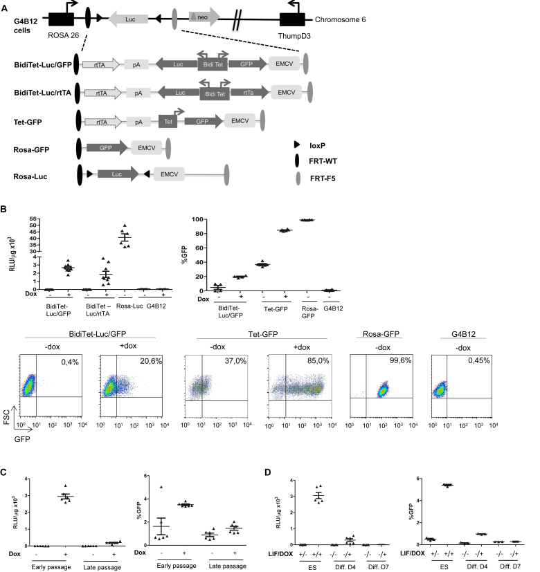
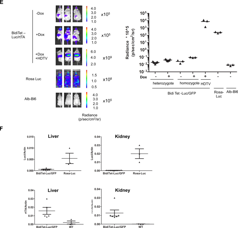
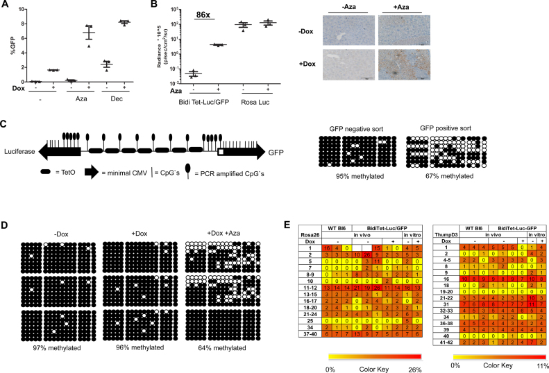
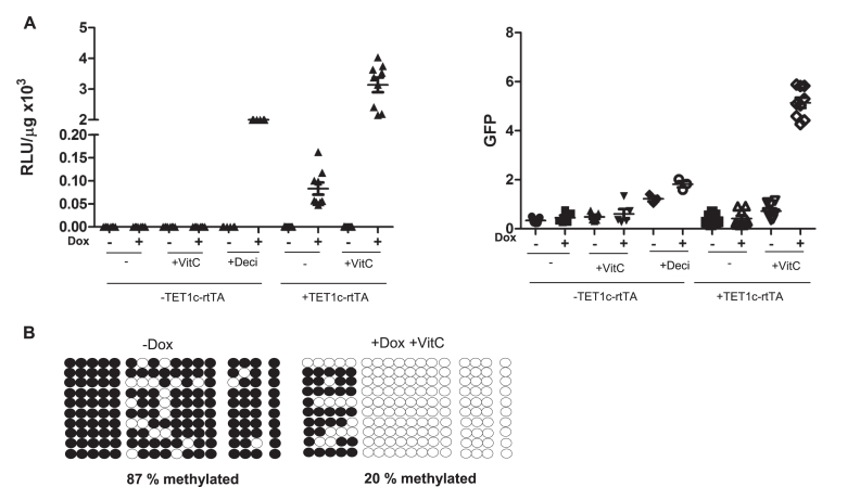
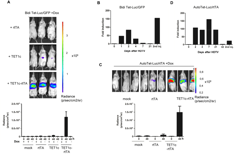
Faithful expression of transgenes in cell cultures and mice is often challenged by locus dependent epigenetic silencing. We investigated silencing of Tet-controlled expression cassettes within the mouse ROSA26 locus. We observed pronounced DNA methylation of the Tet promoter concomitant with loss of expression in mES cells as well as in differentiated cells and transgenic animals. Strikingly, the ROSA26 promoter remains active and methylation free indicating that this silencing mechanism specifically affects the transgene, but does not spread to the host's chromosomal neighborhood. To reactivate Tet cassettes a synthetic fusion protein was constructed and expressed in silenced cells. This protein includes the enzymatic domains of ten eleven translocation methylcytosine dioxygenase 1 (TET-1) as well as the Tet repressor DNA binding domain. Expression of the synthetic fusion protein and Doxycycline treatment allowed targeted demethylation of the Tet promoter in the ROSA26 locus and in another genomic site, rescuing transgene expression in cells and transgenic mice. Thus, inducible, reversible and site-specific epigenetic modulation is a promising strategy for reactivation of silenced transgene expression, independent of the integration site.


Figures

Figure 1.
Figure 1.
Expression from the bidirectional Tet promoter in the Rosa26 locus is silenced in ES cells, upon differentiation and in transgenic mice. (A) Schematic depiction of the RMCE compatible ROSA26 locus in G4B12 ES cells according to (37) and targeted expression cassettes used in this study. The RMCE strategy relies on Flp mediated targeting of the cassettes which are flanked with heterospecific FRT-WT and FRT-F5 recombination sites (dark and gray ovals). Upon chromosomal integration, an IRES element followed by an ATG from the incoming targeting cassette enables expression of the neomycin phosphotransferase gene, thereby rendering successfully recombined cells resistant against G418. Upon targeting of BidiTet-Luc/GFP, Tet-GFP and BidiTet-Luc/rtTA cassettes, the transactivator (rtTA) is controlled by the endogenous ROSA26 promoter. In BidiTet-Luc/rtTA a second copy of the rtTA is controlled by the cognate promoter, resulting in its positive feedback activation. Tet-GFP is a unidirectional Tet promoter driven GFP cassette. Targeting of a promoter-free GFP cassette resulted in Rosa-GFP cells in which GFP is controlled by the ROSA26 promoter. Rosa-Luc cells were derived from G4B12 ES cells upon stable expression of Cre recombinase. (B) Reporter gene expression in targeted ES cells. ES cells carrying the indicated cassettes were cultivated for 48h in the presence and absence of Doxycycline and analysed for expression of luciferase (as relative light units (rlu) per μg of total protein, left) or GFP (as % GFP expressing cells, right). As a negative control untargeted G4B12 ES cells were included. Cumulative data from two to three independent experiments (each performed in duplicates or triplicates) are represented. Representative flow cytometry plots are indicated for the different cell lines. (C) Stability of expression during culture. The BidiTet-Luc/GFP ES cells were cultured in the presence and absence of Doxycycline. Cells were analysed for luciferase and GFP expression early (passage 2–5) and late (passages 17–20) after targeting. Represented data were derived from two independent experiments in triplicate cell cultures. (D) Stability of reporter expression upon differentiation of BidiTet-Luc/GFP ES cells. To induce differentiation, BidiTet-Luc/GFP ES cells were cultivated on gelatinised plates without LIF in the absence and presence of Doxycycline for 4 and 7 days and subsequently analysed for GFP or luciferase expression. Data were generated from triplicates derived from two (luciferase) or one (GFP) experiments. (E) In vivo imaging of BidiTet-Luc/GFP, Rosa-Luc and wild type albino C57Bl/6 (alb-Bl/6) mice. BidiTet-Luc/GFP mice were analyzed for whole body in vivo bioluminescence upon providing 2 μg/ml Doxycycline in the drinking water for 7 days. BidiTet-Luc/GFP animals that received no Doxycycline were used as controls. BidiTet-Luc/GFP mice are also depicted 2 days after HDTV of the BidiTetLuc/GFP plasmid in the presence of Doxycycline. Note that the scale is different in the groups to accommodate different expression levels. Quantification is depicted on the right. Data from independent mice per group are depicted. (F) To assess the mRNA levels of luciferase and rtTA liver and kidney tissues of Doxycycline induced animals presented in E were harvested. Samples from three to seven animals per group were analysed by qRT-PCR. The levels of luciferase and rtTA transcripts were calculated relative to actin mRNA levels.
Figure 1.
Figure 1.
Expression from the bidirectional Tet promoter in the Rosa26 locus is silenced in ES cells, upon differentiation and in transgenic mice. (A) Schematic depiction of the RMCE compatible ROSA26 locus in G4B12 ES cells according to (37) and targeted expression cassettes used in this study. The RMCE strategy relies on Flp mediated targeting of the cassettes which are flanked with heterospecific FRT-WT and FRT-F5 recombination sites (dark and gray ovals). Upon chromosomal integration, an IRES element followed by an ATG from the incoming targeting cassette enables expression of the neomycin phosphotransferase gene, thereby rendering successfully recombined cells resistant against G418. Upon targeting of BidiTet-Luc/GFP, Tet-GFP and BidiTet-Luc/rtTA cassettes, the transactivator (rtTA) is controlled by the endogenous ROSA26 promoter. In BidiTet-Luc/rtTA a second copy of the rtTA is controlled by the cognate promoter, resulting in its positive feedback activation. Tet-GFP is a unidirectional Tet promoter driven GFP cassette. Targeting of a promoter-free GFP cassette resulted in Rosa-GFP cells in which GFP is controlled by the ROSA26 promoter. Rosa-Luc cells were derived from G4B12 ES cells upon stable expression of Cre recombinase. (B) Reporter gene expression in targeted ES cells. ES cells carrying the indicated cassettes were cultivated for 48h in the presence and absence of Doxycycline and analysed for expression of luciferase (as relative light units (rlu) per μg of total protein, left) or GFP (as % GFP expressing cells, right). As a negative control untargeted G4B12 ES cells were included. Cumulative data from two to three independent experiments (each performed in duplicates or triplicates) are represented. Representative flow cytometry plots are indicated for the different cell lines. (C) Stability of expression during culture. The BidiTet-Luc/GFP ES cells were cultured in the presence and absence of Doxycycline. Cells were analysed for luciferase and GFP expression early (passage 2–5) and late (passages 17–20) after targeting. Represented data were derived from two independent experiments in triplicate cell cultures. (D) Stability of reporter expression upon differentiation of BidiTet-Luc/GFP ES cells. To induce differentiation, BidiTet-Luc/GFP ES cells were cultivated on gelatinised plates without LIF in the absence and presence of Doxycycline for 4 and 7 days and subsequently analysed for GFP or luciferase expression. Data were generated from triplicates derived from two (luciferase) or one (GFP) experiments. (E) In vivo imaging of BidiTet-Luc/GFP, Rosa-Luc and wild type albino C57Bl/6 (alb-Bl/6) mice. BidiTet-Luc/GFP mice were analyzed for whole body in vivo bioluminescence upon providing 2 μg/ml Doxycycline in the drinking water for 7 days. BidiTet-Luc/GFP animals that received no Doxycycline were used as controls. BidiTet-Luc/GFP mice are also depicted 2 days after HDTV of the BidiTetLuc/GFP plasmid in the presence of Doxycycline. Note that the scale is different in the groups to accommodate different expression levels. Quantification is depicted on the right. Data from independent mice per group are depicted. (F) To assess the mRNA levels of luciferase and rtTA liver and kidney tissues of Doxycycline induced animals presented in E were harvested. Samples from three to seven animals per group were analysed by qRT-PCR. The levels of luciferase and rtTA transcripts were calculated relative to actin mRNA levels.
Figure 2.
Figure 2.
The bidirectional Tet promoter is methylated in the ROSA26 locus and can be reactivated by DNMTi (A) GFP expression in BidiTet-Luc/GFP ES cells upon cultivation in presence of the DNMTis Decitabine and Azacytidine. BidiTet-Luc/GFP ES cells were cultivated in presence or absence of Doxycycline and Decitabine or Azacytidine for three days and subsequently subjected to flow cytometry. The percentage of GFP expressing cells is depicted. (B) In vivo imaging of induced BidiTet-Luc/GFP mice upon treatment with Azacytidine. Whole body bioluminescence was determined in three independent animals before and three days after the first treatment with Azacytidine. As control, Rosa-Luc animals were used. The bioluminescence of three independent mice per group is depicted on the left. On day 3, the liver was isolated and slices were stained for GFP expression. Depicted are representative sections from mice of the indicated groups. (C) Schematic depiction of the bidirectional Tet promoter in BidiTet-Luc/GFP cells and the position of the CpG's. The bidirectional Tet promoter comprises 30 CpGs, 8 CpG's flank the tetO sequences, 3 CpGs are located in a spacer element and the remaining 19 CpG are in the two opposing CMV promoter sequences. The location of the CpGs within the promoter (lines) and within the amplified PCR fragment (lollipops) are indicated. Note that the 5′ minimal CMV promoter is slightly shorter than the 3′ CMV promoter (72). (D) Methylation status of the Tet promoter in BidiTet-Luc/GFP ES cells. Cells were treated for 3 days with Decitabine. GFP positive and negative cells were sorted, DNA was extracted and subjected to bisulfite analysis. Subsequently, the Tet promoter DNA was PCR amplified and cloned in E. coli. Sequences of eight randomly picked clones representing eight independent cells are depicted. In each line, the circles depict the 17 CpGs of an individual clone/cell. Black circles indicate methylated cytosines and non-filled circles non-methylated cytosines. (E) Methylation status of Tet promoter in BidiTet-Luc/GFP mice. Bisulfite analysis of the Tet promoter based on DNA extracted from mouse livers. The methylation status of the Tet promoter in the non-induced (–Dox), induced (+Dox) and Azacytidine/Dox induced animals (+Dox +Aza) is depicted. Three independent mice per group were analysed. For each mouse, eight independent cells were sequenced and analysed. Black circles indicate methylated cytosines and non-filled circles non methylated cytosines. (F) Epityper based DNA methylation analysis of the ROSA26 and ThumpD3 promoter in cells and mice. The heat maps represent the results of the methylation analysis done on Epityper. The various CpG motifs of the two promoters are indicated on the x-axis of each map and the samples used are indicated on the y-axis. The reference scale is indicated (0.01 = 1% methylation and 0.26 = 26% methylation). Each vertical column represents the same CpG motif analyzed from different gDNA samples while each row represents the different CpGs within the same sample.
Figure 3.
Figure 3.
The silenced Tet promoter can be reactivated by site specific targeted demethylation in fibroblasts. (A) Reactivation of silenced reporter expression in fibroblasts. Immortalized fibroblasts from BidiTet-Luc/GFP mice were transduced with Tet1c-rtTA by lentiviral gene transfer. Infected (+TET1c-rtTA) and non-infected (–TET1c-rtTA) cells were cultured with or without Doxycycline, Vitamin C (+VitC) for three days as indicated and subsequently analysed for luciferase and GFP expression. As control, the non-transduced BidiTet-Luc/GFP fibroblasts were cultured with Decitabine. The data from two or three independent experiments with duplicates or triplicates are depicted. (B) Methylation status of the Tet promoter in Tet1c-rtTA transduced fibroblasts. The genomic DNA was isolated from TET1c-rtTA transduced BidiTet-Luc/GFP fibroblasts previously cultured in absence or presence of Doxycycline and Vitamin C. The DNA was converted with bisulfite. The Tet promoter DNA was PCR amplified, cloned and sequenced. The methylation status of 10 independent clones is depicted. Black circles indicate methylated cytosines and non-filled circles non-methylated cytosines.
Figure 4.
Figure 4.
Reversion of Tet-promoter silencing in vivo. (A) Reactivation of silenced gene expression in vivo. BidiTet-Luc/GFP mice were subjected to HDTV using the indicated plasmid DNA. As negative control the animals were hydrodynamically injected with PBS (mock). After 48 h, bioluminescence was measured by in vivo imaging. Doxycycline feeding was started 7 days before the experiment and was maintained throughout the experiment (upper panel). Control groups were measured in absence of Doxycycline as indicated. Representative in vivo images of mice as well as cumulative data from 5 independent animals per group are shown (lower panel). (B) Kinetics of reactivation. Bioluminescence of BidiTet-Luc/GFP animals was monitored on the indicated days after HDTV injection as described in (A). On day 21, HDTV injection was repeated using the same plasmid DNA and animals were imaged one day later. Bars represent measurement of four animals. The fold induction is related the bioluminescence signal before HDTV injection. Day 2 luciferase measurements were derived from the experiment represented in (A). (C) Reactivation of silenced expression in AutoTet-Luc/rtTA mice. Doxycycline induced AutoTet-Luc/rtTA mice received TET1c-rtTA, rtTA or PBS (mock) via HDTV injection. Bioluminescence of mice 48 h after HDTV injection is depicted on the left. The quantification of bioluminescence before (0) and 48 h after (48) based on three independent animals is depicted in the lower panel. As negative control (mock) AutoTet-Luc/rtTA animals were injected with PBS. (D) Kinetics of reactivation in AutoTet-Luc/rtTA mice. Bioluminescence of AutoTet-Luc/rtTA mice of (C) was monitored on the indicated days after HDTV injection. AutoTet-Luc/rtTA animals received drinking water supplemented with Doxycycline throughout the experiment. The animals were measured on the indicated days before and after HDTV injection. On day 42 HDTV injection was repeated using the same plasmid DNA and animals were analysed one day later. Bars represent measurement of three animals. Day 2 luciferase measurements are derived from the experiment represented in (C). The fold induction is related to the bioluminescence before HDTV injection.

Similar articles

Cited by

References

    1. May T., Butueva M., Bantner S., Markusic D., Seppen J., MacLeod R.A., Weich H., Hauser H., Wirth D.. Synthetic gene regulation circuits for control of cell expansion. Tissue Eng. Part A. 2010; 16:441–452. - PubMed
    1. Saxena P., Heng B.C., Bai P., Folcher M., Zulewski H., Fussenegger M.. A programmable synthetic lineage-control network that differentiates human IPSCs into glucose-sensitive insulin-secreting beta-like cells. Nat. Commun. 2016; 7:11247. - PMC - PubMed
    1. Hockemeyer D., Soldner F., Cook E.G., Gao Q., Mitalipova M., Jaenisch R.. A drug-inducible system for direct reprogramming of human somatic cells to pluripotency. Cell Stem Cell. 2008; 3:346–353. - PMC - PubMed
    1. Vierbuchen T., Ostermeier A., Pang Z.P., Kokubu Y., Sudhof T.C., Wernig M.. Direct conversion of fibroblasts to functional neurons by defined factors. Nature. 2010; 463:1035–1041. - PMC - PubMed
    1. Pfisterer U., Kirkeby A., Torper O., Wood J., Nelander J., Dufour A., Bjorklund A., Lindvall O., Jakobsson J., Parmar M.. Direct conversion of human fibroblasts to dopaminergic neurons. Proc. Natl. Acad. Sci. U.S.A. 2011; 108:10343–10348. - PMC - PubMed

Substances