. 2017 Sep 19;45(16):9679-9693.
doi: 10.1093/nar/gkx608.

The conserved AU dinucleotide at the 5' end of nascent U1 snRNA is optimized for the interaction with nuclear cap-binding-complex

Affiliations

The conserved AU dinucleotide at the 5' end of nascent U1 snRNA is optimized for the interaction with nuclear cap-binding-complex

Chung-Shu Yeh et al. Nucleic Acids Res. .

Abstract

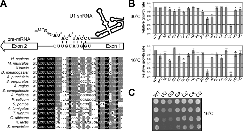
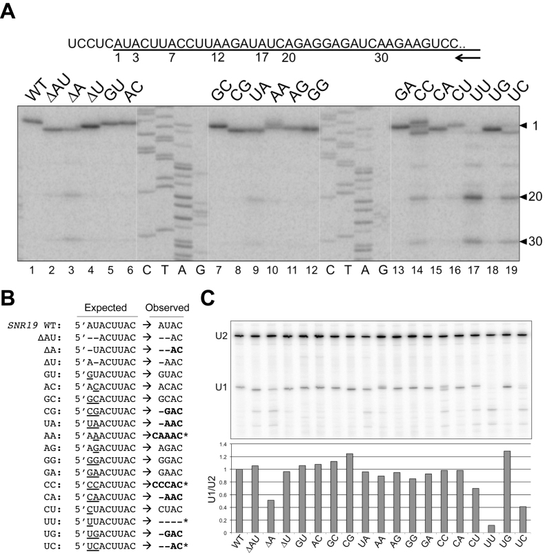
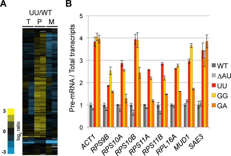
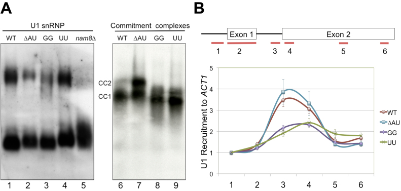
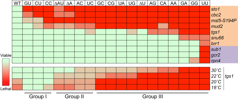
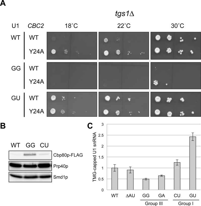
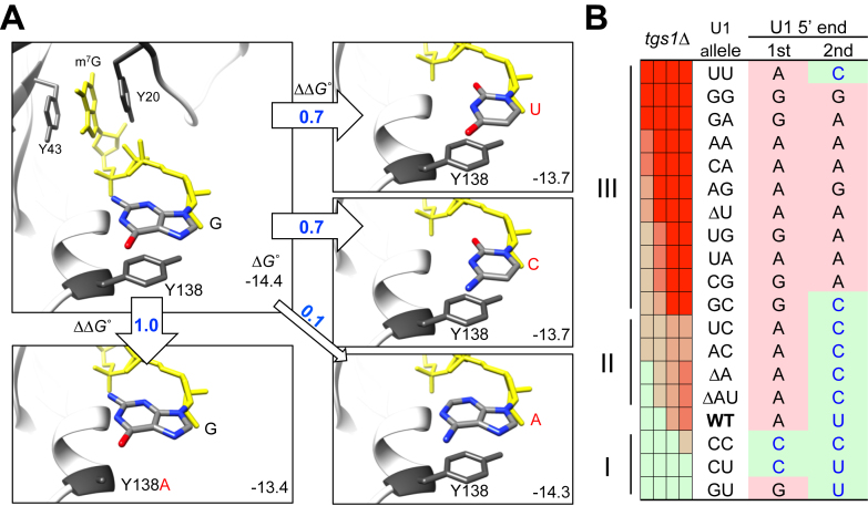
Splicing is initiated by a productive interaction between the pre-mRNA and the U1 snRNP, in which a short RNA duplex is established between the 5' splice site of a pre-mRNA and the 5' end of the U1 snRNA. A long-standing puzzle has been why the AU dincucleotide at the 5'-end of the U1 snRNA is highly conserved, despite the absence of an apparent role in the formation of the duplex. To explore this conundrum, we varied this AU dinucleotide into all possible permutations and analyzed the resulting molecular consequences. This led to the unexpected findings that the AU dinucleotide dictates the optimal binding of cap-binding complex (CBC) to the 5' end of the nascent U1 snRNA, which ultimately influences the utilization of U1 snRNP in splicing. Our data also provide a structural interpretation as to why the AU dinucleotide is conserved during evolution.


Figures

Figure 1.
Figure 1.
Alteration of U1 snRNA 5′-end AU dinucleotide results in growth defect. (A) Base-pairing interaction between U1 snRNA 5′ end and the 5′ss. U1 snRNA is proposed to form a 6-bp interaction with the canonical 5′ss (GUAUGU). The 5′-end AU dinucleotide, although highly conserved (bottom panel), is not predicted to universally interact with 5′ss. Note that the m7G cap is co-transcriptionally added to the 5′ end of U1 snRNA and further modified into m2,2,7G cap. The AU dinucleotide is uniquely missing in S. pombe. (B) The growth rates of yeast strains producing U1 snRNA variants relative to an isogenic wild-type strain were determined by a competitive fitness assay. Strains exhibiting significantly low fitness scores (P < 0.01, two-tailed t-test) at 30°C or 16°C were indicated (*). Data represent the mean ± S.E.M. of three independent biological replicates. (C) Growth phenotypes of strains expressing U1 snRNA variants. Relevant genotypes of the yeast strains are shown on the left of the panel. Cells were grown to saturation at 30°C, serially diluted, and spotted on YPD plates for incubation at 16°C.
Figure 2.
Figure 2.
Alteration of U1 snRNA 5′-end dinucleotide impacts U1 snRNA transcription. (A) Transcription start sites of the U1 snRNA variants were mapped by primer extension. Sequence around the U1 snRNA transcription start is shown in the top panel, with the U1 snRNA sequence underlined. The numbers (1, 3, 7, 12, 17, 20 and 30) below the line indicate the wild-type and variant U1 snRNA start sites determined. The arrow represents the oligonucleotide primer used in the reverse transcription reactions, which is complementary to U1 snRNA positions 266–285. Image of the primer extension results are shown in the bottom panel. The sequencing ladder (C, T, A, G) was generated by Sanger sequencing off a plasmid harboring SNR19 using the same primer. (B) Summary of the transcription start sites used by U1 snRNA variants. Deletions are marked as short dash lines and base substitutions are underlined. Variants that have unexpected start sites are shown in bold, and those which have multiple strong start sites are marked by stars. For example, AA and CC variants have a significant start from the -1 position. We noted the U1 snRNA transcription appears to prefer A and G, although there are three cases of C start (AA, CC, and CU mutants). (C) Quantitation of the full-length U1 snRNA levels in U1 snRNA mutants by primer extension normalized to the U2 snRNA levels.
Figure 3.
Figure 3.
Alteration of U1 snRNA 5′-end dinucleotide impacts on splicing in vivo. (A) Microarrays analysis of the genome-wide splicing phenotype of the UU mutant in comparison to that of the wild-type strain. T, total transcript; P, pre-mRNA; and M, spliced transcript. The two columns under each category, i.e. T, P and M, represent a pair of dye-flipped (Cy3 and Cy5) experiment of the same RNA sample, to which one dataset is inversely presented. (B) Accumulation of intron-containing transcripts in the GG, UU, and GA mutants. Splicing defect (P/T >1) of the nine selected intron-containing genes was quantitated measured by RT-qPCR. Data represent the mean ± S.E.M. of three independent biological replicates.
Figure 4.
Figure 4.
Alteration of U1 snRNA 5′-end dinucleotide impacts on U1 snRNP recruitment to pre-mRNA. (A) Native gel electrophoresis analysis of U1 snRNP and commitment complexes. (Left Panel) Alteration of U1 snRNA 5′-end dinucleotide does not appear to affect U1 snRNP mobility on the native gel. Splicing extracts made from wild-type, ΔAU, UU, GG and nam8Δ strains were electrophoresed on a native polyacrylamide (3%)–agarose (0.5%) gel. The RNAs were transferred to a membrane and probed by a digoxigenin-labeled U1 probe. No apparent differences were detected for the two major forms of the U1 snRNP except for nam8Δ. (Right Panel) Electrophoretic mobilities of CCs are altered in the presence of GG and UU mutations. CC1 and CC2 are two commitment complexes formed in the absence of U2 snRNP with [32P]-labeled RP51A transcript. (B) The GG and UU mutations severely reduce U1 snRNP’s chromatin association. Six pairs of oligonucleotides were used to amplify different regions (short lines at the bottom of the top panel) of the ACT1 gene containing two exons (boxes, top panel) and an intron (connecting thin line, top panel). All data were normalized to the signal of the first oligonucleotide pair from experiments using the wild-type strain. Data represent the mean ± S.E.M. of six independent biological replicates.
Figure 5.
Figure 5.
Genetic analysis identified genes functionally linked to the U1 snRNA 5′-end dinucleotide. Nine candidate genes (except for BBP allele msl5-S194P) marked on the right emerged first from an SGA screen using the UU mutation, among them six are related to splicing (light brown) and three to transcription (light purple). Genetic interaction of these nine candidate genes and msl5-S194P with all the U1 snRNA mutants was further manually analyzed in a pairwise manner. The severities of synthetic lethality, ranging from viable to lethal, are presented in a heat map (top half). Temperature-dependent synthetic lethality between tgs1Δ and all the U1 snRNA mutations is presented in the bottom half. See text for detailed discussion of the biological underpinning of Groups I, II and III.
Figure 6.
Figure 6.
Alteration of U1 snRNA 5′-end dinucleotide impacts on CBC binding to U1 snRNA. (A) Rescue of the GG/tgs1Δ synthetic lethality by CBC2-Y24A mutation at 30°C. Yeast strains bearing tgs1Δ in conjunction with various combinations of SNR19 and CBC2 alleles were examined for their growth phenotypes at different temperatures. The GU allele that can rescue tgs1Δ cs phenotype was included as a control. (B) The U1 snRNA 5′-end GG mutant provides an environment for hyperstable interaction between its m7G cap and CBC. U1 snRNP variants differing at the 5′-end dinucleotide of the U1 snRNA (GG, CU, and wild-type) were affinity purified and probed the abundance of Cbp80p using anti-FLAG M2 antibody. Anti-Prp40p and anti-Smd1p were respectively used to detect the Prp40p and Smd1p, which are integral to U1 snRNP. (C) The identity of the U1 snRNA 5′-end dinucleotide may impact on the TMG cap formation. More U1 snRNA from the CU and GU mutants (Group I) could be precipitated by anti-TMG antibody than that of the control strains (WT and ΔAU). In contrast, less U1 snRNA could be recovered from the GG and GA mutants (Group III). The precipitated U1 snRNA was quantitated by RT-qPCR and normalized against U2 snRNA in the same RNA sample. Data represent the mean ± S.E.M. of at least three independent biological replicates.
Figure 7.
Figure 7.
In silico analysis of stacking between m7GpppG and Y138 residue in CBP20. (A) The G base immediately following the m7Gppp moiety (shown in yellow) was replaced by either U, C or A via molecular docking and the corresponding ΔGo values were computed (see Materials and Methods) and are shown at the lower right corner within each rectangular boxes. The resulting ΔΔGo values in reference to the wild-type state are indicated within or by the empty arrows. The previously experimentally tested Y138A mutation in relationship to m7GpppG is included as a control (box, lower left). (B) Striking correlation of the genetic data with the computationally predicted ΔΔGo (see text for detailed description). The precise 5′ ends of U1 transcripts (U1 5′ end) produced in all U1 snRNA mutants (i.e. U1 allele) are shown for their respective first (1st) and second (2nd) nucleotides in each row (see also Figure 2). Note that the (C/U)-(C/U), (G/A)-(C/U), and (G/A)-(G/A) combinations are predicted to cause weaker, wild-type-like, and excessive binding between U1 snRNA and CBP20, respectively. This pattern parallels to the Group I, II, and III assignments of the tgs1Δ synthetic lethality pattern (see also Figure 5).

Similar articles

Cited by

References

    1. Matera A.G., Wang Z.. A day in the life of the spliceosome. Nat. Rev. Mol. Cell. Biol. 2014; 15:108–121. - PMC - PubMed
    1. Will C.L., Lührmann R.. Spliceosome Structure and Function. Cold Spring Harbor Perspect. Biol. 2011; 3:a003707. - PMC - PubMed
    1. Fabrizio P., Dannenberg J., Dube P., Kastner B., Stark H., Urlaub H., Luhrmann R.. The evolutionarily conserved core design of the catalytic activation step of the yeast spliceosome. Mol. Cell. 2009; 36:593–608. - PubMed
    1. Will C.L., Luhrmann R.. Protein functions in pre-mRNA splicing. Curr. Opin. Cell Biol. 1997; 9:320–328. - PubMed
    1. Chang T.H., Tung L., Yeh F.L., Chen J.H., Chang S.L.. Functions of the DExD/H-box proteins in nuclear pre-mRNA splicing. Biochim. Biophys. Acta. 2013; 1829:764–774. - PubMed

MeSH terms

Substances