. 2017 Sep 19;45(16):9427-9440.
doi: 10.1093/nar/gkx611.

Human DNA polymerase delta double-mutant D316A;E318A interferes with DNA mismatch repair in vitro

Affiliations

Human DNA polymerase delta double-mutant D316A;E318A interferes with DNA mismatch repair in vitro

Dekang Liu et al. Nucleic Acids Res. .

Abstract

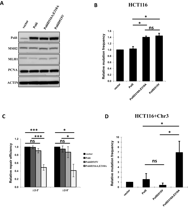
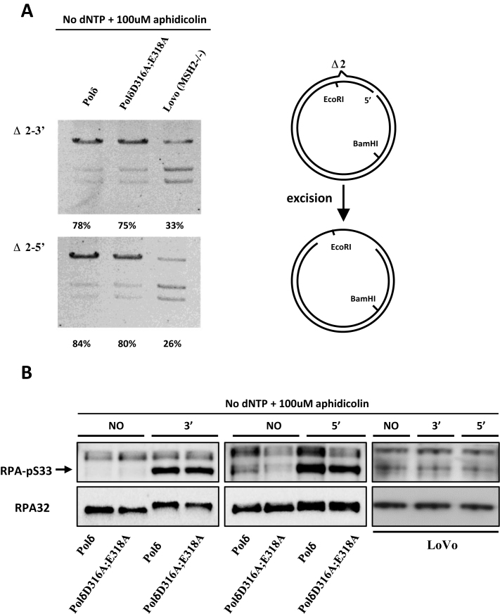
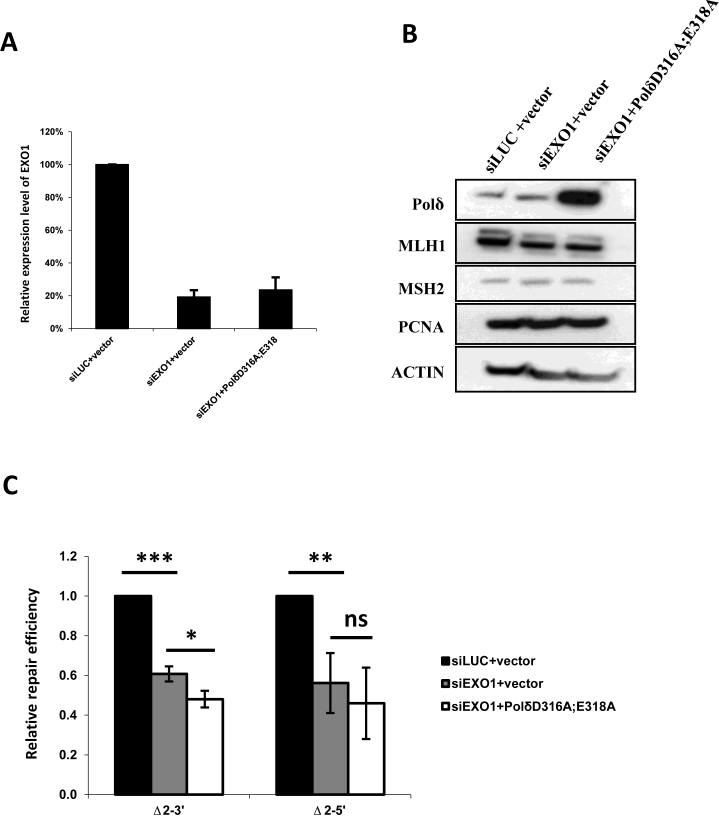
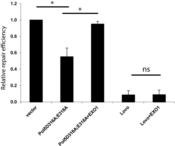
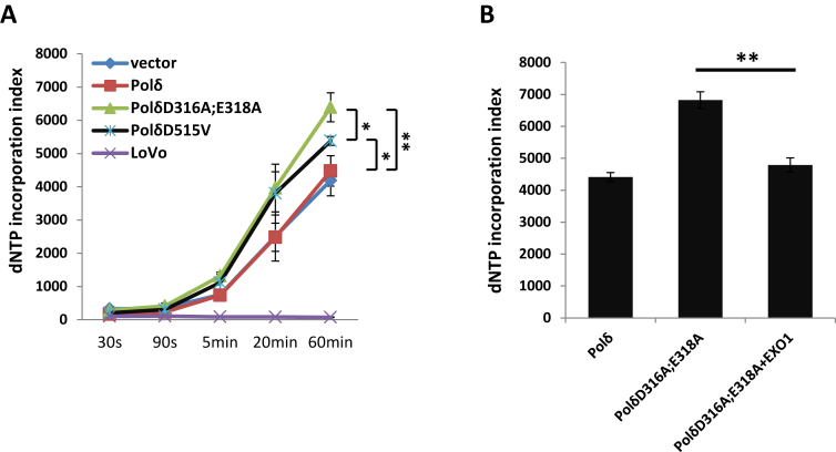
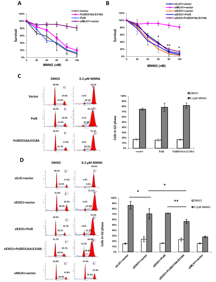
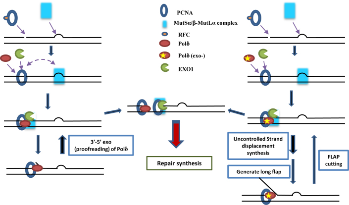
DNA mismatch repair (MMR) is a highly-conserved DNA repair mechanism, whose primary role is to remove DNA replication errors preventing them from manifesting as mutations, thereby increasing the overall genome stability. Defects in MMR are associated with increased cancer risk in humans and other organisms. Here, we characterize the interaction between MMR and a proofreading-deficient allele of the human replicative DNA polymerase delta, PolδD316A;E318A, which has a higher capacity for strand displacement DNA synthesis than wild type Polδ. Human cell lines overexpressing PolδD316A;E318A display a mild mutator phenotype, while nuclear extracts of these cells exhibit reduced MMR activity in vitro, and these defects are complemented by overexpression or addition of exogenous human Exonuclease 1 (EXO1). By contrast, another proofreading-deficient mutant, PolδD515V, which has a weaker strand displacement activity, does not decrease the MMR activity as significantly as PolδD316A;E318A. In addition, PolδD515V does not increase the mutation frequency in MMR-proficient cells. Based on our findings, we propose that the proofreading activity restricts the strand displacement activity of Polδ in MMR. This contributes to maintain the nicks required for EXO1 entry, and in this manner ensures the dominance of the EXO1-dependent MMR pathway.


Figures

Figure 1.
Figure 1.
In vitro MMR. (A) HeLa nuclear extracts from cells carrying empty vector (vector), wild type Polδ, PolδD316A;E318A or PolδD515V were analyzed by western blot. Western blot data are presented for MSH2, MLH1 and PCNA. (B) Mutation frequency in HCT116 (MLH1-/-) cells overexpressing PolδD316A;E318A or PolδD515V. (C)In vitro MMR assay using 2 nt indel heteroduplex DNA substrates Δ2–3′ or Δ2–5′ (see Materials and Methods). (D) As in B except using chromosome 3 complemented HCT116 (HCT116+Chr3) cells. The data are the mean ± SD of three independent experiments. *P< = 0.05; **P< = 0.01; ***P< = 0.005.
Figure 2.
Figure 2.
Mismatch-provoked excision in HeLa cells expressing PolδD316A;E318A. (A) Mismatch provoked excision assay was performed as described in Materials and Methods using the nuclear extracts indicated. The extent of DNA excision was estimated by measuring susceptibility/resistance to cleavage by EcoRI, whose recognition sequence lies in between the DNA excision initiation site and the mismatch (schematic diagram right). Reaction products were digested with EcoRI and BamHI, separated by agarose gel electrophoresis and visualized by staining with ethidium bromide. The intensity of each band was quantified using ImageJ, and the relative excision capacity was calculated as the intensity of the slowest migrating (largest) reaction product per lane /total intensity per lane × 100. Nuclear extracts from MSH2-deficient LoVo cells were used as the negative control. (B) Western blot of pS33-RPA32 was performed to monitor amount of ssDNA generated during in vitro MMR. Briefly, MMR assay was performed as described in Materials and Methods with no substrate (NO), Δ2–3′ (3′) or Δ2–5′ (5′) substrate in the reaction. The MMR was terminated by adding SDS containing loading buffer and boiling at 95°C for 10 min. Western blot was performed as described in Materials and Methods. Aphidicolin was included and dNTPs were omitted as indicated for inhibition of polymerase synthesis of Polδ. The total RPA32 was used as control.
Figure 3.
Figure 3.
Effect of depletion of EXO1 on MMR in cells overexpressing PolδD316A;E318A. (A) Efficiency of EXO1 knockdown was confirmed by qPCR, and (B) overexpression of PolδD316A;E318A mutant was confirmed by western blot. Besides, no alternations in expression levels of MLH1, MSH2 and PCNA was observed among nuclear extracts from cells with Luciferase siRNA and empty vector (siLUC+vector), cells with EXO1 siRNA and empty vector (siEXO1+vector) and cells with EXO1 siRNA and expressing PolδD316A;E318A (siEXO1+ PolδD316A;E318A). (C) in vitro MMR assay was performed as described in Materials and Methods. No strong synergetic effect on MMR activity when EXO1 depletion and expressing PolδD316A;E318A were combined in the nuclear extracts. The data represent the mean ± SD of three independent experiments. *P ≤ 0.05; **P ≤ 0.01.
Figure 4.
Figure 4.
EXO1 complements MMR defect in nuclear extracts expressing PolδD316A;E318A. The in vitro MMR assay was performed as described in Materials and Methods and the nuclear extracts were the same as used in Figure 1. Purified human EXO1 (2.5 nM) was added to the reaction as indicated. The Δ2–3′ heteroduplex was used in the MMR assays. The data represent the mean ± SD of three independent experiments. *P ≤ 0.05; **P ≤ 0.01.
Figure 5.
Figure 5.
Effect of PolδD316A;E318A on incorporation of dNTPs during in vitro MMR. In panel (A) dNTP incorporation was analyzed as described in Methods and Materials. The Δ2–5′ heteroduplex was used as substrate. The asterisk indicates the P value of the pairwise comparison of extracts with PolδD316A;E318A, with PolδD515V, and with wild type Polδ overexpression. In panel (B), dNTP incorporation was measured 60 min after start of the MMR reaction. The data are the mean ± SD of three independent experiments. *P ≤ 0.05; **P ≤ 0.01.
Figure 6.
Figure 6.
Effect of PolδD316A;E318A on susceptibility to killing by MNNG. (A and B) HeLa cells were transfected and seeded as described in Materials and Methods. After one day, cells were treated with 10 μM O6-Benzylguanine and MNNG at the concentration indicated. Colonies were counted after 8 days after crystal violet staining. The percentage of survival for each drug concentration was calculated as number of colonies after MNNG treatment / number of colonies without MNNG treatment. The asterisk above the error bar indicates the P value between group siEXO1+Polδ and group siEXO1+PolδD316A;E318A. (C and D) Cells were seeded into T75 flasks one day after transfection. After 24 hours, cells were treated with 10 μM O6-Benzylguanine and 0.2 μM MNNG and incubated for another two days before subject to FACS analysis. The data represent the mean ± SD of three independent experiments. *P ≤ 0.05.
Figure 7.
Figure 7.
Model of the role of Polδ during MMR. Briefly, PolδD316A;E318A competes with and/or inhibits EXO1-mediated DNA excision during MMR in vitro.

Similar articles

Cited by

References

    1. Modrich P., Lahue R.. Mismatch repair in replication fidelity, genetic recombination, and cancer biology. Annu. Rev. Biochem. 1996; 65:101–133. - PubMed
    1. Li G.M. Mechanisms and functions of DNA mismatch repair. Cell research. 2008; 18:85–98. - PubMed
    1. Jascur T., Boland C.R.. Structure and function of the components of the human DNA mismatch repair system. Int. J. Cancer. 2006; 119:2030–2035. - PubMed
    1. Kolodner R. Biochemistry and genetics of eukaryotic mismatch repair. Genes Dev. 1996; 10:1433–1442. - PubMed
    1. Schofield M.J., Hsieh P.. DNA mismatch repair: molecular mechanisms and biological function. Annu. Rev. Microbiol. 2003; 57:579–608. - PubMed

LinkOut - more resources