. 2017 Sep 19;45(16):9244-9259.
doi: 10.1093/nar/gkx618.

Automatic identification of informative regions with epigenomic changes associated to hematopoiesis

Affiliations

Automatic identification of informative regions with epigenomic changes associated to hematopoiesis

Enrique Carrillo-de-Santa-Pau et al. Nucleic Acids Res. .

Abstract

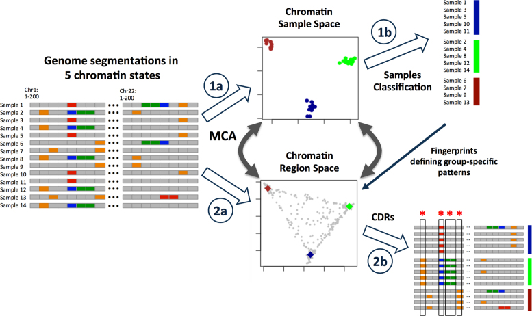
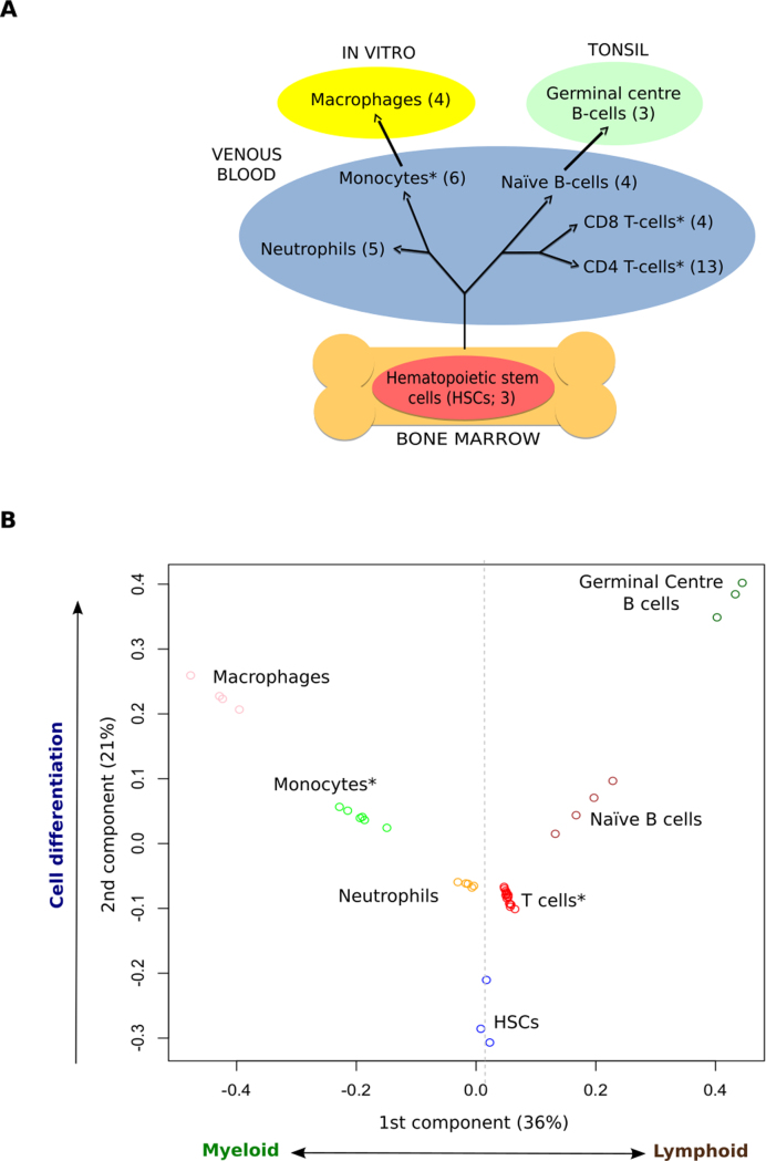
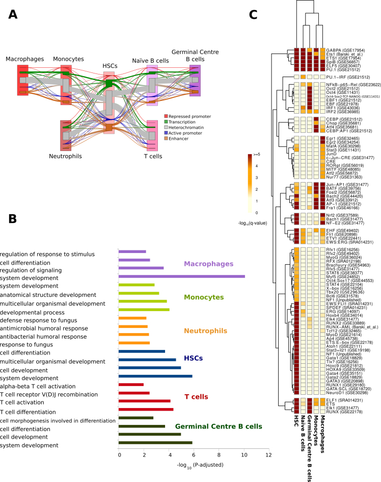
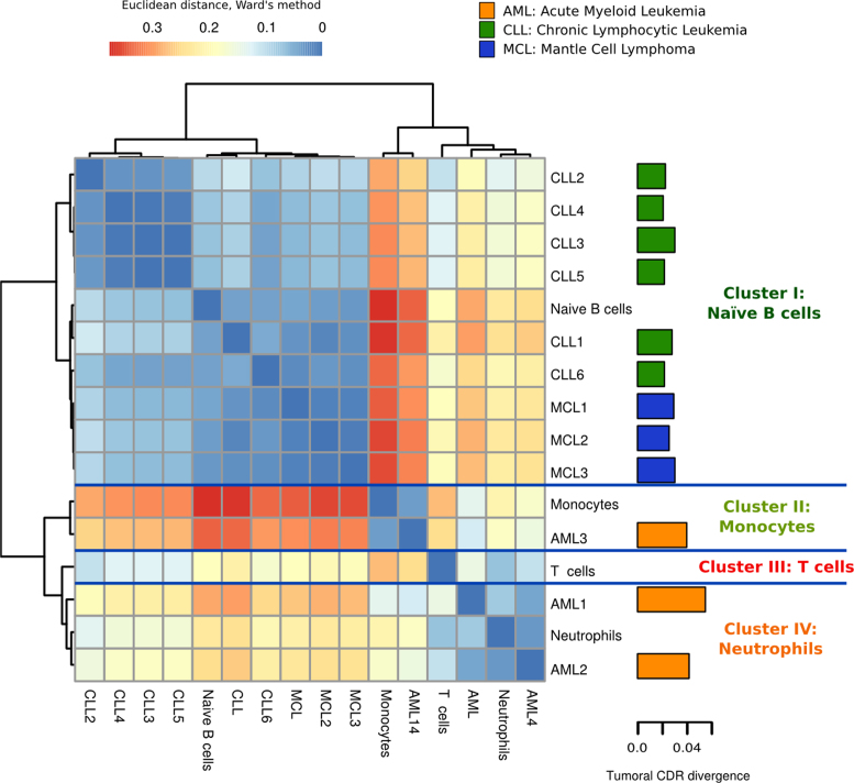
Hematopoiesis is one of the best characterized biological systems but the connection between chromatin changes and lineage differentiation is not yet well understood. We have developed a bioinformatic workflow to generate a chromatin space that allows to classify 42 human healthy blood epigenomes from the BLUEPRINT, NIH ROADMAP and ENCODE consortia by their cell type. This approach let us to distinguish different cells types based on their epigenomic profiles, thus recapitulating important aspects of human hematopoiesis. The analysis of the orthogonal dimension of the chromatin space identify 32,662 chromatin determinant regions (CDRs), genomic regions with different epigenetic characteristics between the cell types. Functional analysis revealed that these regions are linked with cell identities. The inclusion of leukemia epigenomes in the healthy hematological chromatin sample space gives us insights on the healthy cell types that are more epigenetically similar to the disease samples. Further analysis of tumoral epigenetic alterations in hematopoietic CDRs points to sets of genes that are tightly regulated in leukemic transformations and commonly mutated in other tumors. Our method provides an analytical approach to study the relationship between epigenomic changes and cell lineage differentiation. Method availability: https://github.com/david-juan/ChromDet.


Figures

Figure 1.
Figure 1.
Framework to identify CDRs that determine cell or lineage identity based on chromatin state changes. (1a) A chromatin samples space is generated with MCA from the chromatin segmentations by each sample. (1b) Samples are classified depending on clusters derived from the MCA analysis. (2a) A second space is generated with MCA from the chromatin segmentations by each sample. (2b) The CDRs are obtained selecting those genomic regions that overlap with the cluster sample fingerprints, a reference sample representing each cell type cluster. These regions discriminate the different cell types classified in the samples space. (*Regions with chromatin changes among cell types → CDRs). See also Supplementary Figure S1 and Supplementary Data.
Figure 2.
Figure 2.
Hematopoietic cell types cluster based on chromatin states. (A) Schematic differentiation tree of the cell types considered, highlighting the tissue of origin and environment of each sample type. (B) Clustering of the samples in the MCA space recovers ontological relationships among cell types. (*Cell types with samples from different consortia) See also Supplementary Figure S2, Supplementary Table S1 and Supplementary Data.
Figure 3.
Figure 3.
Functional and transcription factor binding motifs characterization of chromatin determinant regions (CDRs). (A) Sankey Plot representation of chromatin state transitions at CDRs during hematopoietic cell differentiation. Nodes for each cell type represent the five ‘collapsed’ chromatin states (see ‘Materials and Methods’ section). For each pair of cell types in the hematopoietic differentiation pathway, flows, represented by line thickness, are proportional to the number of regions that show a transition between a particular pair of states. Changes in chromatin states between two stages of differentiation are shown with lines that change color. The thickness of the lines is proportional to the number of regions that show a transition between a particular pair of states. (B) Enriched ontology terms from the genes related to the CDRs that characterize each cell type. (C) Heatmap and hierarchical clustering based on transcription binding proteins enriched in the CDRs that characterize each cell type (see ‘Materials and Methods’ section). See also Supplementary Figures S3–15, Tables S2–6 and File S1.
Figure 4.
Figure 4.
Hierarchical clustering of leukemias based on CDRs of healthy cell types suggests potential lineage origin of tumors. The healthy cell type clusters are summarized by each fingerprint, a reference sample representing each cell type cluster. The euclidean distances between samples and fingerprints are calculated with the Ward's method. The barplot in the right shows the epigenomic divergence (ratio of chromatin changes in CDRs) of each cancer sample to the healthy states. See also Supplementary Figures S16–20 and Tables S7–9.

References

    1. Rieger M.A., Schroeder T.. Hematopoiesis. Cold Spring Harb. Perspect. Biol. 2012; 4:a008250. - PMC - PubMed
    1. Paul F., Arkin Y., Giladi A., Jaitin D.A., Kenigsberg E., Keren-Shaul H., Winter D., Lara-Astiaso D., Gury M., Weiner A. et al. Transcriptional heterogeneity and lineage commitment in myeloid progenitors. Cell. 2015; 163:1663–1677. - PubMed
    1. Ji H., Ehrlich L.I.R., Seita J., Murakami P., Doi A., Lindau P., Lee H., Aryee M.J., Irizarry R.A., Kim K. et al. Comprehensive methylome map of lineage commitment from haematopoietic progenitors. Nature. 2010; 467:338–342. - PMC - PubMed
    1. Bock C., Beerman I., Lien W.-H., Smith Z.D., Gu H., Boyle P., Gnirke A., Fuchs E., Rossi D.J., Meissner A.. DNA methylation dynamics during in vivo differentiation of blood and skin stem cells. Mol. Cell. 2012; 47:633–647. - PMC - PubMed
    1. Lara-Astiaso D., Weiner A., Lorenzo-Vivas E., Zaretsky I., Jaitin D.A., David E., Keren-Shaul H., Mildner A., Winter D., Jung S. et al. Immunogenetics. Chromatin state dynamics during blood formation. Science. 2014; 345:943–949. - PMC - PubMed

LinkOut - more resources