. 2017 Sep 19;45(16):9547-9557.
doi: 10.1093/nar/gkx638.

Number of inadvertent RNA targets for morpholino knockdown in Danio rerio is largely underestimated: evidence from the study of Ser/Arg-rich splicing factors

Affiliations

Number of inadvertent RNA targets for morpholino knockdown in Danio rerio is largely underestimated: evidence from the study of Ser/Arg-rich splicing factors

Marine Joris et al. Nucleic Acids Res. .

Abstract

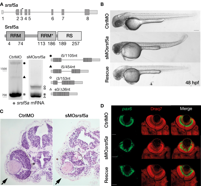
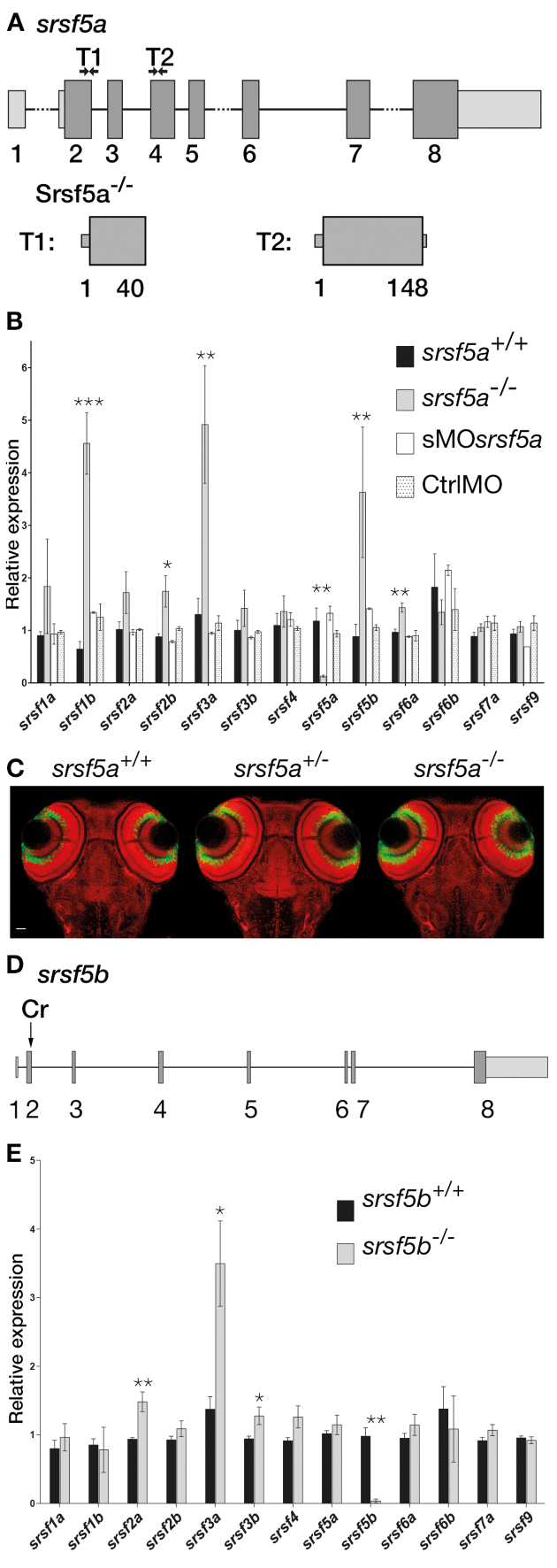
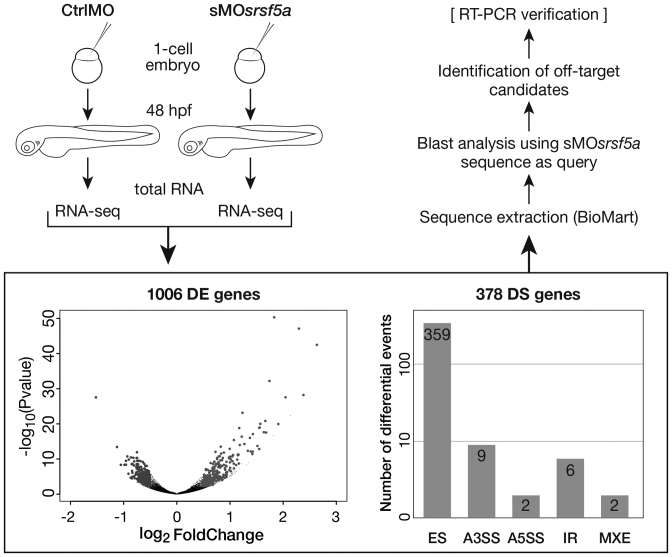
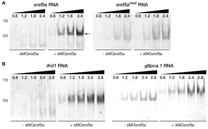
Although the involvement of Ser/Arg-rich (SR) proteins in RNA metabolism is well documented, their role in vertebrate development remains elusive. We, therefore, elected to take advantage of the zebrafish model organism to study the SR genes' functions using the splicing morpholino (sMO) microinjection and the programmable site-specific nucleases. Consistent with previous research, we revealed discrepancies between the mutant and morphant phenotypes and we show that these inconsistencies may result from a large number of unsuspected inadvertent morpholino RNA targets. While microinjection of MOs directed against srsf5a (sMOsrsf5a) led to developmental defects, the corresponding homozygous mutants did not display any phenotypic traits. Furthermore, microinjection of sMOsrsf5a into srsf5a-/- led to the previously observed morphant phenotype. Similar findings were observed for other SR genes. sMOsrsf5a alternative target genes were identified using deep mRNA sequencing. We uncovered that only 11 consecutive bases complementary to sMOsrsf5a are sufficient for binding and subsequent blocking of splice sites. In addition, we observed that sMOsrsf5a secondary targets can be reduced by increasing embryos growth temperature after microinjection. Our data contribute to the debate about MO specificity, efficacy and the number of unknown targeted sequences.


Figures

Figure 1.
Figure 1.
Injection of a splice site blocking MO targeting the srsf5a gene led to developmental defects. (A) srsf5a is composed of eight exons. The protein is encoded by exons 2–8 and consists of two RRM domains responsible for RNA binding and one RS domain, essential for protein–protein interactions. Three different transcripts are produced from srsf5a. Among them, two alternative transcripts retain intron 5 or a part of it (black up-pointing triangle and black dot). sMOsrsf5a was designed to target the exon3–intron3 junction. RT-PCR experiments to amplify srsf5a mRNA in control and morphant embryos at 48 hpf revealed intron3 retention (open hexagon mark), introducing a premature STOP codon into the RRM1 encoding part of the mRNA. Expression of the three normal srsf5a transcripts was strongly reduced in morphants, while MO injection also triggered the use by the splicing machinery of a cryptic splice site located in exon3, and leading to a deletion of 36 bases in the open reading frame of the srsf5a transcript (srsf5a/Δ36, open triangle mark). The resulting protein has a 12-amino acids deletion within the RRM1. The open square mark corresponds to a srsf5a transcript in which intron3 is retained and with a deletion of 36 bases in the exon3 (srsf5a/Δ36-intron3). All PCR products were identified by sequencing. (B) Zebrafish embryos injected with 3 ng of ctrlMO or sMOsrsf5a with or without a rescuing dose of srsf5a mRNA (80 pg) at 48 hpf. The defects in brain, eye and curved tail could be partially rescued by srsf5a mRNA injection. Pigmentation was not visible as embryos were treated with 1-phenyl-2-thiourea to increase their transparency. Bar: 200 μm. (C) Haematoxylin/eosin sections obtained from ctrlMO and sMOsrsf5a injected embryos revealed abnormal organization of cells in the retina and an increase of cell death in the eye and the entire brain at 48 and 72 hpf (data not shown). (D) Fluorescent in situ hybridization using a pax6b probe followed by nuclear staining using draq7® revealed the disorganization of the ganglional cell layer and of the inner nuclear layer in the retina at 72 hpf in morphants compared to control embryos. Rescue experiments allowed us to partially restore the control phenotype. Scale bar: 50 μm.
Figure 2.
Figure 2.
srsf5a −/− and srsf5b −/− did not show any developmental defects, but presented an overexpression of several homologous SR genes. (A) Two TALEN pairs were designed to target exon 2 or exon 4 of the srsf5a locus. TALEN pairs 1 and 2 generated of a deletion of, respectively 11 (Δ11) and 5 nt (Δ5), resulting in the production of a protein truncated in the RRM1 domain. (B) Quantitative RT-PCR to measure mRNA expression of sr genes in wild-type (wt), srsf5a mutants, morphants and ctrlMO microinjected embryos at 24 hpf. A strong decrease of srsf5a mRNA levels was observed in mutants compared to wt, suggesting the loss of Srsf5a protein in the mutant. In contrast, an upregulation of srsf1b, srsf2b, srsf3a, srsf5b and srsf6a was found. No such differences were observed in morphants compared to injected control embryos or wt. The data represent mean ± S.D. expression relative to the ef1alpha reference gene of at least three independent experiments. One-way ANOVA followed by a Tukey's multiple comparison test was used for statistical analysis. *, **, ***Mutants are statistically different from wt (*P ≤ 0.05, **P ≤ 0.01 and ***P ≤ 0.001). (C) Fluorescent in situ hybridization using a pax6b probe followed by nuclear staining using draq7® in srsf5a−/− mutants and wt. No phenotype could be detected. (D) A CRISPr (Cr) was designed to target srsf5b exon2 and allowed us to obtain three different srsf5b mutants presenting 5, 11 or 14 bases deletion. The three mutations led to the production of a truncated protein in the RRM1 domain. (E) Comparison of SR genes expression level between srsf5b homozygous mutants (including the three mutant lines) and wt embryos at 24 hpf showed an overexpression of srsf2a, srsf3a and srsf3b. A drastic decrease of srsf5b expression confirmed its depletion in mutants. All data are expressed as the mean ± SEM. A one-way ANOVA was used for statistical analysis, followed by a multiple comparison Tukey's test. *, **, *** Mutants are statistically different from wt (*P ≤ 0.05, **P ≤ 0.01).
Figure 3.
Figure 3.
Process to determine secondary target RNAs for sMOsrsf5a. CtrlMO and sMOsrsf5a were injected at one cell stage. Total RNAs were extracted at 48 hpf and were processed according to the standard Illumina protocol, including TrueSeq mRNA library construction and sequencing in HiSeq 2000 (2 × 101 nt paired-end sequencing). A volcano plot summarizes RNAseq analysis in which 1006 genes were identified as statistically differentially expressed (DE) between control and morphant embryos. Right gray dots represent overexpressed genes (log2FoldChange > 0.5, padjust < 0.05), while left gray dots represent underexpressed genes in morphants (log2FoldChange < −0.5, padjust < 0.05). The bar plot recapitulates the number of differentially alternative splicing events (DS transcripts) detected by MATS when comparing control and MOsrsf5a transcriptomes. Sequences from these DE genes and DS transcripts were extracted using BioMart and used as subjects in a blast analysis with the sMOsrsf5a sequence as a query. The resulting lists (Supplementary Table S4) were scanned manually to find target regions localized on a splice junction. ES, Exon Skipping; A3SS, 3′ Splice Site; A5SS, 5′ Splice Site; IR, Intron Retention; MXE, Mutually exclusive exons.
Figure 4.
Figure 4.
Injection of sMOsrsf5a disturbed splicing of secondary target genes. (A) Schematic of the ifrd1 splicing junction presenting 15 contiguous bases complementary to the morpholino. RT-PCR analysis using primers targeting exons 6 and 11 of ifrd1 confirmed intron 9 retention due to sMOsrsf5a binding. When the injected embryos were incubated at 33°C, retention of intron 9 was abolished. In srsf5a−/−, splicing is disturbed as observed for wt embryos in response to sMOsrsf5a injection. (B) Splicing of the g6pca.1 gene was also affected by morpholino binding on 12 contiguous bases. Primers used to perform RT-PCR targeted the exon 2 and 6. (C) Amplification of tfpia mRNA (from exon1 to exon5) in control and morphant embryos revealed the existence of an exon skipping event in sMOsrsf5a injected embryos showing 11 bases are sufficient for MO binding. sMOsrsf5a*, embryos were injected with 2 ng of sMOsrsf5a; sMOsrsf5a, embryos were injected with 3 ng of sMOsrsf5a.
Figure 5.
Figure 5.
Detection of sMOsrsf5a–RNAs interactions by EMSA. Various amount of RNAs (from 0.6 to 2.8 pmoles) were incubated with (+sMOsrsf5a) or without (−sMOsrsf5a) 4 pmoles of morpholino. (A) Two 77-nt RNAs containing either the morpholino binding site of srsf5a (srsf5a RNA) or a mutated binding site (srsf5amut RNA) were run on an 11% native polyacrylamide gel. In case of sMOsrsf5a binding to the RNA, a shift was observed due to duplex formation (see arrow). The srsf5a RNA was bound by the morpholino (positive control) while the mutated srsf5a RNA was not (negative control). (B) sMOsrsf5a was able to bind ifrd1 RNA via its 15 bases complementary sequence. Interaction between sMOsrsf5a and the 12 contiguous bases of the g6pca.1 RNA was also assayed.

Similar articles

Cited by

References

    1. Zhong X.Y., Wang P., Han J., Rosenfeld M.G., Fu X.D.. SR proteins in vertical integration of gene expression from transcription to RNA processing to translation. Mol. Cell. 2009; 35:1–10. - PMC - PubMed
    1. Long J.C., Caceres J.F.. The SR protein family of splicing factors: master regulators of gene expression. Biochem. J. 2009; 417:15–27. - PubMed
    1. Howard J.M., Sanford J.R.. The RNAissance family: SR proteins as multifaceted regulators of gene expression. Wiley Interdiscip. Rev. RNA. 2015; 6:93–110. - PMC - PubMed
    1. Manley J.L., Krainer A.R.. A rational nomenclature for serine/arginine-rich protein splicing factors (SR proteins). Genes Dev. 2010; 24:1073–1074. - PMC - PubMed
    1. Fu X.D., Ares M. Jr. Context-dependent control of alternative splicing by RNA-binding proteins. Nat. Rev. Genet. 2014; 15:689–701. - PMC - PubMed