. 2017 Sep 19;45(16):9441-9454.
doi: 10.1093/nar/gkx619.

Phosphorylation regulates human polη stability and damage bypass throughout the cell cycle

Affiliations

Phosphorylation regulates human polη stability and damage bypass throughout the cell cycle

Federica Bertoletti et al. Nucleic Acids Res. .

Abstract

DNA translesion synthesis (TLS) is a crucial damage tolerance pathway that oversees the completion of DNA replication in the presence of DNA damage. TLS polymerases are capable of bypassing a distorted template but they are generally considered inaccurate and they need to be tightly regulated. We have previously shown that polη is phosphorylated on Serine 601 after DNA damage and we have demonstrated that this modification is important for efficient damage bypass. Here we report that polη is also phosphorylated by CDK2, in the absence of damage, in a cell cycle-dependent manner and we identify serine 687 as an important residue targeted by the kinase. We discover that phosphorylation on serine 687 regulates the stability of the polymerase during the cell cycle, allowing it to accumulate in late S and G2 when productive TLS is critical for cell survival. Furthermore, we show that alongside the phosphorylation of S601, the phosphorylation of S687 and S510, S512 and/or S514 are important for damage bypass and cell survival after UV irradiation. Taken together our results provide new insights into how cells can, at different times, modulate DNA TLS for improved cell survival.


Figures

Figure 1.
Figure 1.
DNA polymerase η is phosphorylated even in the absence of DNA damage. (A) 2D-PAGE analysis of XP30RO cells expressing eGFPpolη shows the protein separating at different isoelectric points. Top blot, cell extract processed before incubation with phosphatase buffer. Middle blot, cell extract after incubation in phosphatase buffer at 30°C for 30 min. Bottom blot, cell extract after incubation with λ-phosphatase in phosphatase buffer at 30°C for 30 min. The different isoforms disappear after treatment with λ-phosphatase indicating that they represent phosphorylated forms of the polymerase. The 2D PAGE were aligned by using Vimentin and PCNA as triangulation markers (see Figure 2). The red dotted line is used as reference of the alignment. (B) DNA polymerase η becomes hyper-phosphorylated after damage in an ATR-dependent manner. (C) Densitometric analysis of the blots presented in B.
Figure 2.
Figure 2.
DNA polymerase η phosphorylation changes during an unperturbed cell cycle. XP30RO cells expressing eGFPpolη were synchronized by double thymidine block (0) before being released into S phase in the presence of nocodazole. After 16 h, the cells arrested in anaphase and they were subsequently released into the G1 phase of the next cell cycle (16 + 3). 2D-PAGE shows increasing amounts of phosphorylated polη during the transition from S to G2/M before a reduction in G1. The dotted line is used as reference of the alignment. (B) Densitometric analysis of the blots presented in A, the asterisk marks the reference peak as in A. (C) BrdU analysis of the experiment shown in panel A shows a synchronized progression through the cell cycle.
Figure 3.
Figure 3.
DNA polymerase η phosphorylation is dependent on CDKs. XP30RO cells expressing eGFPpolη were synchronized in anaphase by nocodazole before treatment with CDK inhibitors. (A) 2D-PAGE analysis of polη shows a reduction of phosphorylated forms after treatment with two pan CDK inhibitors (Dinaciclib and roscovitine). Gels were aligned by using Vimentin (shown) and PCNA (not shown) as first dimension migration markers. (B) Densitometric analysis of the blots presented in A, the asterisk marks the peak used as reference. (C) In vitro kinase assay with purified polη and purified CDK1 or CDK2.
Figure 4.
Figure 4.
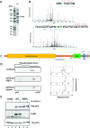
Identification of new phosphorylated residues in Polη. (A) e-Polη was purified and extracted from the gel for MS/MS analysis. (B) MS/MS spectra of the new phosphorylation sites of polη. B ions are marked in blue and Y ions are marked in red, both in the spectra and the peptide sequences. The top spectrum represents the peptide containing P-S687 (*site confidently assigned) while the bottom spectrum represents a peptide (aa 495–533) containing a single phosphate that could not be confidently assigned. Based on the fragmentation pattern the possible location of the phosphate was narrowed down to S510, S512 or S514. (C) Schematic representation of polη domains and alignment of newly identified phospho-residues. (D) 2D-PAGE analysis of XP30RO cells expressing eGFPpolη carrying S687A or S510A, S512A, S514A allele with respective densitometric analysis. The two mutant alleles show a reduced phosphorylation pattern. (E) Characterization of a new antibody against P-S687. Cells were transfected with Flag-polη either WT or S687A and probed with this antibody. Signal is present only in the case of the WT allele.
Figure 5.
Figure 5.
Serine 687 is phosphorylated by CDK and changes during the cell cycle and after UV irradiation. (A) eGFPpolη either WT, S687A or SSSAAA was immunoprecipitated from XP30RO derived cell lines and its phosphorylation status was analyzed with antibodies to P-(S)CDK and pS687. (B) XP30RO cells expressing eGFPpolη were synchronized by double thymidine block and released into the cell cycle in the presence of nocodazole, before IP of eGFPpolη. Polη and P-S687A increase in S phase and G2/M. (C) MRC5 cells were transfected with eGFPpolη and irradiated with 25 J/m2 and followed for the indicated times. (D) Densitometric analysis of polη and P-S687 after UV irradiation. The signal of P-S687 was corrected for the levels of total polη. The plots represent the mean of three experiment ±S.E.M.
Figure 6.
Figure 6.
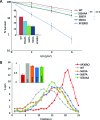
S687 is important for damage bypass and cell survival after UV. (A) Clonogenic survival of XP30RO cells expressing various mutant alleles of polη (n > 3 ± S.E.M). Statistical significance was assessed by t-test: **P = 0.0031, ***P < 0.0001. The inset shows the survival of the cell lines at 6 J/m2 on a linear scale. (B) Damage bypass assay using alkaline sucrose gradient fractionation of newly-synthesized DNA. The inset shows the normalized average molecular weight of DNA from the distributions (n = 3 ± S.E.M).
Figure 7.
Figure 7.
Phosphorylation of polη is important for its stability. (A) Transcript levels of polη after CDK inhibition. (B) Polη protein levels after roscovitine treatment, in the presence of MG132. Relative protein levels normalized to the untreated sample are displayed in red under the eGFPpolη blot. (C) XP30RO cells expressing eGFPpolη WT or S687A were incubated with MG132. Relative protein levels normalized to the 0 h sample of each allele are displayed in bold italic under the eGFPpolη blot. (D) XP30RO cells expressing eGFPpolη WT or S687A were synchronized by double thymidine block and followed in the cell cycle after release in nocodazole for 16 h, before final removal of the drug for 3 h. (E) Densitometric analysis of polη levels, either WT or S687A, during the cell cycle (mean of three experiments ± S.E.M).
Figure 8.
Figure 8.
Proposed model of phosphorylation-controlled TLS. During an unperturbed cell cycle polη becomes phosphorylated by CDK2 on Serine 687 from late S to G2/M. This results in the accumulation of the polymerase, which is available for bypass activity (left panel). After damage, phosphorylated polη is recruited to the chromatin via its UBZ (1) and it is further phosphorylated on S601 by ATR. Once S601 has been phosphorylated, polη can interact with Ubiquitylated PCNA and perform TLS (2). Once the damage has been bypassed P-S687 is removed (3) leading to polη degradation (4).

References

    1. Lehmann A.R., Niimi A., Ogi T., Brown S., Sabbioneda S., Wing J.F., Kannouche P.L., Green C.M.. Translesion synthesis: Y-family polymerases and the polymerase switch. DNA Repair (Amst). 2007; 6:891–899. - PubMed
    1. Biertumpfel C., Zhao Y., Kondo Y., Ramon-Maiques S., Gregory M., Lee J.Y., Masutani C., Lehmann A.R., Hanaoka F., Yang W.. Structure and mechanism of human DNA polymerase eta. Nature. 2010; 465:1044–1048. - PMC - PubMed
    1. Sale J.E., Lehmann A.R., Woodgate R.. Y-family DNA polymerases and their role in tolerance of cellular DNA damage. Nat. Rev. Mol. Cell Biol. 2012; 13:141–152. - PMC - PubMed
    1. Bienko M., Green C.M., Sabbioneda S., Crosetto N., Matic I., Hibbert R.G., Begovic T., Niimi A., Mann M., Lehmann A.R. et al. . Regulation of translesion synthesis DNA polymerase eta by monoubiquitination. Mol. Cell. 2010; 37:396–407. - PubMed
    1. Gohler T., Sabbioneda S., Green C.M., Lehmann A.R.. ATR-mediated phosphorylation of DNA polymerase eta is needed for efficient recovery from UV damage. J. Cell Biol. 2011; 192:219–227. - PMC - PubMed

LinkOut - more resources