. 2017 Sep 19;45(16):9481-9502.
doi: 10.1093/nar/gkx652.

A germline-limited piggyBac transposase gene is required for precise excision in Tetrahymena genome rearrangement

Affiliations

A germline-limited piggyBac transposase gene is required for precise excision in Tetrahymena genome rearrangement

Lifang Feng et al. Nucleic Acids Res. .

Abstract

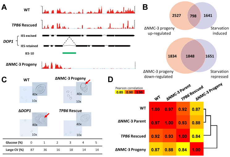
Developmentally programmed genome rearrangement accompanies differentiation of the silent germline micronucleus into the transcriptionally active somatic macronucleus in the ciliated protozoan Tetrahymena thermophila. Internal eliminated sequences (IES) are excised, followed by rejoining of MAC-destined sequences, while fragmentation occurs at conserved chromosome breakage sequences, generating macronuclear chromosomes. Some macronuclear chromosomes, referred to as non-maintained chromosomes (NMC), are lost soon after differentiation. Large NMC contain genes implicated in development-specific roles. One such gene encodes the domesticated piggyBac transposase TPB6, required for heterochromatin-dependent precise excision of IES residing within exons of functionally important genes. These conserved exonic IES determine alternative transcription products in the developing macronucleus; some even contain free-standing genes. Examples of precise loss of some exonic IES in the micronucleus and retention of others in the macronucleus of related species suggest an evolutionary analogy to introns. Our results reveal that germline-limited sequences can encode genes with specific expression patterns and development-related functions, which may be a recurring theme in eukaryotic organisms experiencing programmed genome rearrangement during germline to soma differentiation.


Figures

Figure 1.
Figure 1.
NMC are present in the developing MAC, but absent in the mature MAC. (A) Programmed genome rearrangement, including chromosome fragmentation and excision of internally eliminated sequences (IES), occurs as the somatic MAC is differentiated from the germline MIC-derived zygotic nucleus during sexual reproduction. Addition of telomeres (wavy lines) occurs after chromosome fragmentation at chromosome breakage sequences (CBS). Non-maintained chromosomes (NMC) are present in the developing MAC, but lost in the mature MAC during asexual propagation. (B) Genomic localization of NMC-3: right arm of MIC chromosome 4→MIC genome sequence supercontig_2.75→ CBS: 4R-29 and 4R-30, giving rise to NMC-3 and two adjacent MAC genome sequence scaffolds (scf_8253815 and scf_8254181). (C) DNA sequence coverage of NMC-3 and adjacent regions in the developing MAC and the mature MAC. The developing MAC were isolated from conjugating WT cells at 24 h post-mixing. The mature MAC were purified from the indicated strains of vegetatively growing WT cells. Illumina sequencing results were mapped back to the T. thermophila MIC reference genome (8). (D) DNA sequence coverage of the regions around CBS 4R-29 and 4R-30. Telomere-containing reads (gray) are aligned to and stacked on the reference genome.
Figure 2.
Figure 2.
NMC are generated and amplified in the developing MAC during sexual reproduction, but quickly lost in the mature MAC during asexual propagation. (A) Normalized DNA sequence coverage (RPKM) of NMC-1, 2 and 3 during sexual reproduction and asexual propagation. (B) Comparing coverage of NMC-3 and adjacent MAC-destined sequences (MDS). (C) Comparing coverage of NMC-3 and adjacent internal eliminated sequences (IES). (D) PCR analysis of NMC-3 during conjugation, employing a telomere-specific primer (green arrows) in combination with locus-specific primers (black arrows). Conjugation progress was monitored by a PCR assay for a well-studied IES, the M element. Its processing gives rise to the short PCR product in conjugating CU427 and CU428 cells (red brackets), distinct from the long PCR product from the mature MAC in parental cells (19,41). A MAC gene, JMJ1, was monitored as a loading control. (E) PCR analysis of NMC-3 during asexual propagation. The short PCR product corresponding to the processed M element was monitored as a control for the presence of conjugation progeny (red brackets), which remained at constant levels. JMJ1 was monitored as a loading control.
Figure 3.
Figure 3.
The NMC sequence is retained in the mature MAC when fused to a MAC chromosome. (A) A schematic for deletion of either CBS 4R-29 (CBS-L) or 4R-30 (CBS-R). In the constructs, the corresponding CBS was replaced by the neo4 cassette, conferring paromomycin resistance. (B) Generation of ΔCBS-L cells. The construct was transformed into WT conjugating cells during formation of the developing MAC, but before chromosome fragmentation. Transformed conjugation progeny were selected with paromomycin, passaged asexually for at least 50 generations, and assayed for the presence of NMC-3. DM: the developing MAC; OM: the old MAC. (C) PCR detection of telomere-capped termini of NMC-3 and its adjacent MAC chromosomes in WT, ΔCBS-L, and ΔCBS-R cells. (D) qPCR analysis of the copy number of the NMC-3 sequence in WT (CU427 and CU428), ΔCBS-L and ΔCBS-R cells.
Figure 4.
Figure 4.
TPB6 is expressed from the NMC-3 sequence during formation of the developing MAC. (A) DNA sequence coverage of NMC-3 and adjacent regions in the developing MAC of WT, ΔDCL1, and ΔNMC-3 cells, purified at late conjugation. Positions of NMC-3 (blue box), its flanking CBS (red ovals), and a downstream IES (magenta box) are indicated in the corresponding tracks. (B) mRNA sequence coverage of the same region during formation of the developing MAC. RNA-Seq of polyadenylated (poly(A)) transcripts, isolated from conjugating WT cells (CU427 and CU428) at 3, 6 and 10 h post-mixing (C3, C6 and C10, respectively). A gene model for NMC3-contained TPB6 is shown in a separate track. (C) Generation of ΔNMC-3 strains. The entire region corresponding to NMC-3, including both of its flanking CBS, was replaced by the neo4 cassette. Homozygous heterokaryon strains (ΔNMC-3 parent) were generated by germline transformation and standard genetic manipulations (42,44); they are homozygous for the ΔNMC-3 allele in the MIC, but have a WT MAC. Homozygous homokaryon strains (ΔNMC-3 progeny) were generated by crossing two homozygous heterokaryon strains (ΔNMC-3 parent); they are homozygous for the ΔNMC-3 allele in both the MIC and MAC. (D) Reverse-transcription PCR (RT-PCR) analysis of TPB6 expression during conjugation. RNA was isolated at the indicated hours post-mixing. WT: cross between CU427 and CU428; ΔNMC-3: cross between two ΔNMC-3 homozygous heterokaryon strains (ΔNMC-3 parent). ngoA, which is highly and uniformly expressed during conjugation (Supplementary Figure S17C), was used as a positive control.
Figure 5.
Figure 5.
TPB6 is required for precise excision of a special class of IES. (A) A summary of all 11 TPB6-dependent IES (yellow boxes), together with models of associated MAC genes (exons: black boxes; introns: thin lines) and genes within IES (same as for MAC genes, but in brown). Note that IES-2 and IES-3 are inserted into the same gene. A regular IES (beige box) is nested within IES-11. (B) Excision of IES-4, associated with the coding region of TTHERM_00420400, is abolished in ΔNMC-3 and ΔDCL1 cells. From top to bottom, the tracks represent DNA sequence coverage of the mature MAC from WT and ΔNMC-3 progeny cells, then the developing MAC from WT, ΔNMC-3 and ΔDCL1 cells; directly below is RNA-Seq coverage of poly(A) RNA isolated from conjugating WT cells, at 3 and 10 h post-mixing (C3 and C10, respectively). (C) PCR analysis of excision of all 11 TPB6-dependent IES. Template DNA was prepared from the mature MAC of WT (1), ΔNMC-3 parent (3) and ΔNMC-3 progeny cells (4). It was also prepared from the developing MAC of ΔDCL1 cells (2). Note that the regular IES nested within IES-11 is excised in ΔNMC-3 progeny but not ΔDCL1 cells. (D) PCR analysis of excision of IES-11. Template DNA was prepared from the mature MAC of ΔNMC-3 parent (1) and ΔNMC-3 progeny cells (2). It was also prepared from the developing MAC of WT (3), ΔDCL1 (4), ΔEZL1 (5), and ΔPDD1 cells (6). (E) Highly precise excision of TPB6-dependent IES, compared with imprecise excision of regular IES. (F) Sequence logos for the terminal inverted repeats (TIR) of TPB6-dependent IES. The classical TIR in T. ni piggyBac transposon is above the logos. The left and right correspond to the upstream and downstream TIR (relative to the gene hosting the IES), respectively.
Figure 6.
Figure 6.
TIR flanking TPB6-dependent IES are necessary but not sufficient for excision. (A) Transformation assay for excision of TPB6-dependent IES. IES-4, with its TIR, is inserted into a TTAA sequence within the coding region of the neo4 cassette (neo4::IES-4). Viable transformants were only obtained when the construct was introduced into WT conjugating cells, allowing TPB6 to seamlessly remove IES-4 and restore the neo4 cassette. No viable transformants were obtained with growing cells, in which TPB6 is not expressed. (B) PCR analysis of the neo4 cassette in transformed conjugation progeny (four independent strains). The upper band represents the unprocessed neo4 cassette with IES-4 insertion, while the lower band represents the processed form with IES-4 excised. (C) Factors affecting IES-4 excision. Transformation efficiencies of mutated neo4::IES-4 cassettes were compared to the original cassette. The left TIR consensus sequence was systematically substituted by G, which is not found in any TIR of TPB6-dependent IES. The IES-4 sequence internal to the original TIR was also replaced by heterologous sequences of the same length (UnaG-1 and UnaG-2, two segments of the coding sequence for the UnaG fluorescent protein (111)).
Figure 7.
Figure 7.
Conspicuous phenotypes in ΔNMC-3 progeny are attributable to genes interrupted by retained TPB6-dependent IES. (A) DOP1, encoding a leucine zipper protein potentially localized in the Golgi, is truncated in ΔNMC-3 progeny due to failure to remove IES-10. Shown here are tracks representing RNA-Seq coverage of poly(A) RNA isolated from growing cells of WT, TPB6 rescued, and ΔNMC3 progeny strains. Gene models for both IES-excised and IES-retained forms of DOP1 are also shown, with the insertion of IES-10 (green box and dashed lines) indicated. (B) Overlap between differentially expressed genes from ΔNMC-3 growing cells and starved WT cells. The left circles represent genes up-regulated (top) or down-regulated (bottom) in ΔNMC3 progeny growing cells, compared to WT growing cells. The right circles represent genes up-regulated (top) or down-regulated (bottom) in WT starved cells, compared to WT growing cells. Additional analyses of genes in the overlap are provided in Supplementary Figure S17C-E. (C) Extraordinarily large contractile vacuole in ΔNMC3 progeny and DOP1::IES-10 cells. This phenotype was aggravated in hypotonic media, alleviated in hypertonic media, and rescued by a DNA fragment containing TPB6 delivered into conjugating ΔNMC3 parental cells. (D) Correlations in gene expression profiles. Expression levels (RPKM) are calculated based on RNA-Seq coverage of poly(A) RNA isolated from growing cells of WT, TPB6 rescued, ΔNMC3 parent, and ΔNMC3 progeny strains. Pearson's correlation coefficients (PCC) are calculated for pair-wise comparison and illustrated by the color scale. Cluster analysis, based on PCC, is shown on the right. Note the similarities between the gene expression profiles of WT, TPB6 rescued, and ΔNMC3 parent strains, and their distinction from that of ΔNMC3 progeny.
Figure 8.
Figure 8.
Conservation of TPB6-dependent IES in Tetrahymena species. (A) Conservation of the IES-4 insertion site in four tetrahymenine species. Amino acid (top panel; black shading: identical; gray shading: similar) and nucleotide sequence alignment (bottom panel; black shading: identical) of the corresponding MAC genes containing the IES-4 insertion site (aqua letters). (B) IES-4 equivalent insertions amplified from four Tetrahymena species, using specific primers ending at the TIR. The PCR products were subsequently cloned and sequenced. (C) Summary of TPB6-dependent IES identified in four Tetrahymena species. Left panel: the sizes for all identified IES (their sequences are provided in Supplementary File S4). Note the failure to remove the IES-5 equivalent in the MAC of T. borealis (red asterisk), and the missing of the IES-1, IES-6 and IES-9 equivalents in the MIC of T. malaccensis (green asterisks). Right panel: sequence logos for the left and right boundaries of TPB6-dependent IES in four Tetrahymena species. The left and right boundaries correspond to the upstream and downstream TIR (with adjacent IES sequences), respectively. Note the consensus of the left boundary of IES-5, which deviates from the highly conserved TIR motif, is affected by the mutation in T. borealis that prevents excision of that IES. (D) A single nucleotide mutation (G substitution) in TIR of the IES-5 equivalent is associated with its retention. Using primers flanking the TIR, both processed (blue arrowhead) and unprocessed forms (red arrowhead) of IES-5 were amplified from the T. thermophila, T. malaccensis and T. elliotti samples, while only the unprocessed form was amplified from the T. borealis sample. (E) Conservation of the ORF contained in IES-7.

Similar articles

Cited by

References

    1. Wang J., Davis R.E.. Programmed DNA elimination in multicellular organisms. Curr. Opin. Genet. Dev. 2014; 27:26–34. - PMC - PubMed
    1. Karrer K.M. Nuclear dualism. Methods Cell Biol. 2012; 109:29–52. - PubMed
    1. Bassing C.H., Swat W., Alt F.W.. The mechanism and regulation of chromosomal V(D)J recombination. Cell. 2002; 109(Suppl.):S45–S55. - PubMed
    1. Cervantes M.D., Hamilton E.P., Xiong J., Lawson M.J., Yuan D., Hadjithomas M., Miao W., Orias E.. Selecting one of several mating types through gene segment joining and deletion in Tetrahymena thermophila. PLoS Biol. 2013; 11:e1001518. - PMC - PubMed
    1. Chen X., Bracht J.R., Goldman A.D., Dolzhenko E., Clay D.M., Swart E.C., Perlman D.H., Doak T.G., Stuart A., Amemiya C.T. et al. . The architecture of a scrambled genome reveals massive levels of genomic rearrangement during development. Cell. 2014; 158:1187–1198. - PMC - PubMed

LinkOut - more resources