. 2017 Sep 19;45(16):9611-9624.
doi: 10.1093/nar/gkx649.

Activities of gyrase and topoisomerase IV on positively supercoiled DNA

Affiliations

Activities of gyrase and topoisomerase IV on positively supercoiled DNA

Rachel E Ashley et al. Nucleic Acids Res. .

Abstract

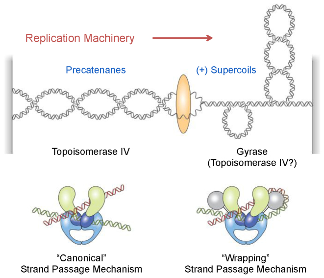
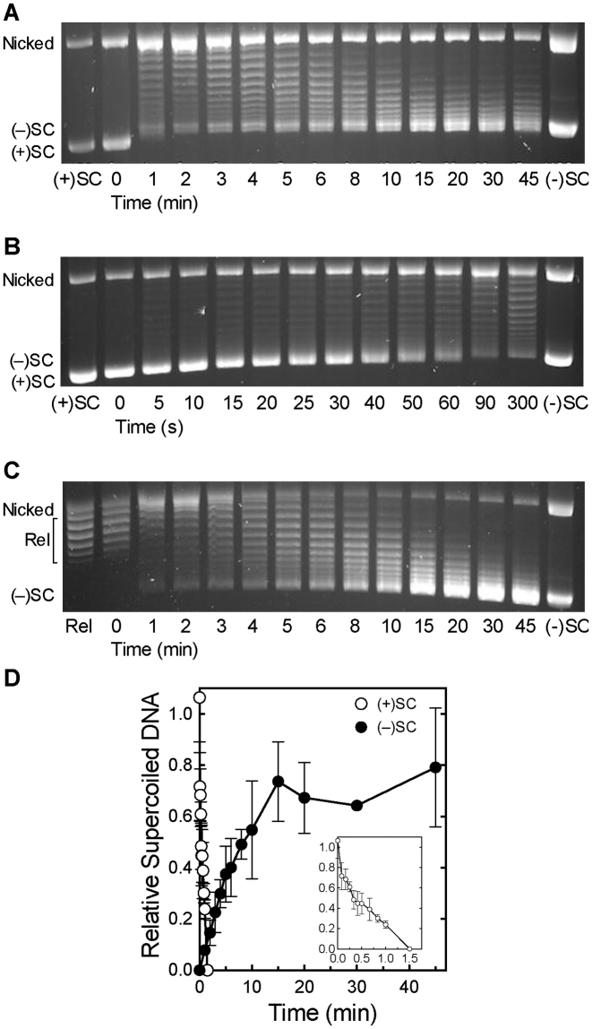
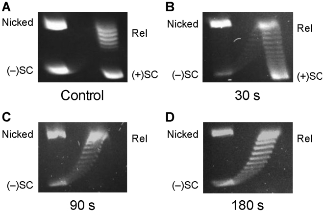
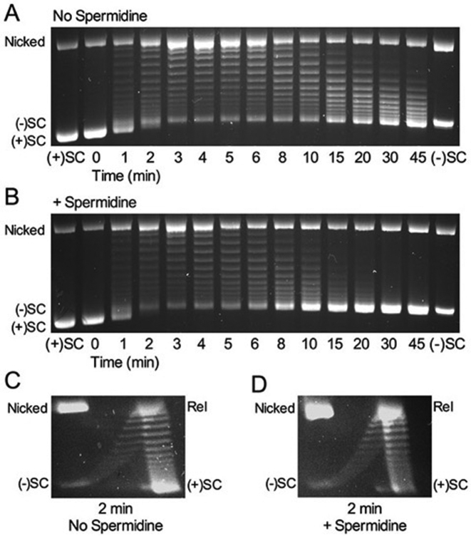
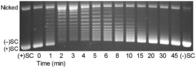
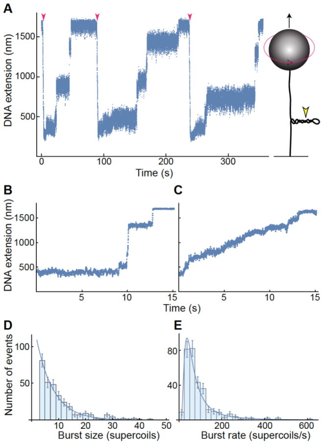
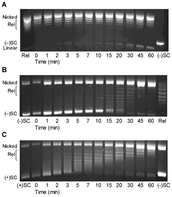
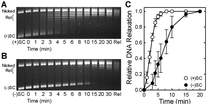
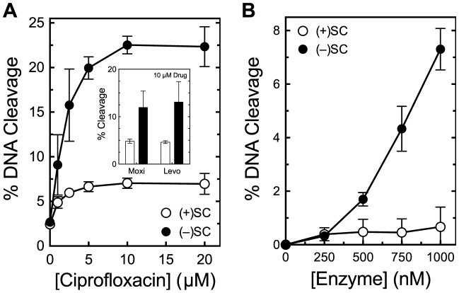
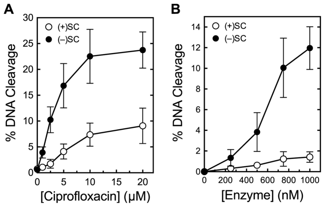
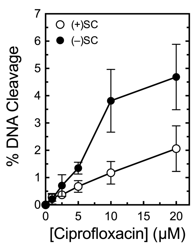
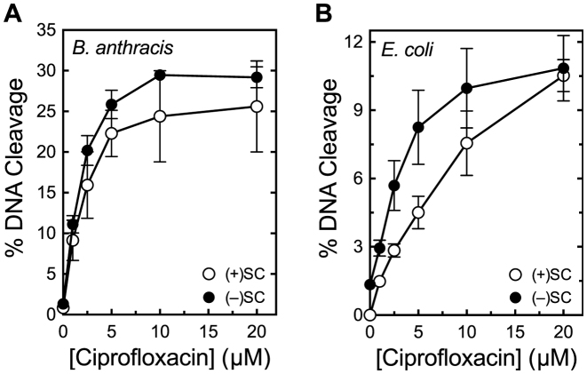
Although bacterial gyrase and topoisomerase IV have critical interactions with positively supercoiled DNA, little is known about the actions of these enzymes on overwound substrates. Therefore, the abilities of Bacillus anthracis and Escherichia coli gyrase and topoisomerase IV to relax and cleave positively supercoiled DNA were analyzed. Gyrase removed positive supercoils ∼10-fold more rapidly and more processively than it introduced negative supercoils into relaxed DNA. In time-resolved single-molecule measurements, gyrase relaxed overwound DNA with burst rates of ∼100 supercoils per second (average burst size was 6.2 supercoils). Efficient positive supercoil removal required the GyrA-box, which is necessary for DNA wrapping. Topoisomerase IV also was able to distinguish DNA geometry during strand passage and relaxed positively supercoiled substrates ∼3-fold faster than negatively supercoiled molecules. Gyrase maintained lower levels of cleavage complexes with positively supercoiled (compared with negatively supercoiled) DNA, whereas topoisomerase IV generated similar levels with both substrates. Results indicate that gyrase is better suited than topoisomerase IV to safely remove positive supercoils that accumulate ahead of replication forks. They also suggest that the wrapping mechanism of gyrase may have evolved to promote rapid removal of positive supercoils, rather than induction of negative supercoils.


Figures

Figure 1.
Figure 1.
Cellular functions and DNA strand passage mechanisms of gyrase and topoisomerase IV. Gyrase removes positively supercoiled DNA ahead of the replication machinery and also introduces negative supercoils into the genome. Topoisomerase IV may assist in the removal of positive supercoils, but primarily acts to resolve precatenanes behind the fork and unlink daughter chromosomes. Topoisomerase IV uses a ‘canonical’ DNA strand passage mechanism and gyrase uses a ‘wrapping’ mechanism to support their decatenation and supercoiling activities, respectively.
Figure 2.
Figure 2.
Bacillus anthracis gyrase removes positive supercoils more rapidly than it introduces negative supercoils into relaxed DNA. (A) Gyrase activity on positively supercoiled DNA. A time course is shown for the relaxation of positive supercoils followed by the introduction of negative supercoils. Positively supercoiled [(+)SC] and negatively supercoiled [(−)SC] standards are shown. (B) Expanded time course for the relaxation of (+)SC DNA by gyrase. (C) Time course for the introduction of negative supercoils into relaxed DNA (Rel) by gyrase. Gel images are representative of at least three independent experiments. (D) Quantification of removal of positive supercoils and introduction of negative supercoils by gyrase shown in parts A–C. The relative amount of (+)SC DNA (white) in each sample was determined by comparison to the (+)SC control. The relative amount of (−)SC DNA (black) in each sample was determined by comparison to the maximum level of (−)SC DNA formed in each experiment. Inset: expanded view of loss of (+)SC DNA. Error bars represent standard deviations of at least three independent experiments.
Figure 3.
Figure 3.
2D gel analysis of Bacillus anthracis gyrase activity on positively supercoiled DNA. (A) Control gel showing mobility of nicked, positively supercoiled [(+)SC], relaxed (Rel) and negatively supercoiled [(−)SC] DNA. (B–D) Activity of gyrase on (+)SC DNA. DNA products generated after 30 s (B), 90 s (C) and 180 s (D) reactions are shown. Gel images are representative of at least three independent experiments.
Figure 4.
Figure 4.
Spermidine enhances the rate of negative supercoiling by Bacillus anthracis gyrase but does not affect the rate of removal of positive supercoils. (A) Gyrase activity on positively supercoiled DNA without spermidine. Positively supercoiled [(+)SC] and negatively supercoiled [(−)SC] standards are shown. (B) Gyrase activity on positively supercoiled DNA in the presence of 5 mM spermidine. (C and D) 2D gel analysis of gyrase activity on positively supercoiled DNA. DNA products generated after 2 min in the absence (C) or presence (D) of 5 mM spermidine. Gel images are representative of at least three independent experiments.
Figure 5.
Figure 5.
Escherichia coli gyrase removes positive supercoils more rapidly than it introduces negative supercoils into relaxed DNA. A time course is shown for the relaxation of positive supercoils followed by the introduction of negative supercoils. Positively supercoiled [(+)SC] and negatively supercoiled [(−)SC] standards are shown. The gel image is representative of at least three independent experiments.
Figure 6.
Figure 6.
Single-molecule measurements of Bacillus anthracis gyrase activity. Data are representative of measurements made on five individual DNA tethers on different days. (A) Representative trace of gyrase activity over time using magnetic tweezers. A cartoon of the experimental setup (not to scale) is shown at right: a single DNA molecule (black line) is torsionally constrained by attachment to a slide and manipulated by the attached magnetic bead (gray sphere). Both the upward force (black arrow) on the bead and its rotation (red arrow) are controlled through an externally applied magnetic field. Counterclockwise bead rotation increases the linking number of the DNA, generating positive supercoils. DNA extension (determined by bead height above slide surface) decreases in proportion to plectoneme supercoiling (yellow arrowhead). Results are shown at left for a single DNA molecule that was extended by a constant upward magnetic force of 1 pN. Upon introduction of 50 positive supercoils (red arrowheads), the DNA extension was reduced as a plectoneme formed. Gyrase removed supercoils in a series of discrete steps and the DNA returned to its initial length, initiating the onset of another measurement cycle. (B and C) Gyrase relaxes positive DNA supercoils in two distinct modes of activity. Representative traces in which the enzyme removed multiple supercoils either in rapid bursts (‘burst mode’ relaxation, B) or at a steady rate (‘steady mode’ relaxation, C), are shown. The DNA was under constant tension of 3.5 and 2.2 pN in B and C, respectively. (D and E) Characterization of burst mode relaxation. Distributions include measurements of 382 individual relaxation cycles in which 50 positive supercoils were removed. In each burst event, gyrase rapidly removed four or more supercoils. The burst size distribution (D) fits a single exponential curve. The burst rate distribution (E) fits an inverse gamma function with shape parameter α = 2.1 ± 0.2 and scale parameter β = 114 ± 11 supercoils/s. Error bars represent the square-root of the number of observed events; these errors were accounted for in the determination of the best-fit parameters (±SE).
Figure 7.
Figure 7.
Bacillus anthracis GyrAAla-box gyrase acts as a canonical type II topoisomerase. (A) GyrAAla-box gyrase does not supercoil relaxed DNA. Relaxed (Rel) and negatively supercoiled [(−)SC] DNA standards are shown. (B) GyrAAla-box gyrase slowly relaxes (−)SC DNA. (C) GyrAAla-box gyrase slowly and distributively relaxes positively supercoiled [(+)SC] DNA. Gel images are representative of at least three independent experiments.
Figure 8.
Figure 8.
Bacillus anthracis topoisomerase IV relaxes positively supercoiled DNA faster than negatively supercoiled DNA. (A and B) Relaxation of positively (A) and negatively (B) supercoiled DNA by topoisomerase IV. Positively supercoiled [(+)SC], negatively supercoiled [(−)SC] and relaxed (Rel) standards are shown. Gel images are representative of at least three independent experiments. (C) Quantification of experiments shown in (A) and (B). Relative amounts of relaxed DNA in each experiment were determined by monitoring the loss of the supercoiled band in comparison to supercoiled DNA present in the 0 min sample. Error bars represent standard deviations for at least three independent experiments.
Figure 9.
Figure 9.
Bacillus anthracis gyrase maintains lower levels of cleavage complexes on positively supercoiled DNA. (A) Levels of cleavage complexes generated by gyrase on positively supercoiled [(+)SC] DNA (white) or negatively supercoiled [(−)SC] DNA (black) in the presence of ciprofloxacin. Inset: levels of cleavage complexes generated by gyrase in the presence of 10 μM moxifloxacin (Moxi) or levofloxacin (Levo). (B) Levels of cleavage complexes generated by varying concentrations of gyrase on (+)SC DNA (white) or (−)SC DNA (black) in the absence of quinolones. Error bars represent standard deviations for at least three independent experiments.
Figure 10.
Figure 10.
Bacillus anthracis GyrAAla-box gyrase maintains lower levels of cleavage complexes on positively supercoiled DNA. (A) Levels of cleavage complexes generated by GyrAAla-box gyrase on positively supercoiled [(+)SC] DNA (white) or negatively supercoiled [(−)SC] DNA (black) in the presence of ciprofloxacin. (B) Levels of cleavage complexes generated by GyrAAla-box gyrase on (+)SC DNA (white) or (−)SC DNA (black) in the absence of quinolones. Error bars represent standard deviations for at least three independent experiments.
Figure 11.
Figure 11.
Escherichia coli gyrase maintains lower levels of cleavage complexes on positively supercoiled DNA. Levels of cleavage complexes generated by gyrase on positively supercoiled [(+)SC] DNA (white) or negatively supercoiled [(−)SC] DNA (black) in the presence of ciprofloxacin are shown. Error bars represent standard deviations for at least three independent experiments.
Figure 12.
Figure 12.
Topoisomerase IV maintains similar levels of cleavage complexes on positively and negatively supercoiled DNA. (A) Levels of cleavage complexes generated by Bacillus anthracis topoisomerase IV on positively supercoiled [(+)SC] DNA (white) or negatively supercoiled [(−)SC] DNA (black) in the presence of ciprofloxacin. (B) Levels of cleavage complexes generated by Escherichia coli topoisomerase IV on (+)SC DNA (white) or (−)SC DNA (black) in the presence of ciprofloxacin. Error bars represent standard deviations for at least three independent experiments.

Similar articles

Cited by

References

    1. Wang J.C. Cellular roles of DNA topoisomerases: a molecular perspective. Nat. Rev. Mol. Cell Biol. 2002; 3:430–440. - PubMed
    1. Koster D.A., Crut A., Shuman S., Bjornsti M.A., Dekker N.H.. Cellular strategies for regulating DNA supercoiling: a single-molecule perspective. Cell. 2010; 142:519–530. - PMC - PubMed
    1. Vos S.M., Tretter E.M., Schmidt B.H., Berger J.M.. All tangled up: how cells direct, manage and exploit topoisomerase function. Nat. Rev. Mol. Cell Biol. 2011; 12:827–841. - PMC - PubMed
    1. Bates A.D., Maxwell A.. DNA Topology. 2005; NY: Oxford University Press.
    1. Liu Z., Deibler R.W., Chan H.S., Zechiedrich L.. The why and how of DNA unlinking. Nucleic Acids Res. 2009; 37:661–671. - PMC - PubMed

MeSH terms

LinkOut - more resources