. 2017 Sep 1;40(9):zsx121.
doi: 10.1093/sleep/zsx121.

Traveling Slow Oscillations During Sleep: A Marker of Brain Connectivity in Childhood

Affiliations

Traveling Slow Oscillations During Sleep: A Marker of Brain Connectivity in Childhood

Salome Kurth et al. Sleep. .

Abstract

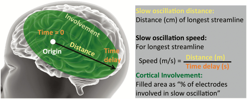
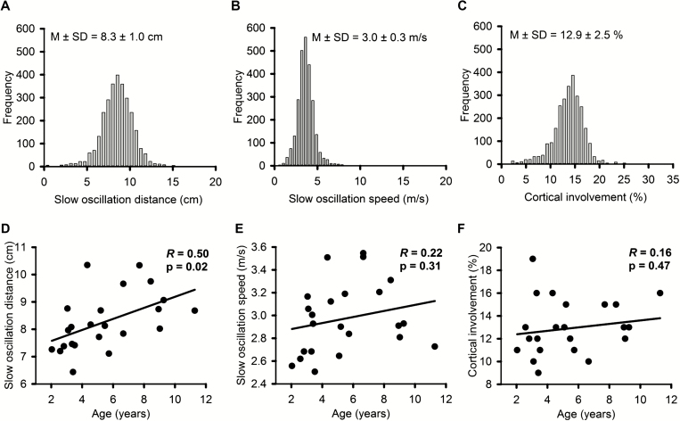
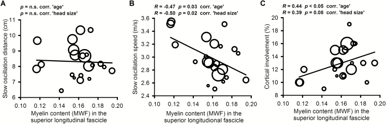
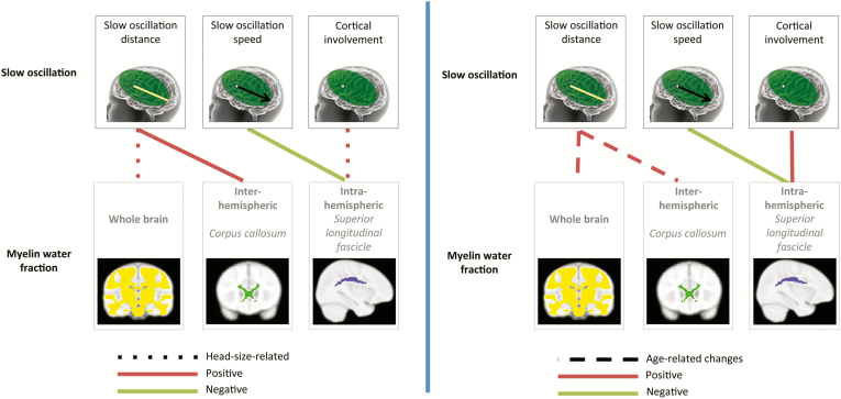
Slow oscillations, a defining characteristic of the nonrapid eye movement sleep electroencephalogram (EEG), proliferate across the scalp in highly reproducible patterns. In adults, the propagation of slow oscillations is a recognized fingerprint of brain connectivity and excitability. In this study, we (1) describe for the first time maturational features of sleep slow oscillation propagation in children (n = 23; 2-13 years) using high-density (hd) EEG and (2) examine associations between sleep slow oscillatory propagation characteristics (ie, distance, traveling speed, cortical involvement) and white matter myelin microstructure as measured with multicomponent Driven Equilibrium Single Pulse Observation of T1 and T2-magnetic resonance imaging (mcDESPOT-MRI). Results showed that with increasing age, slow oscillations propagated across longer distances (average growth of 0.2 cm per year; R(21) = 0.50, p < .05), while traveling speed and cortical involvement (ie, slow oscillation expanse) remained unchanged across childhood. Cortical involvement (R(20) = 0.44) and slow oscillation speed (R(20) = -0.47; both p < .05, corrected for age) were associated with myelin content in the superior longitudinal fascicle, the largest anterior-posterior, intrahemispheric white matter connectivity tract. Furthermore, slow oscillation distance was moderately associated with whole-brain (R(21) = 0.46, p < .05) and interhemispheric myelin content, the latter represented by callosal myelin water fraction (R(21) = 0.54, p < .01, uncorrected). Thus, we demonstrate age-related changes in slow oscillation propagation distance, as well as regional associations between brain activity during sleep and the anatomical connectivity of white matter microstructure. Our findings make an important contribution to knowledge of the brain connectome using a noninvasive and novel analytic approach. These data also have implications for understanding the emergence of neurodevelopmental disorders and the role of sleep in brain maturation trajectories.

Keywords: brain maturation; high-density EEG; mcDESPOT; myelination; neurodevelopment; traveling waves; white matter.


Figures

Figure 1
Figure 1
Wave detection. The EEG signal of all channels overlaid for a representative 10-second time segment in NREM sleep stage 3; reference wave is plotted in black. The detection algorithm identified the waves indicated with black dots. Waves surviving the criteria targeting stereotyped large-amplitude waves are indicated with red dots (see Methods).
Figure 2
Figure 2
Slow oscillation propagation metrics. Slow oscillation parameters (see also Methods section and Figure 1).
Figure 3
Figure 3
Slow oscillation propagation distance but not speed or cortical involvement increases with age. Histograms based on data distribution for each participant and then averaged across participants for (A) slow oscillation distance (skewness 0.2–0.5, kurtosis 2.3–2.5), (B) slow oscillation speed (skewness 1.4–2.2, kurtosis 4.8–8.8), and (C) cortical involvement (skewness 0.3–0.7, kurtosis 2.2–2.7). M ± SD indicate mean and standard deviation. Pearson correlations between age and (D) slow oscillation distance, (E) slow oscillation speed, and (F) cortical involvement; df = 21. Partial correlations reveal R(20) = 0.44, p = .04 for age versus slow oscillation distance, factor “head size”, R(20) = 0.19, p = .40 for age versus slow oscillation speed, factor “head size”, and R(20) = 0.09, p = .71 for age versus cortical involvement, factor “head size”.
Figure 4
Figure 4
Relationship between slow oscillation propagation and myelin water fraction (MWF) in the superior longitudinal fascicle. (A) Slow oscillation distance, (B) slow oscillation speed, and (C) cortical involvement. Statistics indicate partial correlations with factor “age” and “head size” (df = 20). Correlations survive controlling in most cases (MWF in superior longitudinal fascicle vs. slow oscillation speed; MWF in superior longitudinal fascicle vs. cortical involvement). Circle size is age coded with larger circles signifying older participants.
Figure 5
Figure 5
Summarized relationships between slow oscillation propagation and myelin water fraction (MWF). Connections coded for corrections (dotted lines when relationships did not survive controlling for “head size”, left side, dashed line when relationships did not survive controlling for “age”, right side) represent correlative associations between slow oscillation parameters (distance, speed, cortical involvement) and MWF (data are presented in Figures 3 and 4, and Tables 1 and 2). MWF was included for the whole brain, the superior longitudinal fascicle, which is the primary intrahemispheric fiber tract of anterior-posterior connectivity, and, respectively, the corpus callosum, representative of interhemispheric connectivity.

Similar articles

Cited by

References

    1. Marx V. Neurobiology: brain mapping in high resolution. Nature. 2013; 503(7474): 147–152. - PubMed
    1. Kurth S, Ringli M, Geiger A, LeBourgeois M, Jenni OG, Huber R. Mapping of cortical activity in the first two decades of life: a high-density sleep electroencephalogram study. J Neurosci. 2010; 30(40): 13211–13219. - PMC - PubMed
    1. Deoni SC, Matthews L, Kolind SH. One component? Two components? Three? The effect of including a nonexchanging “free” water component in multicomponent driven equilibrium single pulse observation of T1 and T2. Magn Reson Med. 2013; 70(1): 147–154. - PMC - PubMed
    1. Belaroussi B, Milles J, Carme S, Zhu YM, Benoit-Cattin H. Intensity non-uniformity correction in MRI: existing methods and their validation. Med Image Anal. 2006; 10(2): 234–246. - PubMed
    1. Massimini M, Huber R, Ferrarelli F, Hill S, Tononi G. The sleep slow oscillation as a traveling wave. J Neurosci. 2004; 24(31): 6862–6870. - PMC - PubMed