

doi: 10.1164/rccm.201707-1446LE.
Endobronchial Optical Coherence Tomography for Low-Risk Microscopic Assessment and Diagnosis of Idiopathic Pulmonary Fibrosis In Vivo
Affiliations
- PMID: 28934552
- PMCID: PMC6020407
- DOI: 10.1164/rccm.201707-1446LE
Item in Clipboard
Endobronchial Optical Coherence Tomography for Low-Risk Microscopic Assessment and Diagnosis of Idiopathic Pulmonary Fibrosis In Vivo
Am J Respir Crit Care Med.
.
No abstract available
Figures
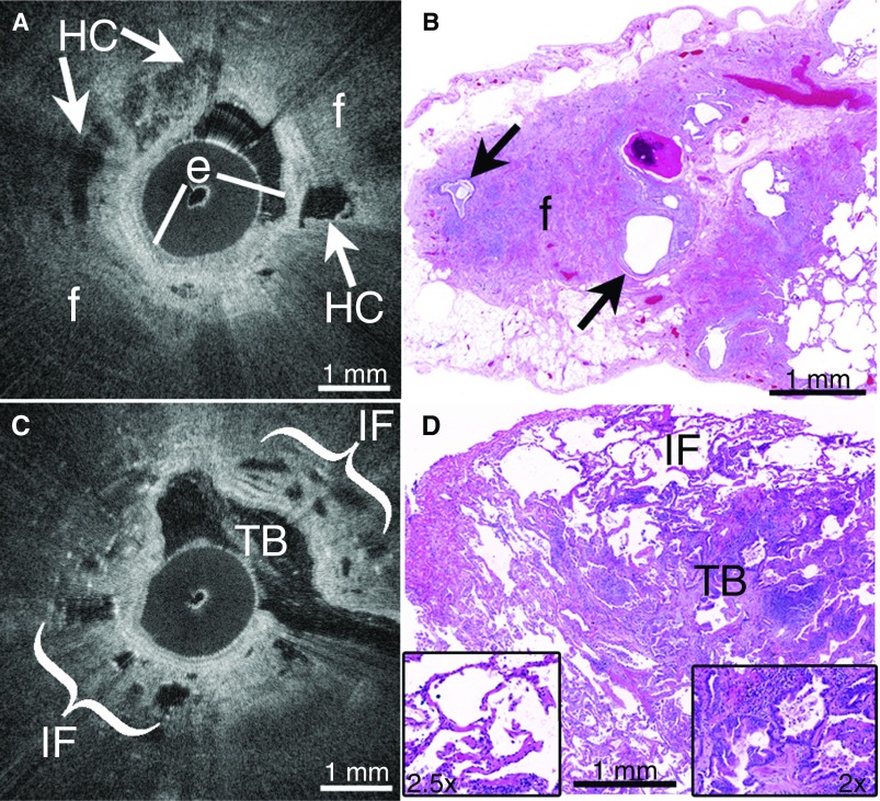
(A) In vivo endobronchial optical coherence tomography (OCT) accurately identified features of usual interstitial pneumonitis (UIP)/idiopathic pulmonary fibrosis (IPF), including multifocal microscopic honeycombing (HC, arrows) as layered cystic structures embedded within peripheral, destructive fibrosis (f) beyond the bronchiolar epithelium (e). (B) Subsequent surgical lung biopsies were independently diagnosed as UIP, and confirmed the presence of peripheral fibrosis (f) and microscopic honeycombing (arrows). (C) OCT accurately identified traction bronchiectasis (TB) as cystic airway branchpoints, and nondestructive interstitial fibrosis (IF) in alveolar walls in non-UIP/IPF interstitial lung disease (ILD). (D) Subsequent surgical lung biopsy independently confirmed all features visualized on OCT, including traction bronchiectasis (TB, right inset) and interstitial fibrosis (IF) in alveolar walls (left inset). No honeycombing or other UIP features were seen, and the biopsy was diagnosed as a non-UIP/IPF ILD.
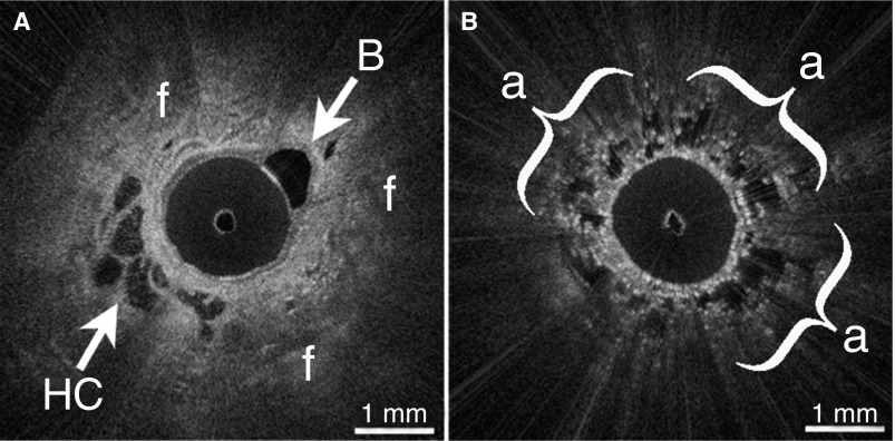
(A) In vivo endobronchial optical coherence tomography (OCT) identified microscopic honeycombing (HC) and destructive peripheral fibrosis (f) in a patient with nondiagnostic high-resolution computed tomography and indeterminate lung biopsy. A branching bronchiole (B) is also visible. (B) OCT visualized spatial heterogeneity as regions of preserved alveoli (a) adjacent to fibrosis.
Comment in
-
Seeing Deeply into the Lung in Interstitial Lung Disease.Am J Respir Crit Care Med. 2018 Apr 1;197(7):857-858. doi: 10.1164/rccm.201709-1798ED. Am J Respir Crit Care Med. 2018. PMID: 28957640 No abstract available.
-
Reply to Wijmans et al.: Optical Coherence Tomography: A Valuable Novel Tool for Assessing the Alveolar Compartment in Interstitial Lung Disease?Am J Respir Crit Care Med. 2018 May 1;197(9):1232-1233. doi: 10.1164/rccm.201711-2347LE. Am J Respir Crit Care Med. 2018. PMID: 29244521 Free PMC article. No abstract available.
-
Optical Coherence Tomography: A Valuable Novel Tool for Assessing the Alveolar Compartment in Interstitial Lung Disease?Am J Respir Crit Care Med. 2018 May 1;197(9):1231-1232. doi: 10.1164/rccm.201711-2152LE. Am J Respir Crit Care Med. 2018. PMID: 29244526 No abstract available.
References
-
- Raghu G, Collard HR, Egan JJ, Martinez FJ, Behr J, Brown KK, et al. ATS/ERS/JRS/ALAT Committee on Idiopathic Pulmonary Fibrosis. An official ATS/ERS/JRS/ALAT statement: idiopathic pulmonary fibrosis: evidence-based guidelines for diagnosis and management. Am J Respir Crit Care Med. 2011;183:788–824. - PMC - PubMed
-
- Richeldi L, du Bois RM, Raghu G, Azuma A, Brown KK, Costabel U, et al. INPULSIS Trial Investigators. Efficacy and safety of nintedanib in idiopathic pulmonary fibrosis. N Engl J Med. 2014;370:2071–2082. - PubMed
-
- King TE, Jr, Bradford WZ, Castro-Bernardini S, Fagan EA, Glaspole I, Glassberg MK, et al. ASCEND Study Group. A phase 3 trial of pirfenidone in patients with idiopathic pulmonary fibrosis. N Engl J Med. 2014;370:2083–2092. - PubMed
Publication types
MeSH terms
Grants and funding
LinkOut - more resources
Full Text Sources
Other Literature Sources
