. 2017 Sep 1;125(9):097001.
doi: 10.1289/EHP1443.

Neural Mechanisms Underlying the Disruption of Male Courtship Behavior by Adult Exposure to Di(2-ethylhexyl) Phthalate in Mice

Affiliations

Neural Mechanisms Underlying the Disruption of Male Courtship Behavior by Adult Exposure to Di(2-ethylhexyl) Phthalate in Mice

Carlos Dombret et al. Environ Health Perspect. .

Abstract

Background: Courtship behavior plays a critical role in attracting females and reproduction success. However, the effects of exposure to a ubiquitous contaminant di(2-ethylhexyl) phthalate (DEHP) on these behaviors and, in particular, on courtship vocalizations have not been examined.

Objective: The effects of adult exposure to DEHP on courtship and mating behaviors and gonadotropic axis and neural mechanisms involved in DEHP-induced effects were analyzed in male mice.

Methods: Adult C57BL/6J males were orally exposed to DEHP (0, 0.5, 5, and 50μg/kg/d) for 4 wk. Olfactory preference, ultrasonic vocalizations (USVs), partner preference and mating, as well as locomotor activity and motor coordination, were measured. The kisspeptin system and testosterone levels were analyzed. Proteomic and molecular studies were conducted on the hypothalamic preoptic nucleus, the key region involved in sexual motivation to vocalize and mate.

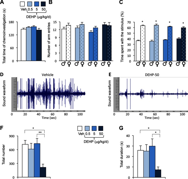
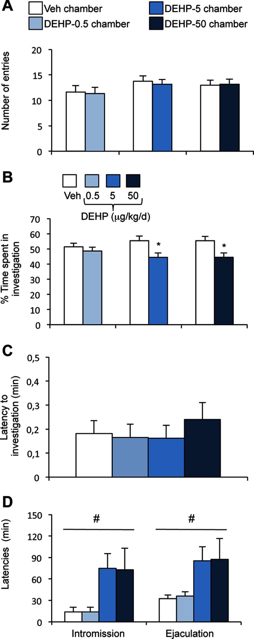
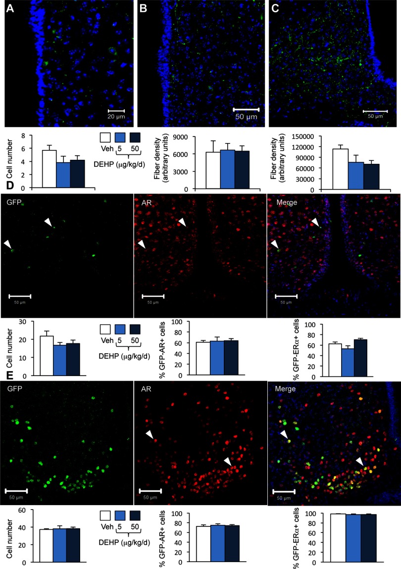
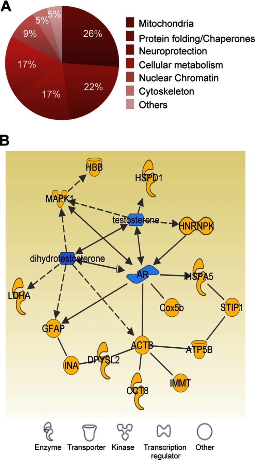
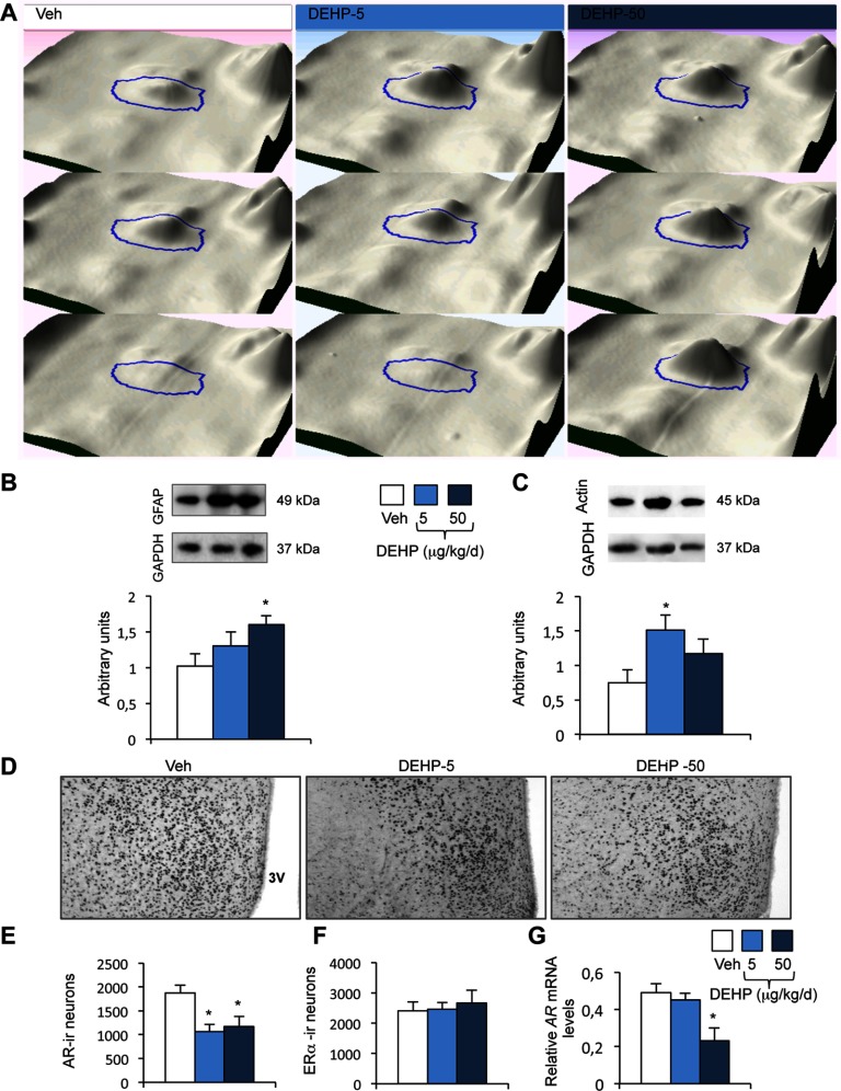
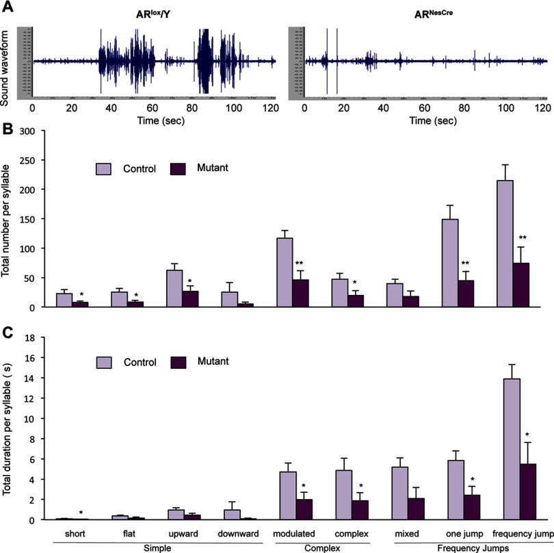
Results: DEHP at 50μg/kg/d reduced the emission of USVs, whereas lower doses changed the ratio of syllable categories. This was associated with diminished sexual interest of female partners toward males exposed to 5 or 50μg/kg/d and increased latency to mate, despite normal olfactory preference. The kisspeptin system and circulating testosterone levels were unaffected. In DEHP-exposed males, proteomic analysis of the preoptic nucleus identified differentially expressed proteins connected to the androgen receptor (AR). Indeed, exposure to 5 or 50μg/kg/d of DEHP induced selective AR downregulation in this nucleus and upstream chemosensory regions. The involvement of AR changes in the observed alterations was further supported by the reduced emission of courtship vocalizations in males with disrupted neural AR expression.

Conclusions: These data demonstrate the critical role of neural AR in courtship vocalizations and raises the possibility that the vulnerability of this signaling pathway to exposure to endocrine disrupters may be detrimental for courtship communication and mating in several species. https://doi.org/10.1289/EHP1443.


Figures

Figure 1A, 1F, and 1G are bar graphs with standard errors of mean plotting total time of chemoinvestigation, total number, and total duration in seconds, respectively, (y-axis) for the Veh and 0.5, 5, and 50 micrograms per kilogram per day DEHP groups (x-axis). Figures 1B and 1C are bar graphs with standard errors of mean plotting number of arm entries and percent time spent with the stimulus, respectively, for male and female components of the exposure groups (x-axis). Figures 1D and 1E illustrate sound waveforms (y-axis) for the duration of exposure to for Vehicle and DEHP-50), respectively, in seconds (x-axis).
Figure 1.
Effects of phthalate di(2-ethylhexyl) phthalate (DEHP) on olfactory preference and courtship vocalizations in male mice. (A) Total time spent in the chemoinvestigation of male (♂) and sexually receptive female (♀) stimuli by males exposed to the vehicle (Veh) or DEHP (0.5, 5, or 50μg/kg/d). (B) Number of entries into the male or female arm of the Y-maze. (C) Percentage of time spent in investigating males (♂) vs. sexually receptive females (♀). Data are expressed as the means ±SEM of 11–12 males per treatment group, *p<0.05 vs. the male arm. (DE) Representative sound waveform for males exposed to vehicle (D) or DEHP at 50μg/kg/d (E) in the presence of a sexually receptive female. (FG) Total number (F) and duration (G) of ultrasonic vocalizations (USVs) produced during the 4-min test by males exposed to the vehicle or DEHP at 0.5, 5, or 50μg/kg/d. Data are expressed as the means ±SEM of 11–12 males per treatment group; *p<0.05, **p<0.01.
Figure 2A is a bar graph with standard errors of mean plotting total number (y-axis) of short, flat, upward, and downward (simple group); modulated and complex (complex group); and mixed, one jump, and frequency jump (frequency jump group) syllables (x-axis) for the Veh and 0.5, 5, and 50 micrograms per kilogram per day DEHP exposure groups. Figure 2B comprises four pie charts showing the proportion of simple, complex, and frequency jump syllables in the Veh (29, 24, and 47 percent) and DEHP 0.5 (18, 25, 57 and percent), DEHP 5 (18, 28, and 54 percent), and DEHP 50 (29.5, 26, and 44.5 percent) exposure groups. Figure 2C is a bar graph with standard errors of mean plotting total duration (y-axis) of short, flat, upward, and downward (simple group); modulated and complex (complex group); and mixed, one jump, and frequency jump (frequency jump group) syllables (x-axis) for the Veh and 0.5, 5, and 50 micrograms per kilogram per day DEHP exposure groups].
Figure 2.
Quantitative and qualitative analyses of syllables emitted by males exposed to di(2-ethylhexyl) phthalate (DEHP). (A) Total number of each syllable type produced in the presence of a sexually receptive female during the 4-min recording for males exposed to the vehicle (Veh) or DEHP at 0.5, 5, or 50μg/kg/d. One-way analysis of variance (ANOVA) showed an effect of DEHP treatment (#) on short (p<0.0001), flat (p=0.0008), downward (p=0.007), modulated (p=0.03), complex (p=0.01), mixed (p=0.05), one-jump (p=0.013), and frequency jump (0.019) syllables. Post hoc comparisons are indicated: *p<0.05, **p<0.01, ***p<0.001. Data are expressed as the means ±SEM of 11–12 males per treatment group. (B) Percentage of courtship vocalizations per category (simple, complex, and with frequency jumps). (C) Total duration of each syllable type. One-way ANOVA showed an effect of DEHP treatment (#) on short (p=0.0003), modulated (p=0.047), complex (p=0.035), one-jump (p=0.035), and frequency jump (0.05) syllables. Post hoc comparisons are indicated: *p<0.05, **p<0.01, ***p<0.001.
Figure 3A is a bar graph with standard errors of mean plotting number of entries of sexually receptive females (y-axis) for the Veh and 0.5, 5, and 50 micrograms per kilogram per day DEHP exposure groups. Figure 3B is a bar graph with standard errors of mean plotting percentage of time spent in investigation (y-axis) for the Veh and 0.5, 5, and 50 micrograms per kilogram per day DEHP exposure groups. Figure 3C is a bar graph with standard errors of mean plotting latency to investigation in minutes (y-axis) for the Veh and 0.5, 5, and 50 micrograms per kilogram per day DEHP exposure groups. Figure 3D is a bar graph with standard errors of mean plotting latencies in minutes (y-axis) to first intromission and ejaculation for the Veh and 0.5, 5, and 50 micrograms per kilogram per day DEHP exposure groups.
Figure 3.
Effects of di(2-ethylhexyl) phthalate (DEHP) treatment on female interest in males and latency to intromission and ejaculation. (AB) The partner preference test was performed in a three-chamber paradigm. (A) The number of entries of sexually receptive females into the chamber of the vehicle (Veh) vs. the chamber of the DEHP-exposed male. (B) The percentage of time spent investigating the vehicle (Veh) vs. the DEHP-exposed male. Data are expressed as the means ±SEM of 11–12 males per treatment group, *p<0.05 vs. the vehicle male. (CD) In the mating test, one-way analysis of variance (ANOVA) showed no effect of DEHP treatment on the latency to the first anogenital chemoinvestigation towards sexually receptive females (C), but an effect (#) on the latencies to first intromission and ejaculation (D). Data are expressed as the means ±SEM of 11–12 males per treatment.
Figures 4A, 4B, and 4C comprise microscopic images and three bar graphs with standard errors of mean. Graph A plots cell number and Graphs B and C plot fiber density (arbitrary units) for the Veh and 5 and 50 micrograms per kilogram per day DEHP exposure groups. Figure 4D comprises three microscopic images for GFP, AR, and both merged, as well as three bar graphs with standard errors of mean plotting cell number, percentage of GFP-AR plus cells, and percentage of GFP-ER alpha plus cells for the Veh and 5 and 50 micrograms per kilogram per day DEHP exposure groups. Figure 4C comprises three microscopic images for GFP, AR, and both merged, as well as three bar graphs with standard errors of mean plotting cell number, percentage of GFP-AR plus cells, and percentage of GFP-ER alpha plus cells for the Veh and 5 and 50 micrograms per kilogram per day DEHP exposure groups.
Figure 4.
Effects of di(2-ethylhexyl) phthalate (DEHP) on kisspeptin, green fluorescent protein (GFP)/androgen receptor (AR) and GFP/estrogenreceptor(ER)α immunoreactivity in wild type and Kiss1-creGFP male mice. Mice were treated with DEHP (0.5, 5, or 50μg/kg/d) or the vehicle (Veh). Analyses were performed in the rostral periventricular area of the third ventricle (RP3V) and arcuate nucleus. (AC) Representative immunolabelling (upper panels) and quantification (lower panels) of the number of kisspeptin-immunoreactive neurons in the RP3V (A) and fiber density in the RP3V (B) and arcuate nucleus (C) in wild type mice. Data are expressed as the means ±SEM of six males per treatment group. (D) Upper panels: Representative immunolabelling of GFP- (left), AR-immunoreactivity (middle), and the merge (right) in the RP3V of Kiss1-creGFP males. Lower panels: quantification of the number of GFP-cells (left), GFP/AR- (middle), and GFP/ERα-coexpressing cells (right). Data are expressed as the means ±SEM of five to six males per treatment group. (E) Upper panels: Representative immunolabelling of GFP- (left), AR-immunoreactivity (middle), and the merge (right) in the arcuate nucleus of Kiss1-creGFP males. Lower panels: quantification of the number of GFP-cells (left), GFP/AR-(middle), and GFP/ERα-coexpressing cells (right). Data are expressed as the means ±SEM of five to six males per treatment group.
Figure 5A is a pie chart showing the proportion of mitochondria (26 percent), protein folding or chaperones (22 percent), neuroprotection (17 percent), cellular metabolism (17 percent), nuclear chromatin (9 percent), cytoskeleton (5 percent), and others (5 percent). Figure 5B is an ingenuity pathway analysis.
Figure 5.
Proteome analysis of the medial preoptic nucleus (MPN) in di(2-ethylhexyl) phthalate (DEHP)–exposed males. (A) Classification of differentially expressed proteins between the exposed groups into six major categories based on cellular localization and function. The percentages of proteins in each category are indicated. (B) Androgen-related protein interaction network generated by Ingenuity Pathway Analysis.
Figure 6A comprises three 3D images for Veh, DEHP-F, and DEHP-50. Figure 6B comprises two Western blots of GFAP and GAPDH as well as a bar graph with standard errors of mean plotting arbitrary units of GFAP (y-axis) for the Veh and 5 and 50 micrograms per kilogram per day DEHP exposure groups. Figure 6C comprises two Western blots of GAPDH and Actin as well as a bar graph with standard errors of mean plotting arbitrary units of Actin (y-axis) for the Veh and 5 and 50 micrograms per kilogram per day DEHP exposure groups. Figure 6D comprises three software images for Veh, DEHP-5 and DEHP-50. Figures 6E, 6F, and 6G are bar graphs with standard error plotting AR-ir neurons, ER-alpha neurons, and relative AR mRNA levels, respectively, (y-axis) for the Veh and 5 and 50 micrograms per kilogram per day DEHP exposure groups.
Figure 6.
Validation of proteomic data and characterization of androgen receptor (AR) and estrogenreceptor(ER)α expression in the medial preoptic nucleus (MPN). (A) 3-D view of the spot ID:0656, corresponding to GFAP, shown for three animals exposed to the vehicle (Veh) or DEHP at 5 or 50μg/kg/d. (BC) Upper panels: Representative Western blots of GFAP (B), β-actin (C), and GAPDH used as a protein reference, in the MPN of the Veh, DEHP-5, and DEHP-50 groups. Lower panels: quantification of the protein levels normalized to GAPDH. Data are expressed as the means ±SEM of four males per treatment group, *p<0.05 compared to the vehicle group. (DF) Representative AR-immunolabeling of the medial preoptic nucleus of males exposed to the vehicle (Veh) and DEHP at 5 or 50μg/kg/d (D) and quantitative data for AR- (E) and ERα-immunoreactive cells (F). 3V: Third ventricle. Data are expressed as the means ±SEM of four to six males per treatment group, *p<0.05 vs. the vehicle group. (G) AR gene expression in males exposed to the vehicle and DEHP at 5 or 50μg/kg/d. Data are expressed as the means ±SEM of six to eight males per treatment group, *p<0.05 compared to the vehicle group.
Figure 7A illustrates two sound waveforms (y-axis) for AR super lox virgule Y and AR super NesCre during exposure of a sexually receptive female. Figure 7B is a bar graph with standard errors of mean plotting total number (y-axis) of short, flat, upward, and downward (simple group); modulated and complex (complex group); and mixed, one jump, and frequency jump (frequency jump group) syllables (x-axis) for the controls and mutants. Figure 7C is a bar graph with standard errors of mean plotting total duration (y-axis) of short, flat, upward, and downward (simple group); modulated and complex (complex group); and mixed, one jump, and frequency jump (frequency jump group) syllables (x-axis) for control and mutants.
Figure 7.
Effect of neural androgen receptor (AR) invalidation on ultrasonic vocalizations (USVs). (A) Representative sound waveform for a control (ARlox/Y, left) and mutant male (ARNesCre, right) in the presence of a sexually receptive female. (BC) Number (B) and duration (C) of syllable types by category produced during the 4-min recording in the presence of a sexually receptive female. Data are expressed as the means ±SEM of nine males per treatment group, *p<0.05, **p<0.01 compared to control littermates.

Comment in

Similar articles

Cited by

References

    1. Agarwal DK, Eustis S, Lamb JC, Reel JR, Kluwe WM. 1986. Effects of di(2-ethylhexyl) phthalate on the gonadal pathophysiology, sperm morphology, and reproductive performance of male rats. Environ Health Perspect 65:343–350, PMID: 3709461, 10.1289/ehp.8665343. - DOI - PMC - PubMed
    1. Ågmo A. 1997. Male rat sexual behavior. Brain Res Protoc 1(2):203–209, 10.1016/S1385-299X(96)00036-0. - DOI - PubMed
    1. Akingbemi BT, Youker RT, Sottas CM, Ge R, Katz E, Klinefelter GR, et al. 2001. Modulation of rat Leydig cell steroidogenic function by di(2-ethylhexyl)phthalate. Biol Reprod 65(4):1252–1259, PMID: 11566751, 10.1095/biolreprod65.4.1252. - DOI - PubMed
    1. Alward BA, Balthazart J, Ball GF. 2013. Differential effects of global versus local testosterone on singing behavior and its underlying neural substrate. Proc Natl Acad Sci USA 110(48):19573–19578, PMID: 24218603, 10.1073/pnas.1311371110. - DOI - PMC - PubMed
    1. Andrade AJM, Grande SW, Talsness CE, Gericke C, Grote K, Golombiewski A, et al. 2006. A dose response study following in utero and lactational exposure to di-(2-ethylhexyl) phthalate (DEHP): reproductive effects on adult male offspring rats. Toxicology 228(1):85–97, PMID: 16996189, 10.1016/j.tox.2006.08.020. - DOI - PubMed

Publication types

Substances

LinkOut - more resources