. 2017 Oct 1;67(5):407-415.

Spontaneous Development of Cutaneous Squamous Cell Carcinoma in Mice with Cell-specific Deletion of Inhibitor of κB Kinase 2

Affiliations

Spontaneous Development of Cutaneous Squamous Cell Carcinoma in Mice with Cell-specific Deletion of Inhibitor of κB Kinase 2

Kelly S Kirkley et al. Comp Med. .

Abstract

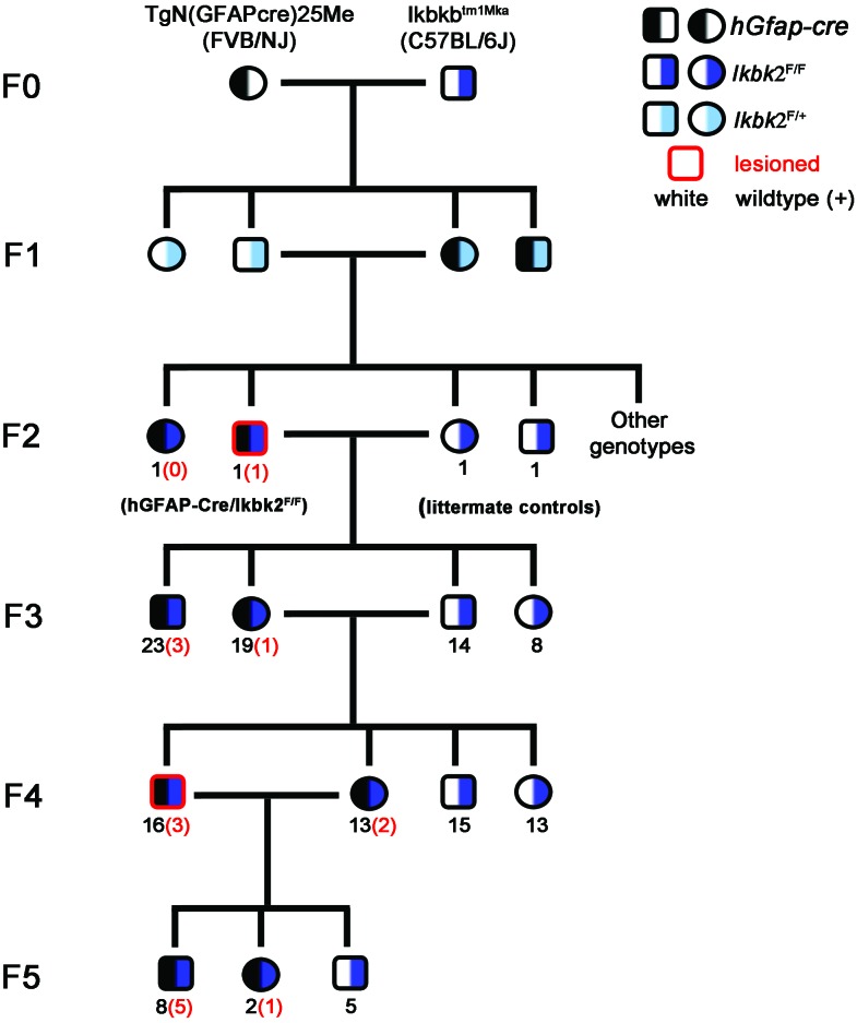
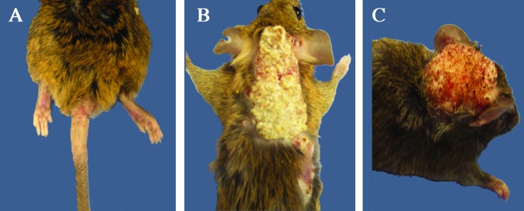
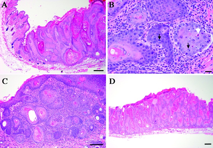
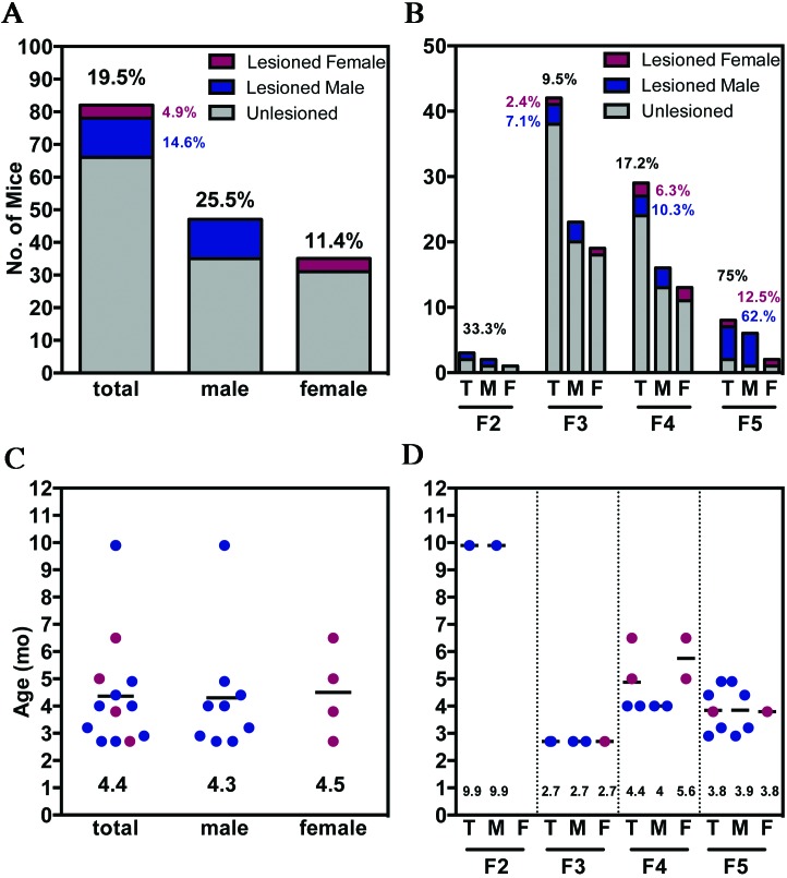
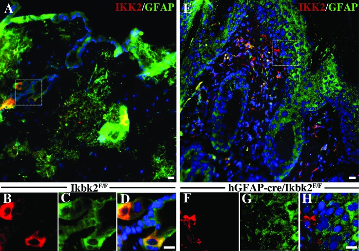
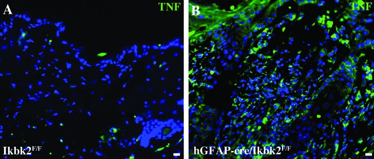
The deletion of NFκB in epithelial tissues by using skin-specific promoters can cause both tumor formation and severe inflammatory dermatitis, indicating that this signaling pathway is important for the maintenance of immune homeostasis in epithelial tissues. In the present study, we crossed mice transgenic for loxP-Ikbk2 and human Gfap-cre to selectively delete IKK2 in CNS astrocytes. Unexpectedly, a subset of mice developed severe and progressive skin lesions marked by hyperplasia, hyperkeratosis, dysplasia, inflammation, and neoplasia with a subset of lesions diagnosed as squamous cell carcinoma (SCC). The development of lesions was monitored over a 3.5-y period and over 4 filial generations. Average age of onset of was 4 mo of age with 19.5% of mice affected with frequency increasing in progressive generations. Lesion development appeared to correlate not only with unintended IKK2 deletion in GFAP expressing cells of the epidermis, but also with increased expression of TNF in lesioned skin. The skins changes described in these animals are similar to those in transgenic mice with an epidermis-specific deletion of NFκB and thus represents another genetic mouse model that can be used to study the role of NFκB signaling in regulating the development of SCC.


Figures

Figure 1.
Figure 1.
Pedigree of hGfap-cre /Ikk2F/F mice over 6 filial generations (F0 through F5). Half black indicates presence of the hGfap-cre transgene, whereas half-navy blue or half-light blue indicates homozygozity or heterozygozity for the floxed Ikk2 allele, respectively. Black numerals indicate the number of mice of that genotype and sex in the indicated generation, whereas red numbers in parentheses indicate the number of lesioned hGfap-cre/Ikk2F/F mice of each sex. Red highlighted shapes indicate a lesioned mouse was used as a breeder for the subsequent generation.
Figure 2.
Figure 2.
Gross pathology of skin lesions in hGfap-cre/Ikbk2F/F mice. (A) Lesion on tail base and perianal region of with areas of hypotrichosis, hyperkeratosis, and ulceration. (B) Severe hyperkeratotic lesion with multifocal ulcerations at the periphery on scruff and lateral thorax. (C) Discrete, hyperkeratotic lesion on the dorsal head, causing displacement of the right pinna laterally. In all images, the background was changed from white to blue to enhance lesion visualization.
Figure 3.
Figure 3.
Histopathology of skin lesions from hGfap-cre/Ikbk2F/F mice. (A) Hematoxylin and eosin staining shows the transition from unaffected skin to a region of increasing epidermal hyperplasia and dysplasia. Scale bar, 100 μm. (B) Hematoxylin and eosin staining reveals nests of neoplastic epithelial cells, with numerous mitotic figures (black arrows) and apoptosis of individual cells (white arrow). Scale bar, 20 μm. (C) Hematoxylin and eosin staining shows marked inflammatory cell infiltration with the tumor. Scale bar, 100 μm. (D) Hematoxylin and eosin staining reveals marked expansion of the epidermis by multifocal nests of epithelial cells. Scale bar, 100 μM.
Figure 4.
Figure 4.
Frequency and age of onset of SCC in hGfap-cre/Ikbk2F/F mice. (A) Overall lesion development frequency of hGfap-cre/Ikbk2F/F total (T, black), male (M; blue) and female (F; red) mice, (B) with increasing percentage of mice affected through progressive generations. Gray indicates nononlesioned mic. Scatter plots depicting the age (m, months) of hGfap-cre/Ikbk2F/F total (T, black), male (M; blue) and female (F; red) mice at lesion development (C) overall and (D) by generation. Mean age is indicated by a black line and written at the bottom of the scatter plot.
Figure 5.
Figure 5.
Immunofluorescent analysis of IKK2 expression in Ikbk2F/F and hGfap-cre/Ikbk2F/F mice. (A) Normal skin from Ikbk2F/F mice (n = 12) has sporadic IKK2 (red) expression with colocalization found in GFAP- (green) expressing cells including keratinocytes, sebaceous glands, and dermal fibroblasts. Higher magnification images obtained from the dotted white line showing (B) IKK2 and (C) GFAP (D) colocalization in cells from an Ikbk2F/F mouse. (E) IKK2 (red) fluorescence is absent from GFAP- (green) positive cells in SCC lesions in hGfap-cre-Cre/Ikbk2F/F mice (n = 11). Higher magnification images obtained from the dotted white line show of (F) IKK2 and (G) GFAP (H) colocalization in cells from an hGfap-cre-Cre/Ikbk2F/F mouse. Nuclei are shown in blue. Scale bars, 20 μm.
Figure 6.
Figure 6.
Immunofluorescent analysis of TNF expression in Ikbk2F/F and hGfap-cre/Ikbk2F/F mice. (A) Normal skin from Ikbk2F/F mice (n = 12) has low to absent TNF fluorescence (green). (B) TNF (green) fluorescence is increased in all skin layers in SCC lesions and surrounding nonlesioned skin from hGfap-cre/Ikbk2F/F mice (n = 11). Nuclei are shown in blue. Scale bars, 20 μm.

Similar articles

Cited by

References

    1. Abel TW, Clark C, Bierie B, Chytil A, Aakre M, Gorska A, Moses HL. 2009. GFAP-Cre-mediated activation of oncogenic K-ras results in expansion of the subventricular zone and infiltrating glioma. Mol Cancer Res 7:645–653. - PMC - PubMed
    1. Banno T, Gazel A, Blumemberg M. 2005. Pathway-specific profiling identifies the NFκB-dependent tumor necrosis factor α-regulated genes in epidermal keratinocytes. J Biol Chem 280: 18973­–18980. - PubMed
    1. Blake JA, Eppig JT, Kadin JA, Richardson JE, Smith CL, Bult CJ. 2017. Mouse genome database (MGD)—2017: community knowledge resource for the laboratory mouse. Nucleic Acids Res 45:D723–D729. - PMC - PubMed
    1. Bonizzi G, Karin M. 2004. The 2 NFκB activation pathways and their role in innate and adaptive immunity. Trends Immunol 25:280–288. - PubMed
    1. Cornish GH, Tung SL, Marshall D, Ley S, Seddon BP. 2012. Tissue specific deletion of inhibitor of κB kinase 2 with OX40-Cre reveals the unanticipated expression from the OX40 locus in skin epidermis. PLoS ONE 7:1–10. - PMC - PubMed