. 2017 Dec;187(12):2726-2743.
doi: 10.1016/j.ajpath.2017.08.017. Epub 2017 Sep 19.

Human Pancreatic Acinar Cells: Proteomic Characterization, Physiologic Responses, and Organellar Disorders in ex Vivo Pancreatitis

Affiliations

Human Pancreatic Acinar Cells: Proteomic Characterization, Physiologic Responses, and Organellar Disorders in ex Vivo Pancreatitis

Aurelia Lugea et al. Am J Pathol. 2017 Dec.

Abstract

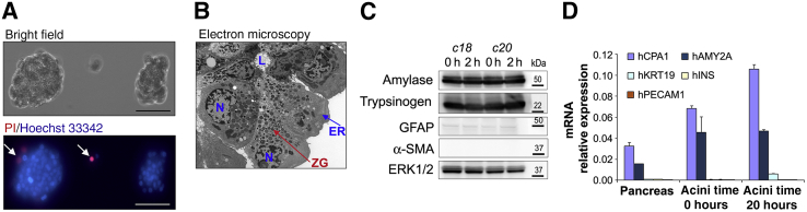
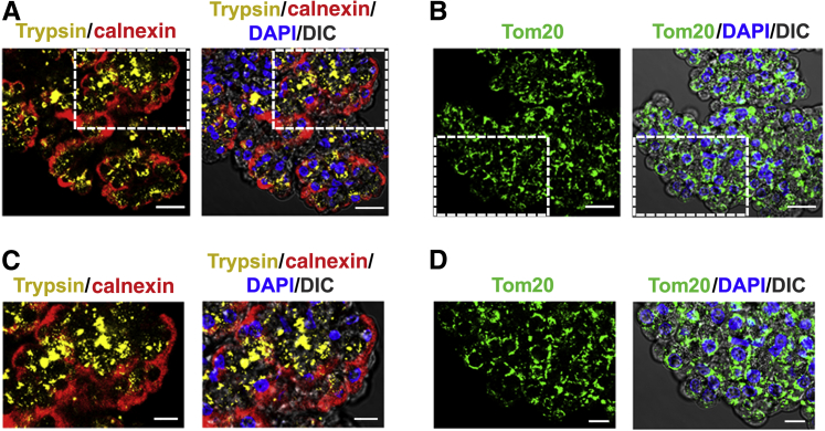
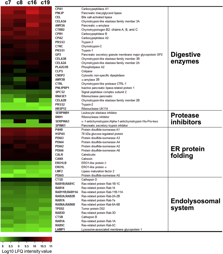
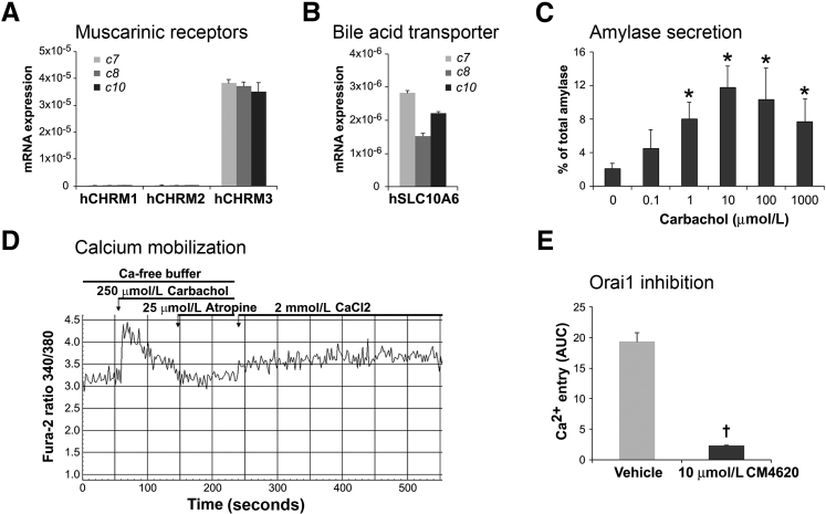
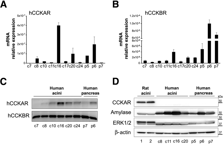
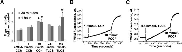
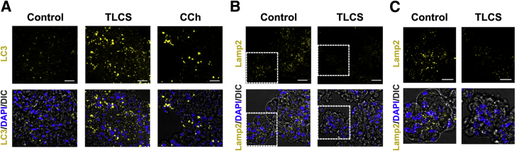
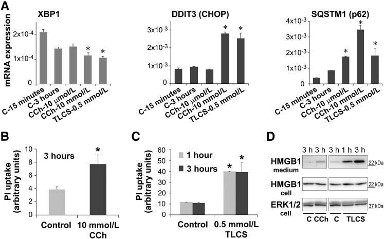
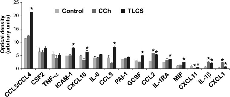
Knowledge of the molecular mechanisms of acute pancreatitis is largely based on studies using rodents. To assess similar mechanisms in humans, we performed ex vivo pancreatitis studies in human acini isolated from cadaveric pancreata from organ donors. Because data on these human acinar preparations are sparse, we assessed their functional integrity and cellular and organellar morphology using light, fluorescence, and electron microscopy; and their proteome by liquid chromatography-tandem mass spectrometry. Acinar cell responses to the muscarinic agonist carbachol (CCh) and the bile acid taurolithocholic acid 3-sulfate were also analyzed. Proteomic analysis of acini from donors of diverse ethnicity showed similar profiles of digestive enzymes and proteins involved in translation, secretion, and endolysosomal function. Human acini preferentially expressed the muscarinic acetylcholine receptor M3 and maintained physiological responses to CCh for at least 20 hours. As in rodent acini, human acini exposed to toxic concentrations of CCh and taurolithocholic acid 3-sulfate responded with trypsinogen activation, decreased cell viability, organelle damage manifest by mitochondrial depolarization, disordered autophagy, and pathological endoplasmic reticulum stress. Human acini also secreted inflammatory mediators elevated in acute pancreatitis patients, including IL-6, tumor necrosis factor-α, IL-1β, chemokine (C-C motif) ligands 2 and 3, macrophage inhibitory factor, and chemokines mediating neutrophil and monocyte infiltration. In conclusion, human cadaveric pancreatic acini maintain physiological functions and have similar pathological responses and organellar disorders with pancreatitis-causing treatments as observed in rodent acini.


Figures

Figure 1
Figure 1
Characterization of isolated human pancreatic acini. A: Untreated acini were placed in a cell culture fluorodish with a glass bottom, cultured for 2 hours, stained with propidium iodide (PI) and Hoechst 33342, and observed under a fluorescence inverted microscope to assess cell morphology and viability. Arrows indicate PI-positive cells. B: Representative electron micrograph of untreated human acini showing typical acinar cell ultrastructure and acinar lumen (L), with nuclei (N) in basal position, endoplasmic reticulum (ER) network surrounding the nucleus, and abundant zymogen granules (ZG). C and D: Untreated human acini were cultured for up to 20 hours. Panels show representative Western blot images (C) and real-time quantitative PCR analysis (D) for markers of acinar, stellate, ductal, endothelial, and β cells. mRNA isolated from pancreatic tissues samples (Pancreas) from organ donors was used for comparison. Data are expressed as means ± SD (D). n = 3 (D). Scale bars = 100 μm (A). Original magnification, ×1500 (B). c, organ donor case (patient coding) used as a source of human acinar preparation; ERK, extracellular signal regulated kinase; GFAP, glial fibrillary acidic protein; hAMY2A, human pancreatic α-amylase; hCPA1, human carboxypeptidase A1; hINS, human insulin; hKRT19, human cytokeratin-19; hPECAM1, human platelet endothelial cell adhesion molecule-1; α-SMA, α-smooth muscle actin; h, hours.
Figure 2
Figure 2
The distribution of intracellular organelles retains its polarity in isolated human acini. A and B: Untreated freshly isolated human acini were formaldehyde fixed and immunostained with antibodies against trypsinogen (Trypsin; zymogen granules; yellow stain), calnexin (endoplasmic reticulum; red stain), and translocase of outer membrane 20 (Tom20; mitochondria; green stain). The signals were detected with Alexa Fluor 555–, Alexa Fluor 633–, and Alexa Fluor 488–conjugated secondary antibodies, respectively. Nuclei were stained with DAPI (blue). Images were analyzed under a confocal microscope. C and D: Higher magnifications of the boxed areas in A and B, respectively. Scale bars: 10 μm (A and B); 20 μm (C and D). DIC, differential interference contrast.
Figure 3
Figure 3
Gene Ontology (GO) analysis of the human pancreatic acini proteome. Protein extracts from untreated, freshly isolated, human acini from four organ donors (Table 3 provides organ donor information) were analyzed by high-precision liquid chromatography–tandem mass spectrometry in an Orbitrap Elite analyzer; data were quantified using MaxQuant version 1.3.0.5. Of the total proteins identified, 70% were common to all samples and used for further analysis. GO information was retrieved from the DAVID annotation tool. Diagram shows percentage of total genes analyzed corresponding to the indicated GO biological processes (BPs). A P value threshold of < 0.05 was used to confidently predict enriched GO terms among the proteins.
Figure 4
Figure 4
Enriched Kyoto Encyclopedia of Genes and Genomes (KEGG) pathways of the human acinar proteome. The data sets originating from the human acinar proteome indicated in Figure 3 were used to interrogate the DAVID analysis tool for the significantly enriched KEGG pathways. The diagram shows the percentage of total enriched genes that fall in each metabolic pathway or molecular cellular process. TCA, tricarboxylic acid.
Figure 5
Figure 5
The relative abundance of selected proteins in the human pancreatic acinar proteome is similar across donor tissue sources. The human acinar proteomic data sets originating from the four organ donors (c7, c8, c16, and c19 [c indicates organ donor case (patient coding) used as a source of human acinar preparation]) indicated in Figure 3 and Table 3 were analyzed to determine the interdonor variability. The heat map shows relative amounts of the indicated selected proteins determined by label-free quantification (LFQ). Selected proteins include all identified digestive enzymes, protease inhibitors, abundant protein biosynthetic components (endoplasmic reticulum chaperones and foldases), and proteins in the endolysosomal system.
Figure 6
Figure 6
Human acini express the human muscarinic acetylcholine receptor M3 (hCHRM3) and exhibit a biphasic secretory response to cholinergic stimulation. A and B: mRNA extracts from human acini isolated from three organ donor pancreata (c7, c8, and c10 [c indicates organ donor case (patient coding) used as a source of human acinar preparation]) were analyzed by real-time quantitative PCR to determine the expression of the indicated muscarinic receptors (A) and the bile acid transporter solute carrier family 10 member 6 (SLC10A6; B). All three human acinar preparations analyzed preferentially express the muscarinic receptor CHRM3 and the SLC10A6 transporter. C: Human acini were stimulated for 30 minutes with the cholinergic agonist carbachol at the indicated concentrations, and amylase release was measured using the Phadebas test. As shown, human acinar cells respond to carbachol stimulation by releasing amylase in a typical biphasic pattern also reported for rodent acini. D and E: Characterization of carbachol-induced calcium response in human acini. Acini were loaded with Fura-2 AM and then stimulated with 250 μmol/L carbachol in the absence of free Ca2+ in the extracellular buffer. As shown, carbachol causes a rapid, transit elevation of cytosolic Ca2+ in Fura-loaded cells. Atropine was then added to terminate the Ca2+-releasing signal. Adding Ca2+ to the extracellular media (2 mmol/L CaCl2) leads to Ca2+ entry, and this effect is blocked by preincubation with CM4620, a specific inhibitor of the store-operated Ca2+ channel, Orai (E). Data are expressed as means ± SD (C and E). n = 3 independent experiments (C and E). P < 0.05 versus basal (no carbachol); P < 0.05 versus vehicle (dimethyl sulfoxide). AUC, area under the curve.
Figure 7
Figure 7
Human acini express low levels of the cholecystokinin-A receptor (CCKAR). A and B: mRNA extracts from untreated human acini isolated from eight organ donor pancreata (batches c7, c8, c10, c11, c16, c17, c20, and c24 [c indicates organ donor case (patient coding) used as a source of human acinar preparation]), cadaveric pancreas tissues from an organ donor (p5; normal parenchyma), and surgically resected pancreas tissues from two cancer patients (p6 and p7; normal parenchyma distant from tumor margins) were analyzed by real-time quantitative PCR (qPCR) to determine the expression of hCCKAR (A) and human CCK-B receptor (CCKBR; B). Expression levels are relative to those of 18S. C: qPCR products from studies illustrated in A and B. cDNA (50 ng) was used per reaction, and reactions were run for 40 cycles. D: Panels show immunoblot images of protein levels of CCKAR and amylase in human acini and pancreatic tissue specimens. Extracellular signal regulated kinase (ERK) 1/2 and β-actin were used as loading controls.
Figure 8
Figure 8
Carbachol (CCh) or taurolithocholic acid 3-sulfate (TLCS) at a high concentration induces trypsinogen activation and mitochondrial depolarization in human acini. A: Human acini were incubated for the indicated times in the absence (control) or presence of CCh or TLCS. Trypsin activity was measured in cell homogenates by enzymatic assay using a specific fluorogenic substrate. Graph indicates trypsin activity in stimulated cells relative to control values (set at 1.0). B and C: Mitochondrial membrane potential was measured in acini loaded with the fluorescent probe tetramethylrhodamine methyl ester (TMRM) and treated with CCh (B) or TLCS (C). p-Trifluoromethoxy-phenylhydrazone (FCCP) was used to completely dissipate the mitochondrial membrane potential. Data are expressed as means ± SD (A). n = 3 independent experiments (A). P < 0.05 versus control. AU, arbitrary units.
Figure 9
Figure 9
Autophagic puncta accumulate, and the lysosomal membrane protein lysosome-associated membrane protein (LAMP)-2 decreases in ex vivo pancreatitis using human acini. A and B: Immunofluorescence analysis of light chain 3 (LC3; A) and LAMP-2 (B) in human acini incubated for 3 hours without (Control) or with 0.5 mmol/L taurolithocholic acid 3-sulfate (TLCS) or 1 mmol/L carbachol (CCh). Nuclei were stained with DAPI (blue). C: Higher magnification of the boxed areas in B. Scale bars: 20 μm (A and B); 10 μm (C). DIC, differential interference contrast.
Figure 10
Figure 10
Dysregulation of endoplasmic reticulum (ER) stress pathways in ex vivo pancreatitis is associated with acinar cell death. RNA was extracted from untreated human acini after 15 minutes of incubation (C-15 minutes) or from acini incubated for 3 hours in the absence (C-3 hours) or presence of carbachol (CCh) or taurolithocholic acid 3-sulfate (TLCS) at the indicated concentrations. A: The ER stress regulators spliced X box protein-1 (XBP1) and DNA damage inducible transcript 3 (DDIT3; CHOP) and the cellular stress marker sequestosome 1 (SQSTM1; p62) were measured by real-time quantitative PCR. B and C: Human acini were treated with toxic concentrations of CCh or TLCS for the indicated times. Cell death was assessed by propidium iodide (PI) uptake. D: Cells were kept untreated [control (C)] or treated for the indicated times with 10 mmol/L CCh or 0.5 mmol/L TLCS, and then conditioned media were collected. Panels show representative immunoblot images of protein levels of the high-mobility group protein 1 (HMGB1) in conditioned media and cell homogenates. Extracellular signal regulated kinase (ERK) 1/2 was used as loading control. Data are expressed as means ± SD (A) or means ± SEM (B and C). n = 3 independent experiments (A); n = 4 independent experiments (B and C). P < 0.05 versus control. h, hours.
Figure 11
Figure 11
Select cytokine/chemokine secretion is up-regulated in ex vivo pancreatitis using human acini. Cells were kept untreated (Control) or treated for 16 hours with 1 mmol/L carbachol (CCh) or 0.5 mmol/L taurolithocholic acid 3-sulfate (TLCS). Cytokines and chemokines released into the media were measured by a membrane-based human cytokine/chemokine array (36 cytokines/chemokines; catalog number ARY005B; R&D Systems). Optical density (arbitrary units) for all 15 detected cytokines/chemokines is shown. The following cytokines/chemokines were tested, but not detected, in conditioned media: IL-1α, IL-2, IL-4, IL-5, IL-8, IL-10, IL-12, IL-13, IL-16, IL-17A, IL-17E, IL-21, IL-32a, IL-18, IL-27, interferon-γ, C5/C5A, chemokine (C-C motif) ligand (CCL) 1, CXCL12, CD40 ligand, and TREM-1. Data are expressed as means ± SD. n = 2 independent experiments. P < 0.05 versus control. CSF, colony-stimulating factor; GCSF, granulocyte CSF; ICAM, intercellular adhesion molecule 1; IL-1RA, IL-1 receptor antagonist; MIF, macrophage inhibitory factor; PAI, plasminogen activator inhibitor; TNF-α, tumor necrosis factor-α; TREM, triggering receptor expressed on myeloid cells 1.

Similar articles

Cited by

References

    1. Yadav D., Lowenfels A.B. The epidemiology of pancreatitis and pancreatic cancer. Gastroenterology. 2013;144:1252–1261. - PMC - PubMed
    1. Peery A.F., Dellon E.S., Lund J., Crockett S.D., McGowan C.E., Bulsiewicz W.J., Gangarosa L.M., Thiny M.T., Stizenberg K., Morgan D.R., Ringel Y., Kim H.P., Dibonaventura M.D., Carroll C.F., Allen J.K., Cook S.F., Sandler R.S., Kappelman M.D., Shaheen N.J. Burden of gastrointestinal disease in the United States: 2012 update. Gastroenterology. 2012;143 1179–1187.e1–1187.e3. - PMC - PubMed
    1. Afghani E., Pandol S.J., Shimosegawa T., Sutton R., Wu B.U., Vege S.S., Gorelick F., Hirota M., Windsor J., Lo S.K., Freeman M.L., Lerch M.M., Tsuji Y., Melmed G.Y., Wassef W., Mayerle J. Acute pancreatitis-progress and challenges: a report on an International Symposium. Pancreas. 2015;44:1195–1210. - PMC - PubMed
    1. Pandol S.J., Saluja A.K., Imrie C.W., Banks P.A. Acute pancreatitis: bench to the bedside. Gastroenterology. 2007;132:1127–1151. - PubMed
    1. Lerch M.M., Gorelick F.S. Models of acute and chronic pancreatitis. Gastroenterology. 2013;144:1180–1193. - PubMed