. 2018 Jan 1;314(1):L69-L82.
doi: 10.1152/ajplung.00058.2017. Epub 2017 Sep 21.

Phagocytosis of microparticles by alveolar macrophages during acute lung injury requires MerTK

Affiliations

Phagocytosis of microparticles by alveolar macrophages during acute lung injury requires MerTK

Michael P Mohning et al. Am J Physiol Lung Cell Mol Physiol. .

Abstract

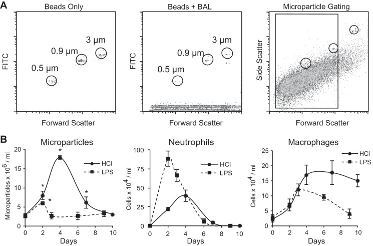
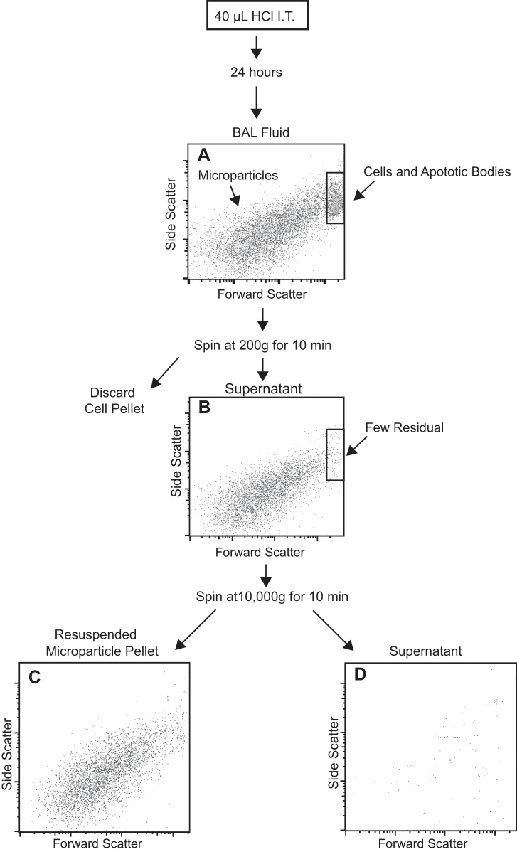
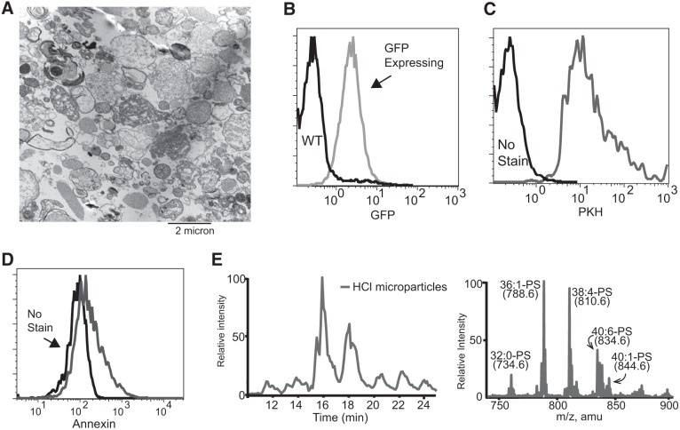
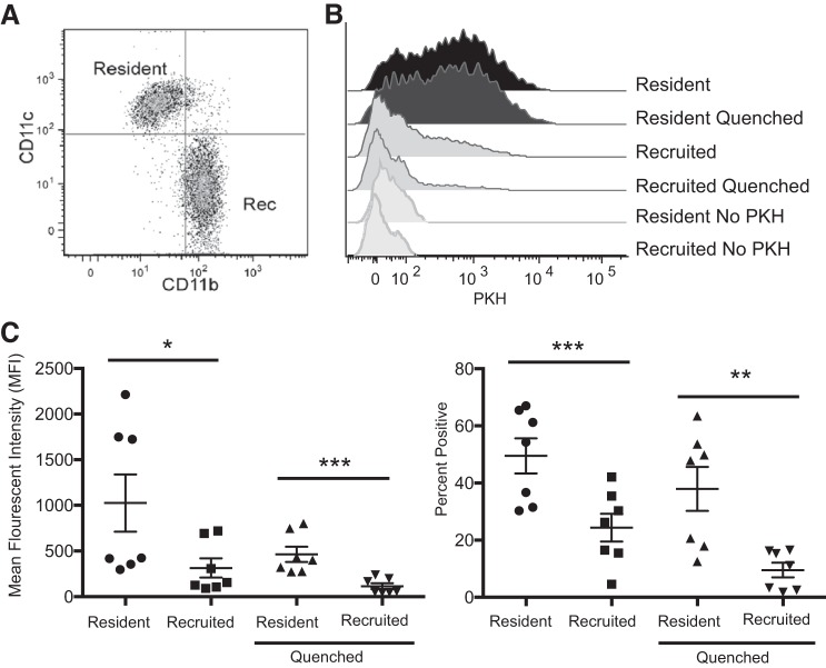
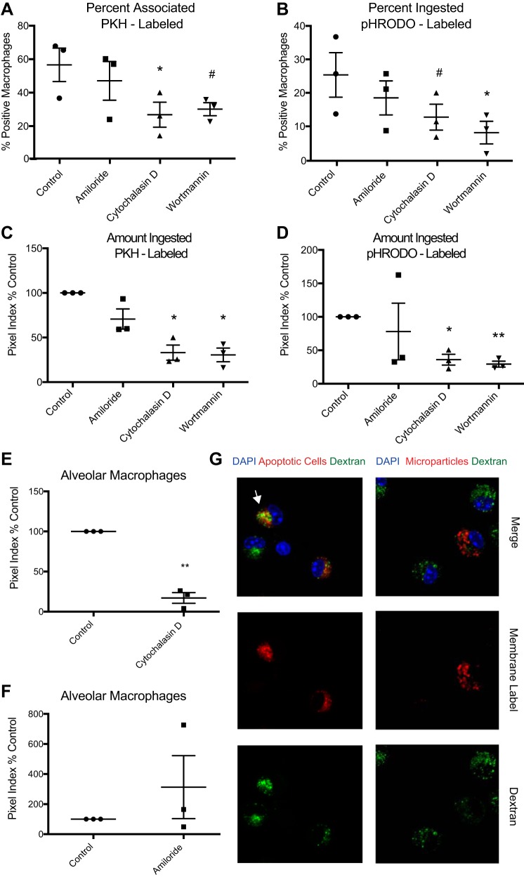
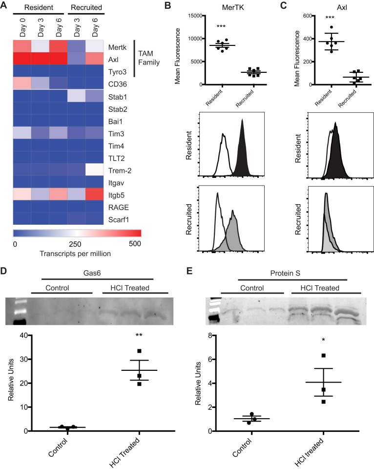
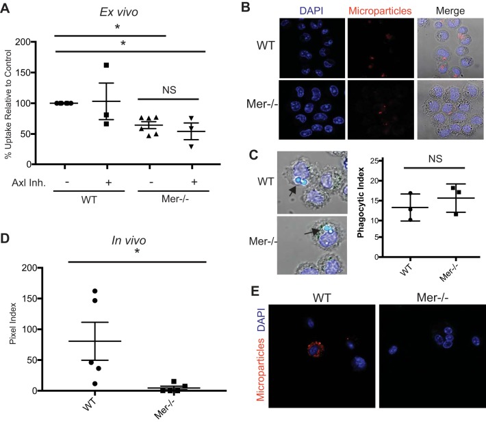
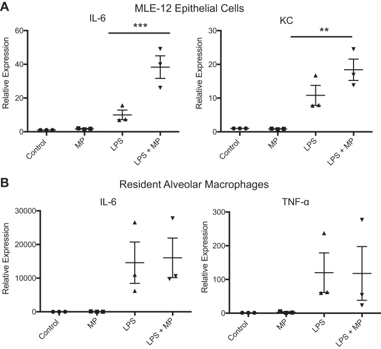
Microparticles are a newly recognized class of mediators in the pathophysiology of lung inflammation and injury, but little is known about the factors that regulate their accumulation and clearance. The primary objective of our study was to determine whether alveolar macrophages engulf microparticles and to elucidate the mechanisms by which this occurs. Alveolar microparticles were quantified in bronchoalveolar fluid of mice with lung injury induced by LPS and hydrochloric acid. Microparticle numbers were greatest at the peak of inflammation and declined as inflammation resolved. Isolated, fluorescently labeled particles were placed in culture with macrophages to evaluate ingestion in the presence of endocytosis inhibitors. Ingestion was blocked with cytochalasin D and wortmannin, consistent with a phagocytic process. In separate experiments, mice were treated intratracheally with labeled microparticles, and their uptake was assessed though microscopy and flow cytometry. Resident alveolar macrophages, not recruited macrophages, were the primary cell-ingesting microparticles in the alveolus during lung injury. In vitro, microparticles promoted inflammatory signaling in LPS primed epithelial cells, signifying the importance of microparticle clearance in resolving lung injury. Microparticles were found to have phosphatidylserine exposed on their surfaces. Accordingly, we measured expression of phosphatidylserine receptors on macrophages and found high expression of MerTK and Axl in the resident macrophage population. Endocytosis of microparticles was markedly reduced in MerTK-deficient macrophages in vitro and in vivo. In conclusion, microparticles are released during acute lung injury and peak in number at the height of inflammation. Resident alveolar macrophages efficiently clear these microparticles through MerTK-mediated phagocytosis.

Keywords: Mer tyrosine kinase; alveolar macrophage; lung injury; microparticle; phagocytosis.


Figures

Fig. 1.
Fig. 1.
Microparticles accumulate in the alveolus during acute lung injury. A: flow cytometry gating strategy showing initial gating on size-calibrated FITC fluorescent microbeads and microparticles from bronchoalveolar lavage (BAL). B: C57BL/6 mice were treated with either lipopolysaccharide (LPS; 20 μg) or hydrochloric acid (1 N, pH 1.1) via intratracheal instillation. BAL was performed at serial time points. Cell counts and differentials were performed on the lavage specimens. Microparticles present in the BAL were quantified using flow cytometry and a known quantity of microbeads; each value represents means ± SE (n = 5). *P < 0.05 for microparticles compared with naïve mice.
Fig. 2.
Fig. 2.
Microparticle purification using serial centrifugation. BAL was performed on HCl acid-treated mice at 24 h. A: whole BAL before centrifugation. Microparticles and cells are present. B: supernatant after BAL was centrifuged at 200 g for 10 min to pellet cells. Cells are no longer present; remaining events are microparticles predominantly below 1 μm in diameter. A second centrifuge step was performed at 10,000 g for 10 min to pellet microparticles. C: resuspended pellet demonstrates that microparticles are contained in this fraction. D: supernatant remaining after the second centrifugation step. No microparticles are present. IT, intratracheal.
Fig. 3.
Fig. 3.
Microparticle characterization. A: microparticles isolated from HCl-treated mice were fixed and imaged using electron microscopy. B: representative flow cytometry of microparticles isolated from green fluorescent protein (GFP)-expressing mice (gray line) and wild-type (WT) mice (black line) treated with HCl. C: microparticles either stained with PKH (gray line) for lipid membranes or unstained (black line). D: microparticles either stained with annexin V (gray line) or unstained (black line). E: diacyl-phosphatidylserine (PS) is enriched in microparticles from HCl-treated mice. Left: total ion chromatography from 10 to 25 min from the liquid chromatography-mass spectrometry/mass spectrometry chromatogram of diacyl-PS. Right: corresponding total ion mass spectrum of diacyl-PS monitoring neutral loss of 87 atomic mass units in negative ion mode.
Fig. 4.
Fig. 4.
Microparticles are engulfed by resident alveolar macrophages during acute lung injury. A: dating strategy to isolate resident and recruited (Rec) alveolar macrophages in BAL fluid. BAL macrophages were identified as CD45+, F4/80+, Ly6G negative, and CD64+ and then classified as shown into resident (CD11chi, CD11blo) or Rec (CD11bhi, CD11clo) macrophages. B: PKH-labeled microparticles were instilled into LPS-treated mice. BAL was performed 1 h later, and macrophages were assessed for PKH intensity. Immediately after initial analysis, cells were quenched with trypan blue to eliminate fluorescence of bound but not internalized microparticles. C: microparticle uptake quantified using mean fluorescence intensity (left) and %macrophages ingesting microparticles (right); n = 7. Data represent means ± SE. *P < 0.05; **P < 0.01; ***P < 0.001.
Fig. 5.
Fig. 5.
Microparticles are phagocytosed by macrophages in vitro. Peritoneal macrophages were cultured with fluorescently labeled microparticles in the presence of endocytosis inhibitors. A: %macrophages associated with PKH-labeled microparticles. Uptake quantified by confocal microscopy. B: %macrophages engulfing pHRODO-labeled microparticles. C and D: magnitude of engulfment of PKH-labeled (C) and pHRODO-labeled (D) microparticles. Fluorescent pixels per macrophage were calculated (pixel index) and normalized to WT control. Data are expressed as %control. E: endocytosis of PKH-labeled microparticles by resident alveolar macrophages with or without cytochalasin D. Engulfment assessed after 1 h by confocal microscopy. F: membrane-labeled (cellvue maroon) microparticles or apoptotic thymocytes (positive control for macropinocytosis) and FITC-labeled dextran were added in coculture with resident alveolar macrophages. Dextran colocalizes with apoptotic cells (white arrow) but not microparticles. G: membrane-labeled microparticles or apoptotic cells were added in coculture with resident alveolar macrophages in the presence of amiloride. Data represent means ± SE; n = 3 independent experiments. #P < 0.1; *P < 0.05; **P < 0.01. Symbols are replicates in the following groups: ●, control; ■, amiloride; ▲, cytochalasin; ▼, wortmannin.
Fig. 6.
Fig. 6.
Alveolar macrophages express Mer tyrosine kinase (MerTK) and Axl. A: expression of phagocytic receptors in resident and recruited alveolar macrophages following LPS-induced lung injury, as measured with RNA-seq. Gene expression shown as mean transcripts per million; n = 3 experiments. B and C: MerTK (B) and Axl (C) expression on resident (black) and recruited (gray) alveolar macrophages isolated from LPS-treated mice (day 3). White histogram represents fluorescence minus one for MerTK or Axl. D and E: Western blotting for Gas6 and protein S in microparticle pellets isolated from naïve and hydrochloric acid-treated mice. Equivalent volumes of BAL fluid were used to recover microparticles; one-quarter of the total microparticle protein was loaded for each sample. Representative blots are shown with corresponding densitometry. Data represent means ± SE; n = 3. *P < 0.05; **P < 0.01; ***P < 0.001.
Fig. 7.
Fig. 7.
MerTK is required for optimal microparticle uptake by alveolar macrophages. A: resident alveolar macrophages from MerTK−/− or B6.129 control mice were cultured on microscope slides with or without an Axl inhibitor. Membrane-labeled microparticles were placed in coculture with the macrophages. One hour later, cells were washed to remove noningested microparticles and then fixed with paraformaldehyde. Fluorescent pixels per macrophage were calculated (pixel index) and normalized to WT controls; n = 3–6. Data represent means ± SE. B: representative images of microparticle endocytosis. C: alveolar macrophages from WT and MerTK−/− mice cultured with 2-μm latex beads (arrows). Endocytosis of beads by MerTK−/− mice is not impaired. D: membrane-labeled microparticles were intratracheally instilled into naïve MerTK−/− and WT. Cytospins were performed on BAL, and endocytosis was quantified with confocal microscopy; n = 5. E: representative images of in vivo microparticle uptake by alveolar macrophages from WT and MerTK−/− mice. *P < 0.05. Symbols are replicates in the following groups: ●, control; ■, amiloride; ▲, cytochalasin; ▼, wortmannin.
Fig. 8.
Fig. 8.
Microparticles are pro-inflammatory toward alveolar epithelial cells in vitro but not alveolar macrophages. A: MLE-12 cells were treated with microparticles (MP), LPS, or LPS and microparticles. RNA was isolated at 4 h, and RT-PCR was performed to quantify the proinflammatory cytokines KC and IL-6. B: resident alveolar macrophages obtained from C57/bl6 mice were treated with microparticles, LPS, or LPS and microparticles. RNA was isolated at 4 h, and RT-PCR was performed to quantify IL-6 and TNFα. Data represent means ± SE; n = 3. **P < 0.01; ***P < 0.001.

References

    1. Aggarwal NR, King LS, D’Alessio FR. Diverse macrophage populations mediate acute lung inflammation and resolution. Am J Physiol Lung Cell Mol Physiol 306: L709–L725, 2014. doi: 10.1152/ajplung.00341.2013. - DOI - PMC - PubMed
    1. Barrès C, Blanc L, Bette-Bobillo P, André S, Mamoun R, Gabius HJ, Vidal M. Galectin-5 is bound onto the surface of rat reticulocyte exosomes and modulates vesicle uptake by macrophages. Blood 115: 696–705, 2010. doi: 10.1182/blood-2009-07-231449. - DOI - PubMed
    1. Bastarache JA, Fremont RD, Kropski JA, Bossert FR, Ware LB. Procoagulant alveolar microparticles in the lungs of patients with acute respiratory distress syndrome. Am J Physiol Lung Cell Mol Physiol 297: L1035–L1041, 2009. doi: 10.1152/ajplung.00214.2009. - DOI - PMC - PubMed
    1. Buesing KL, Densmore JC, Kaul S, Pritchard KA Jr, Jarzembowski JA, Gourlay DM, Oldham KT. Endothelial microparticles induce inflammation in acute lung injury. J Surg Res 166: 32–39, 2011. doi: 10.1016/j.jss.2010.05.036. - DOI - PMC - PubMed
    1. Densmore JC, Signorino PR, Ou J, Hatoum OA, Rowe JJ, Shi Y, Kaul S, Jones DW, Sabina RE, Pritchard KA Jr, Guice KS, Oldham KT. Endothelium-derived microparticles induce endothelial dysfunction and acute lung injury. Shock 26: 464–471, 2006. doi: 10.1097/01.shk.0000228791.10550.36. - DOI - PubMed

Publication types

MeSH terms

Substances