. 2018 Jan 1;314(1):L118-L126.
doi: 10.1152/ajplung.00296.2017. Epub 2017 Sep 21.

Isolation and characterization of endothelial-to-mesenchymal transition cells in pulmonary arterial hypertension

Affiliations

Isolation and characterization of endothelial-to-mesenchymal transition cells in pulmonary arterial hypertension

Toshio Suzuki et al. Am J Physiol Lung Cell Mol Physiol. .

Abstract

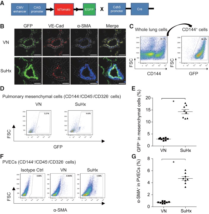
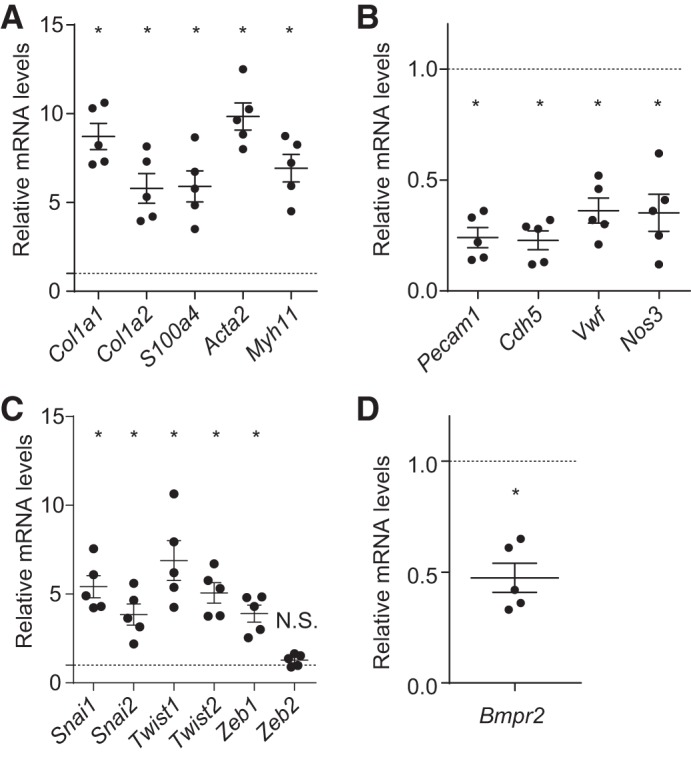
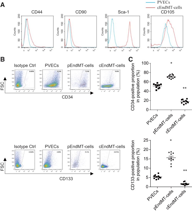
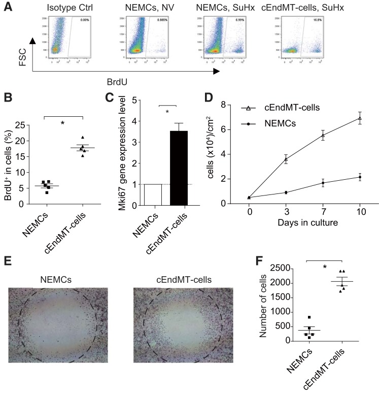
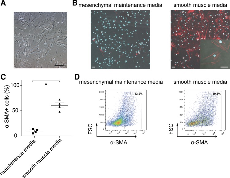
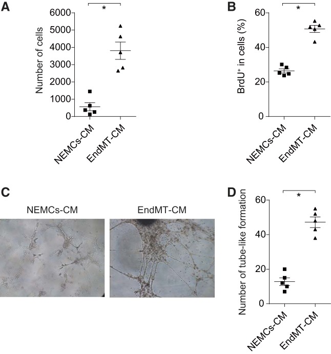
Endothelial-to-mesenchymal transition (EndMT) is a process in which endothelial cells lose polarity and cell-to cell contacts, and undergo a dramatic remodeling of the cytoskeleton. It has been implicated in initiation and progression of pulmonary arterial hypertension (PAH). However, the characteristics of cells which have undergone EndMT cells in vivo have not been reported and so remain unclear. To study this, sugen5416 and hypoxia (SuHx)-induced PAH was established in Cdh5-Cre/Gt(ROSA)26Sortm4(ACTB-tdTomato,EGFP)Luo/J double transgenic mice, in which GFP was stably expressed in pan-endothelial cells. After 3 wk of SuHx, flow cytometry and immunohistochemistry demonstrated CD144-negative and GFP-positive cells (complete EndMT cells) possessed higher proliferative and migratory activity compared with other mesenchymal cells. While CD144-positive and α-smooth muscle actin (α-SMA)-positive cells (partial EndMT cells) continued to express endothelial progenitor cell markers, complete EndMT cells were Sca-1-rich mesenchymal cells with high proliferative and migratory ability. When transferred in fibronectin-coated chamber slides containing smooth muscle media, α-SMA robustly expressed in these cells compared with cEndMT cells that were grown in maintenance media. Demonstrating additional paracrine effects, conditioned medium from isolated complete EndMT cells induced enhanced mesenchymal proliferation and migration and increased angiogenesis compared with conditioned medium from resident mesenchymal cells. Overall, these findings show that EndMT cells could contribute to the pathogenesis of PAH both directly, by transformation into smooth muscle-like cells with higher proliferative and migratory potency, and indirectly, through paracrine effects on vascular intimal and medial proliferation.

Keywords: cell transformation; endothelial-to-mesenchymal transition; paracrine effects; pulmonary arterial hypertension; vascular remodeling.


Figures

Fig. 1.
Fig. 1.
Generation of Cdh5-Cre/GFP double transgenic mice and endothelial-to-mesenchymal transition (EndMT) in sugen5416/hypoxia (SuHx)-induced pulmonary arterial hypertension (PAH) model. A: schematic description of gene constructs in Cdh5-Cre/GFP mice. B: triple immunostaining for GFP (green), VE-cadherin (red), and α-smooth muscle actin (α-SMA; blue) was performed for lung tissues from control and SuHx mice. Scale bars, 50 μm. VN, vehicle + normoxia; SuHx, sugen + hypoxia. C: representative flow cytometry (FCM) panel with CD144+-gated whole lung cells and GFP+-gated CD144+ cells. D: representative FCM panels with GFP+-gated mesenchymal cells (CD144/CD45/CD326 cells) are shown. E: percentage of GFP+-gated mesenchymal cells significantly increased in SuHx-treated mice, indicating complete EndMT was observed in PAH model (*P < 0.05, n = 8). Values are means ± SE. F: representative FCM panels with α-SMA+-gated pulmonary vascular endothelial cells (PVECs: CD144+/CD45/CD326 cells). G: percentage of α-SMA+-gated PVECs significantly increased in SuHx-treated mice, indicating partial EndMT was observed in PAH model (*P < 0.05, n = 8). Values are means ± SE.
Fig. 2.
Fig. 2.
Gene expression in complete EndMT (cEndMT) cells. Gene expression analysis of isolated PVECs and cEndMT cells was performed by quantitative RT-PCR. The expression level in cEndMT cells is presented relative to the expression in PVECs (normalized to 1, dotted line). A: gene expression of mesenchymal-specific markers significantly increased in cEndMT cells. B: gene expression of endothelial-specific markers significantly decreased in cEndMT cells. C: gene expression of transcription factors related to EndMT was significantly increased in cEndMT cells. D: bone morphogenetic protein receptor type II (Bmpr2) expression was decreased in cEndMT cells (*P < 0.05, no. of mice from which cEndMT cells and PVECs were isolated = 5). Values are means ± SE.
Fig. 3.
Fig. 3.
Stem/progenitor cell marker expressions in SuHx-induced EndMT cells. A: FCM results are expressed as MFI in arbitrary units (x-axis) vs. number of cells (y-axis). Blue histograms represent endothelial cells; red histograms represent complete EndMT (cEndMT) cells. Percentage of mesenchymal stem cell (MSC) markers such as CD105 and Sca-1 significantly increased in cEndMT cells (GFP+/CD144/CD45/CD326 cells) than in endothelial cells (CD144+/CD45/CD326 cells). B: representative FCM panels with EPC marker+-gated partial EndMT cells and PVECs. C: percentage of endothelial progenitor cell (EPC) markers such as CD133 and c-kit significantly increased in partial EndMT (pEndMT) cells (α-SMA+/CD144+/CD45/CD326 cells) than in PVECs (CD144+/CD45/CD326 cells). Interestingly, expression of these markers significantly decreased in cEndMT cells (*P < 0.05 vs. PVECs, **P < 0.05 vs. PVECs and pEndMT cells, n = 10). Values are means ± SE.
Fig. 4.
Fig. 4.
Characterization of SuHx-induced cEndMT cells. A: representative FCM panels with BrdU+-gated cEndMT cells and non-endothelium-derived mesenchymal cells (NEMCs). B: percentage of BrdU was significantly higher in cEndMT cells than in NEMCs in SuHx-induced PAH model (*P < 0.05, n = 5). C: expression of Mki67 was also significantly higher in cEndMT cells than in NEMCs (*P < 0.05, n = 5). Values are means ± SE. D: growth curves of cEndMT cells and NEMCs over 10 days in culture (n = 3). Values are means ± SE. E: migration of cEndMT cells and NEMCs. Cells that migrated into the circle were measured in a wound healing assay. F: wound healing assays demonstrated that cEndMT cells significantly enhanced migration activity compared with NEMCs (*P < 0.05, n = 5). Values are means ± SE.
Fig. 5.
Fig. 5.
Differentiation of cEndMT cells. A: cEndMT cells proliferated in mesenchymal cell maintenance medium. Cells display morphological features of mesenchymal cells (long, thin, and stellate appearance). Scale bar, 50 μm. B: cultured in smooth muscle medium for 5 days, α-SMA was robustly expressed, wereas expression was small in mesenchymal maintenance medium. Red, α-SMA; Blue, DAPI. Scale bars, 50 μm. C: quantification of immunocytochemistry data (*P < 0.05, n = 4). Values are means ± SE. D: FCM analyses also revealed higher α-SMA expression in cEndMT cells cultured with smooth muscle medium.
Fig. 6.
Fig. 6.
Paracrine effect of cEndMT cells. A: cEndMT cells conditioned medium (EndMT-CM) or NEMCs-CM was added to isolated NEMCs (GFP/CD144−/CD45/CD326 cells), and wound healing assays were performed. EndMT-CM significantly promoted migration activity of NEMCs. Values are means ± SE (*P < 0.05, n = 5). B: BrdU incorporation was analyzed by FCM. Proliferation activity of mesenchymal cells was also upregulated by EndMT-CM. Values are means ± SE (*P < 0.05, n = 5). C: EndMT-CM was added to endothelial cells, and tube formation was investigated by microscopy. D: EndMT-CM significantly promoted angiogenesis (*P < 0.05, n = 5). Data are given as means of tube length in µm ± SE.

References

    1. Alva JA, Zovein AC, Monvoisin A, Murphy T, Salazar A, Harvey NL, Carmeliet P, Iruela-Arispe ML. VE-Cadherin-Cre-recombinase transgenic mouse: a tool for lineage analysis and gene deletion in endothelial cells. Dev Dyn 235: 759–767, 2006. doi:10.1002/dvdy.20643. - DOI - PubMed
    1. Arciniegas E, Frid MG, Douglas IS, Stenmark KR. Perspectives on endothelial-to-mesenchymal transition: potential contribution to vascular remodeling in chronic pulmonary hypertension. Am J Physiol Lung Cell Mol Physiol 293: L1–L8, 2007. doi:10.1152/ajplung.00378.2006. - DOI - PubMed
    1. Ciuclan L, Bonneau O, Hussey M, Duggan N, Holmes AM, Good R, Stringer R, Jones P, Morrell NW, Jarai G, Walker C, Westwick J, Thomas M. A novel murine model of severe pulmonary arterial hypertension. Am J Respir Crit Care Med 184: 1171–1182, 2011. doi:10.1164/rccm.201103-0412OC. - DOI - PubMed
    1. Dai Z, Li M, Wharton J, Zhu MM, Zhao YY. Prolyl-4 hydroxylase 2 (PHD2) deficiency in endothelial cells and hematopoietic cells induces obliterative vascular remodeling and severe pulmonary arterial hypertension in mice and humans through hypoxia-inducible factor-2alpha. Circulation 133: 2447–2458, 2016. doi:10.1161/CIRCULATIONAHA.116.021494. - DOI - PMC - PubMed
    1. Dixit R, Ai X, Fine A. Derivation of lung mesenchymal lineages from the fetal mesothelium requires hedgehog signaling for mesothelial cell entry. Development 140: 4398–4406, 2013. doi:10.1242/dev.098079. - DOI - PMC - PubMed

Publication types