. 2017 Sep 21;7(1):12052.
doi: 10.1038/s41598-017-12324-4.

Trap diversity and character evolution in carnivorous bladderworts (Utricularia, Lentibulariaceae)

Affiliations

Trap diversity and character evolution in carnivorous bladderworts (Utricularia, Lentibulariaceae)

Anna Sofia Westermeier et al. Sci Rep. .

Abstract

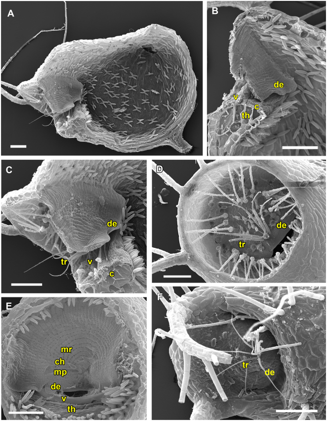
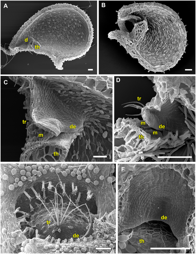
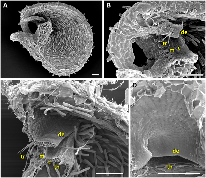
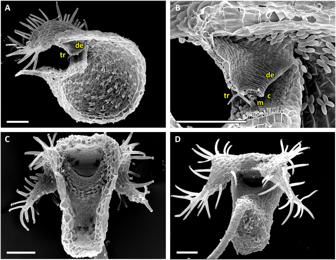
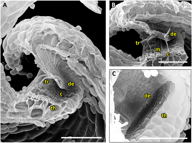
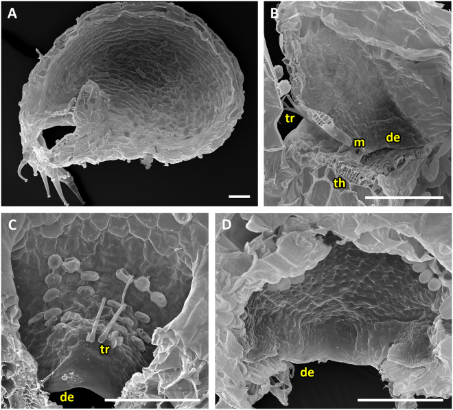
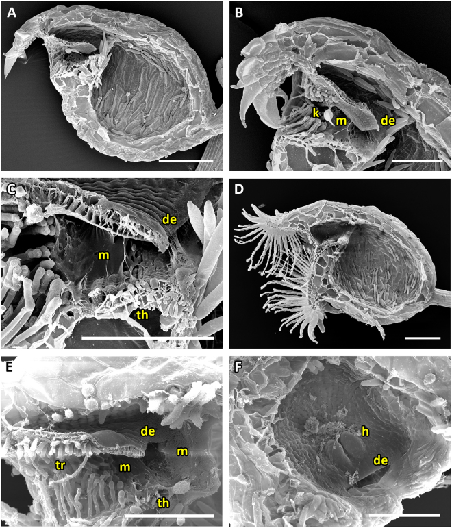
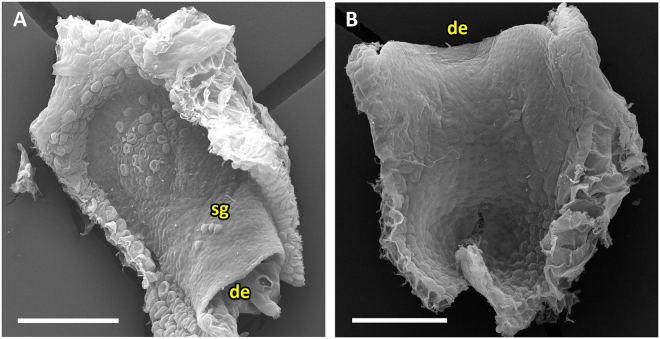
Bladderworts (Utricularia, Lentibulariaceae, Lamiales) constitute the largest genus of carnivorous plants but only aquatic species (about one fifth of the genus) have so far been thoroughly studied as to their suction trap functioning. In this study, we comparatively investigated trap biomechanics in 19 Utricularia species to examine correlations between life-forms, trapping mechanisms, and functional-morphological traits. Our investigations show the existence of two functional trap principles (passive trap in U. multifida vs. active suction traps), and - in active suction traps - three main trapdoor movement types (with several subtypes). The trapdoor movement types and their corresponding functional-morphological features most presumably represent adaptations to the respective habitat. We furthermore give insights into fluid dynamics during suction in three representatives of the main types of trapdoor movement. The results on functional morphology and trapdoor movement were mapped onto a new phylogenetic reconstruction of the genus, derived from the rapidly evolving chloroplast regions trnK, rps16 and trnQ-rps16 and a sampling of 105 Utricularia species in total. We discuss potential scenarios of trap character evolution and species radiation, highlighting possible key innovations that enable such a unique carnivorous lifestyle in different habitats.


Conflict of interest statement

The authors declare that they have no competing interests.

Figures

Figure 1
Figure 1
Utricularia trapdoor movement types in active traps. Schematic drawings in column ‘set position’ depict sagittal sections of trap entrances, highlighting the initial door postures when the traps are ready to fire. Columns right from ‘set position’ depict different movement steps observed for respective species. Timescales and standard deviations are given for species written in boldface (see Table 2 for detailed results on all species). For species indicated by an asterisk (*) see ref.. The threshold (th) as well as the progression of the median door axis (d) and the movement of trigger hairs (tr) are outlined. In the U. vulgaris trapdoor type (UVTT), the trapdoor performs a curvature inversion from convex to concave prior to opening (Movie S1). The UVTT1 is the trapdoor type as present in the ‘Utricularia vulgaris trap type’ with the obtuse door-to-threshold angle (90°) (see Introduction). In the UVTT2, the trigger hair movement is different to the UVTT1 (Movie S2). In the U. praelonga trapdoor type (UPTT), the door is in acute angle in relation to the threshold and, like in the UVTT, performs curvature inversion prior to opening (Movie S3). Doors of the U. uniflora trapdoor type (UUTT) do not perform curvature inversions prior to opening (Movies S4–S7). They slowly detach from the threshold and then swing open. The different subtypes (UUTT1-4) are subdivided according to the divergent door postures in the set positions and to the occurrence of trigger hairs.
Figure 2
Figure 2
SEM images showing morphological trap characteristics of U. gibba (representing the UVTT 1) and of U. resupinata (representing the UVTT 2). (A) Sagittal section of a trap of U. gibba. The entrance forms a short tube. The trapdoor, the threshold, and the different glands covering the trap inside are clearly visible. (B) Sagittal section of an U. resupinata trap entrance. Note the cavity on the threshold, the free door edge, the trigger hairs, the velum and the threshold. (C) Exterior view on trap entrance of U. resupinata. The trigger hairs, the free door edge and a multitude of glandular structures are visible. (D) Sagittal section through the short trap entrance of U. gibba with the trigger hairs, the door edge and distinct velum and cavity on the threshold clearly visible. (E) Exterior view on trap entrance of U. gibba. (F) Concentric cellular constrictions on the inner door surface of U. gibba, with different door regions sensu [15] indicated. Scale bars 100 µm. Abbreviations: c = cavity, ch = central hinge, de = door edge, mp = middle piece, mr = middle region, th = threshold, tr = trigger hairs, v = velum.
Figure 3
Figure 3
SEM images showing morphological trap characteristics of U. reniformis, U. alpina and U. flaccida, all representing the UPTT. (A) Sagittal section through an U. reniformis trap with the tubular trap entrance visible. (B) Sagittal section of an U. alpina trap. The narrow trap entrance is tube-shaped with the appendages covering the entrance. (C) Sagittal section through the U. reniformis trap entrance. Clearly visible are the trigger hairs protruding from the trapdoor, the free door edge, the threshold and the mucilage on the threshold. (D) Sagittal section through an U. flaccida trap entrance. (E) View on the outer surface of an U. reniformis trapdoor with trigger hairs. (F) View on the inner surface of the trapdoor of U. reniformis with a weak concentric cellular constriction pattern. Scale bars = 100 µm. Abbreviations: de = door edge, m = mucilage, th = threshold, tr = trigger hairs.
Figure 4
Figure 4
SEM images of U. alpina and U. aureomaculata, representing the UPTT. (A) Sagittal section of a trap of U. alpina. The narrow trap entrance is shaped like a tube, the appendages covering the entrance are clearly visible. (B) Sagittal section through trap entrance of U. alpina. Clearly visible are the trigger hairs, the free door edge, the cavity, and mucilage sticking to the threshold. (C) Sagittal section through a trap entrance with a rather tubular shape of U. aureomaculata. Clearly visible are the trigger hairs, the free door edge, the threshold and the mucilage on the threshold. A shallow cavity on the threshold can be observed. (D) View on the inner surface of trap door of U. aureomaculata. A distinct cellular constriction pattern cannot be identified. Scale bars = 100 µm. Abbreviations: c = cavity, de = door edge, m = mucilage, th = threshold, tr = trigger hairs.
Figure 5
Figure 5
SEM images of a trap, the trap entrance and trapdoor of U. calycifida, representing the UPTT. (A) Sagittal section of a trap. The tubular shape of the trap entrance, the free door edge and the protruding trigger hairs are visible. (B) Sagittal section through the trap entrance. Visible are the free door edge, the trigger hairs, a weak cavity on the threshold and mucilage between the door and the threshold. (C) Interior view on the front part of a bladder. (D) Frontal, exterior view on an U. calycifida trap. Note the filamentous appendages covering the obscured, narrow trap entrance. Scale bars = 200 µm. Abbreviations: c = cavity, de = door edge, m = mucilage, th = threshold, tr = trigger hairs.
Figure 6
Figure 6
SEM images of trap entrance and trapdoor of U. longifolia representing the UPTT. (A and B) Sagittal sections through the tubular trap entrance. Clearly visible are the trigger hairs, the free door edge and a weak cavity on the threshold. In B) also mucilage sticking to the threshold can be seen. (C) View on the inner door surface. A pattern of concentric constrictions is faintly visible. Scale bars = 100 µm. Abbreviations: c = cavity, de = door edge, m = mucilage, th = threshold, tr = trigger hairs.
Figure 7
Figure 7
SEM images of a trap, trap entrance and trapdoor of U. praelonga representing the UPTT. (A) Sagittal section through a trap with the tubular trap entrance visible. (B) Sagittal section through the trap entrance. Note the trigger hairs and mucilage sticking to the free door edge and the threshold. (C) View on the outer surface of the trapdoor with trigger hairs. (D) View on the inner surface of the trapdoor. A distinct concentric cellular constriction pattern is not visible. Scale bars = 100 µm. Abbreviations: de = door edge, m = mucilage, th = threshold, tr = trigger hairs.
Figure 8
Figure 8
SEM and light microscopy (LM) images showing morphological trap characteristics of U. uniflora (representing the UUTT1), U. menziesii (representing the UUTT2), U. warburgii and U. cornuta (both representing the UUTT3). (A) Sagittal section of a trap of U. menziesii with the free door edge resting on the inner region of the threshold, reaching far into the trap lumen. The entrance is narrow and tubular, and covered by appendages on the outside. (B) Sagittal section of a trap of U. warburgii. (C) View on the inner door surface of U. uniflora. No concentric constrictions are visible. (D) View on the outer door surface of U. uniflora. Sessile glands in the upper part of the door as well as a pad of unknown function and teeth-like-structures are visible. (E) Detail of the inner door surface and the threshold with conspicuous teeth-like structures on the door edge as well as mucilage adhering to the teeth and the threshold. (F) LM sagittal thin-section (toluidine blue staining) of the trap entrance of U. cornuta. Perceptible are the cavity on the threshold and a thread of mucilage between the free door edge and the cavity. Scale bars A,B = 500 µm, C–F = 100 µm. Abbreviations: c = cavity, d = door, de = door edge, m = mucilage, p = pad, sg = sessile glands, t = teeth, th = threshold.
Figure 9
Figure 9
SEM and LM images of traps, trap entrances and trapdoors of U. cornuta, U. warburgii and U. prehensilis representing the UUTT3. (A) Sagittal section through the narrow, tubular trap entrance of U. cornuta (scale bar = 50 µm). (B) Sagittal section of a trap entrance of U. warburgii (scale bar = 50 µm). Note the free door edge, the fringe of long sessile glands presumably functioning as trigger hairs and the conspicuous pad on the door. (C) Sagittal section of a trap of U. prehensilis (scale bar = 500 µm). The trap entrance is tubular. On the trapdoor, sessile glands are observable and on the threshold a cavity. (D) View on the inner trapdoor surface. No pattern of concentric constrictions is visible (scale bar = 50 µm). Abbreviations: c = cavity, de = door edge, m = mucilage, p = pad, sg = sessile glands, th = threshold.
Figure 10
Figure 10
SEM pictures of traps, trap entrances and trapdoors of U. welwitschii and U. livida, representing the UUTT4. (A) Sagittal section through a trap of U. welwitschii (scale bar = 200 µm). Note the tubular trap entrance. (B) Sagittal section through a trap entrance of U. welwitschii (scale bar = 100 µm). Clearly visible are the free door edge, mucilage sticking to door and threshold and the noticeable, species-specific trigger hair, the so-called kriss-trichome. (C) Detail of mucilage sticking to the door of U. welwitschii (scale bar = 100 µm). (D) Sagittal section of a trap of U. livida (scale bar = 200 µm). Numerous appendages covering the tubular trap entrance can be seen. (E) Sagittal section through the trap entrance of U. livida (scale bar = 100 µm). A trigger hair protruding from the trapdoor is visible as well as mucilage adhering to the threshold and the free door edge. (F) View on the inner surface of U. livida with two humps visible on the trapdoor (scale bar = 100 µm). Abbreviations: de = door edge, h = hump, k = kriss-trichome, m = mucilage, th = threshold, tr = trigger hairs.
Figure 11
Figure 11
Trap door of U. menziesii (UUTT2). (A) View on outer door surface. (B) View on the inner door surface. The door maintains a S-shaped curvature though freed from the trap entrance. Abbreviations de = door edge, sg = sessile glands. Scale = 100 µm.
Figure 12
Figure 12
SEM images showing morphological trap characteristics of U. multifida. (A) Sagittal section through a trap (scale bar = 500 µm). The long, tubular and narrow trap entrance is densely covered with glands pointing towards the trapdoor. The free door edge and the threshold are visible. (B) Sagittal section through the trap entrance with intact trapdoor (scale bar = 100 µm). Note the small sessile glandular structures on the trapdoor. (C) Detail view of the sessile glandular structures (scale bar = 20 µm). (D) View on the inner surface of the trapdoor (scale bar = 100 µm). Concentric cellular constrictions could not be detected. (E) Posterior part of a bladder filled with caught copepods (scale bar = 100 µm). Note the characteristic triangular shape of the U. multifida bladder and its thick and putatively stiff trap walls. Abbreviations: cp = copepod, de = door edge, sg = sessile glands, th = threshold.
Figure 13
Figure 13
Fluid velocities and aspiration zones in three Utricularia species representing the three main trapdoor movement types. (A) The top fluid speeds among the three tested species do not differ statistically (Kruskal-Wallis test, χ2 (2) = 0.24698, p = 0.884). (B–D) Lateral views on trap entrances showing the aspiration zones (dotted lines). The set positions of the doors (white lines) with trigger hairs (where present, yellow lines) are indicated. Also visible are the hair used for manually triggering the traps (arrows), as well as a multitude of tracer particles used to study the flow of water during suction. (B) Aquatic U. gibba (UVTT). (C) Terrestrial U. praelonga (UPTT). (D) Terrestrial U. prehensilis (UUTT3). Scale bar (applies for BD) = 100 µm.
Figure 14
Figure 14
Phylogeny of Utricularia as inferred with Maximum Likelihood, Maximum Parsimony and Bayesian Inference of trnK/matK and trnL-F sequences. Tree topology was taken from the ML analysis, numbers above branches indicate ML bootstrap percentages (bold italics; right), and MP bootstrap percentages (plain, left), numbers below branches are posterior probabilities from Bayesian Inference.
Figure 15
Figure 15
U. subgenus Polypompholyx, as inferred with Maximum Likelihood, Maximum Parsimony and Bayesian Inference of trnK/matK sequences. Tree topology was taken from the ML analysis, numbers above branches indicate ML bootstrap percentages (bold italics; right), and MP bootstrap percentages (plain, left), numbers below branches are posterior probabilities from Bayesian Inference. Classification according to ref., as modified by ref., is indicated to the right of the tree.
Figure 16
Figure 16
Phylogeny of U. subgenus Bivalvaria as inferred with Maximum Likelihood, Maximum Parsimony and Bayesian Inference of trnK/matK sequences. Tree topology was taken from the ML analysis, numbers above branches indicate ML bootstrap percentages (bold italics; right), and MP bootstrap percentages (plain, left), numbers below branches are posterior probabilities from Bayesian Inference. Classification according to ref., as modified by ref., is indicated to the right of the tree.
Figure 17
Figure 17
U. subgenus Utricularia, as inferred with Maximum Likelihood, Maximum Parsimony and Bayesian Inference of trnK/matK sequences. Tree topology was taken from the ML analysis, numbers above branches indicate ML bootstrap percentages (bold italics; right), and MP bootstrap percentages (plain, left), numbers below branches are posterior probabilities from Bayesian Inference. Classification according to ref., as modified by ref., is indicated to the right of the tree.
Figure 18
Figure 18
Relative state probabilities of trap characters at ancestral nodes. Positions of characters on branches (three above branches, three below branches) and colours of individual character states in the pie chart labels as indicated in the legend (top left). Full circle (all possible states; probability = 1.0, 100%) is divided into sectors (representing states) that each visualize the probability (<=0) of their state being the ancestral state at a given node. In the legend, all states have equal probability to indicate all colours/states.

Similar articles

Cited by

References

    1. Schäferhoff B, et al. Towards resolving Lamiales relationships: insights from rapidly evolving chloroplast sequences. BMC Evol. Biol. 2010;10:352–374. doi: 10.1186/1471-2148-10-352. - DOI - PMC - PubMed
    1. Greilhuber J, et al. Smallest angiosperm genomes found in Lentibulariaceae, with chromosomes of bacterial size. Plant Biol. 2006;6:770–777. doi: 10.1055/s-2006-924101. - DOI - PubMed
    1. Ibarra-Laclette E, et al. Architecture and evolution of a minute plant genome. Nature. 2013;498:94–98. doi: 10.1038/nature12132. - DOI - PMC - PubMed
    1. Veleba A, et al. Genome size and genomic GC content evolution in the miniature genome-sized family Lentibulariaceae. New Phytol. 2014;203:22–28. doi: 10.1111/nph.12790. - DOI - PubMed
    1. Taylor, P. The genus Utricularia: a taxonomic monograph. Kew Bulletin Additional Series XIV, London, (1989).

Publication types

LinkOut - more resources