. 2017 Sep 21;7(1):12131.
doi: 10.1038/s41598-017-12409-0.

The crystal structure of the regulatory domain of the human sodium-driven chloride/bicarbonate exchanger

Affiliations

The crystal structure of the regulatory domain of the human sodium-driven chloride/bicarbonate exchanger

Carolina M Alvadia et al. Sci Rep. .

Abstract

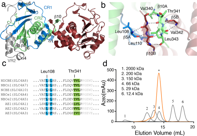
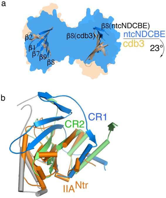
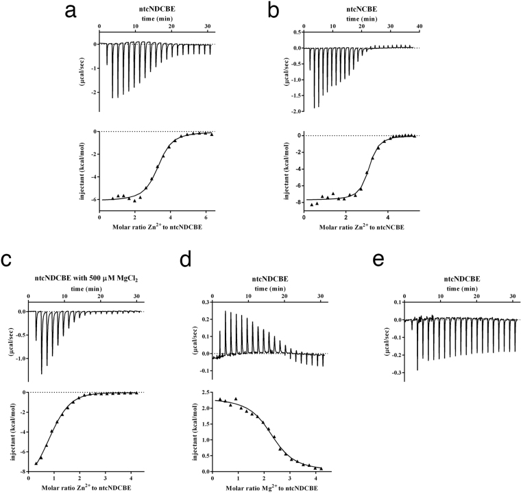
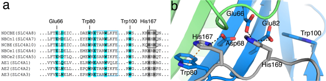
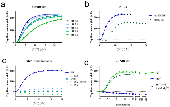
The sodium-driven chloride/bicarbonate exchanger (NDCBE) is essential for maintaining homeostatic pH in neurons. The crystal structure at 2.8 Å resolution of the regulatory N-terminal domain of human NDCBE represents the first crystal structure of an electroneutral sodium-bicarbonate cotransporter. The crystal structure forms an equivalent dimeric interface as observed for the cytoplasmic domain of Band 3, and thus establishes that the consensus motif VTVLP is the key minimal dimerization motif. The VTVLP motif is highly conserved and likely to be the physiologically relevant interface for all other members of the SLC4 family. A novel conserved Zn2+-binding motif present in the N-terminal domain of NDCBE is identified and characterized in vitro. Cellular studies confirm the Zn2+ dependent transport of two electroneutral bicarbonate transporters, NCBE and NBCn1. The Zn2+ site is mapped to a cluster of histidines close to the conserved ETARWLKFEE motif and likely plays a role in the regulation of this important motif. The combined structural and bioinformatics analysis provides a model that predicts with additional confidence the physiologically relevant interface between the cytoplasmic domain and the transmembrane domain.


Conflict of interest statement

The authors declare that they have no competing interests.

Figures

Figure 1
Figure 1
Dimerization determinants of ntcNDCBE. (a) ntcNDCBE is a dimer with 2-fold symmetry. The core domain of ntcNDCBE contains CR1 (light blue), VR2 (grey) and CR2 (light green). The 58 unstructured residues from Lys173 to Lys232 in VR2 are not visible in the electron density. Monomer B is shown in dark red. (b) Zoomed in view of the dimerization β-sheet, around 90 ° rotation along the x-axis from (a). The backbone of the residues of the dimerization β-sheet is shown as sticks. Only the side-chain residues from monomer A are shown. Hydrogen bonds are represented with black dotted lines. (c) Sequence alignment of the human N-terminal cytoplasmic domain of NBCs and AEs. Residues involved in the dimerization motif of ntcNDCBE are colour coded as in (a). The residues not present in ntcNDCBE are coloured grey. (d) Size-exclusion chromatography of 1 mg of ntcNDCBE (orange) on a Superdex 200 10/300 GL (GE Healthcare). ntcNDCBE eluted at 14.2 mL, corresponding to a dimer with an apparent MW of 80.6 kDa compared with molecular weight standards (grey).
Figure 2
Figure 2
Structural comparisons of ntcNDCBE. (a) Superimposition of the central β-sheet of ntcNDCBE (chain A, blue) with the corresponding amino acids from cdb3 (chain P, light brown) with an RMSD of 0.82 Å. The relative movement of chain B of ntcNDCBE compared with chain Q of cdb3, showing a pivotal rotation of 23°. Only the central β-sheet of both structures is shown. The surface representation of each structure is shown in the background. (b) Superimposition of CR2 from ntcNDCBE chain A with E. coli IIANtr (PDB ID: 1A6J, Chain A), showing an RMSD of 2.25 Å. IIANtr is coloured orange.
Figure 3
Figure 3
The cytoplasmic domain of NBCs has divalent metal-binding properties. (ae) Top: the heat released/absorbed upon metal injection. Bottom: the integrated heat data and corresponding binding isotherm (black line). ITC measurement of ZnCl2 titration into (a) ntcNDCBE. Binding model: one set of sites (n = 3.2, K D = 2.3 µM). (b) ntcNCBE. Binding model: one set of sites (n = 3.0, K D = 0.7 µM). (c) ntcNDCBE in the presence of 500 µM MgCl2. Binding model: sequential binding sites (n = 2, K D = 1.1 µM and 14.5 µM). (d) ITC measurement of MgCl2 titration into ntcNDCBE. Binding model: one set of sites (n = 2.3, K D = 4.4 µM). (e) ITC measurement of CaCl2 titration into ntcNDCBE. No binding isotherm could be fitted.
Figure 4
Figure 4
Putative Zn2+-binding site in ntcNDCBE. (a) Fraqmented sequence alignment of the N-terminal cytoplasmic domain of NBCs and AEs. The conserved ETARWLKFEE motif, proposed to be part of a substrate channel, is marked by a blue box. The residues are coloured as in Fig. 1a. (b) The ntcNDCBE structure: The H167-X-H169 Zn2+-binding motif, the secondary shell of negative residues and the three Trp residues of the construct are shown as sticks. Hydrogen bonds are represented as black dotted lines and π-stacking interactions in orange.
Figure 5
Figure 5
Effect of Zn2+ on the tryptophan fluorescence yield of NBCs. (a) Zn2+ titration into ntcNDCBE at different pH values. Individual runs are shown in Supplementary Figure S2. (b) Zn2+ titration into, ntcNCBE at pH 7.2. The Trp fluorescence yield increases faster in ntcNDCBE when exposed to Zn2+ as compared with ntcNCBE. Individual runs are shown in Supplementary Figure S4. (c) Zn2+ titration into the different ntcNDCBE constructs at pH 7.2. The ntcNDCBE mutants show little response to Zn2+ titration. Individual runs are shown in Supplementary Figure S2. (d) Mg2+ and Zn2+ (in the absence and presence of 1 mM Mg2+) titration into ntcNDCBE at pH 7.2. Individual runs are shown in Supplementary Figure S2. The fits (lines) were calculated with equation (2).
Figure 6
Figure 6
pH measurements performed in slc4a10-transfected cells. (a) Na+ and HCO3 dependent pHi recovery following intracellular acidification is reduced by 10 uM ZnCl2. Left: Intracellular pH recordings in slc4a10 transfected cells. After a baseline pHi measurement in HBS cells were acidified using an NH4Cl prepulse (i) followed by a washout in a Na+ free BBS (ii). After a short stabilization, the pHi recovery was determined after the addition of Na+ containing BBS (iii). The recordings were performed in the absence (black line) and presence (grey line) of 10 µM ZnCl2 added to the BBS. (b) Mean values ± SEM for net acid extrusion rate in the slc4a10 transfected cells.
Figure 7
Figure 7
Orientation of ntcNDCBE towards the membrane. (a) Top: Sequence conservation of the transmembrane domain of NBCs and AEs mapped onto the transmembrane domain structure of AE1 (PDB ID: 4YZF), as viewed from the cytoplasm. Dark blue indicates high conservation. Bottom: Sequence conservation of the NBC and AE N-terminal cytoplasmic domains mapped onto the structure of ntcNDCBE, as viewed from the membrane. (b) Model of the structure of NDCBE. The ntcNDCBE is shown as a grey cartoon in either a blue (monomer A) or a pink (monomer B) surface. The ETARWLKFEE motif is coloured yellow. The dimerization β-sheet and the identified Zn2+-binding site are coloured as in Fig. 1a. Resudies in the identified Zn2+-binding site is shown as sticks. The transmembrane domain of AE1 is shown as a grey cartoon in either a pink (monomer A) or a blue (monomer B) surface. The first two residues of the transmembrane domain are shown as green coloured spheres, according to which N-terminal cytoplasmic monomer is bound. The intrinsically disordered VRs are represented as dotted lines. The cytoplasmic domain is not represented.

Similar articles

Cited by

References

    1. Boron, W. F. & Boulpaep, E. L. In Medical Physiology (Saunders, Philadelphia, 2009).
    1. Parker MD, Boron WF. The divergence, actions, roles, and relatives of sodium-coupled bicarbonate transporters. Physiol. Rev. 2013;93:803–959. doi: 10.1152/physrev.00023.2012. - DOI - PMC - PubMed
    1. Praetorius J, Nejsum LN, Nielsen S. A SCL4A10 gene product maps selectively to the basolateral plasma membrane of choroid plexus epithelial cells. Am. J. Physiol. Cell. Physiol. 2004;286:C601–10. doi: 10.1152/ajpcell.00240.2003. - DOI - PubMed
    1. Sassani P, et al. Functional characterization of NBC4: a new electrogenic sodium-bicarbonate cotransporter. Am. J. Physiol. Cell. Physiol. 2002;282:C408–16. doi: 10.1152/ajpcell.00409.2001. - DOI - PubMed
    1. Parker MD, Bouyer P, Daly CM, Boron WF. Cloning and characterization of novel human SLC4A8 gene products encoding Na+ -driven Cl-/HCO3(−) exchanger variants NDCBE-A, -C, and -D. Physiol. Genomics. 2008;34:265–276. doi: 10.1152/physiolgenomics.90259.2008. - DOI - PMC - PubMed

Publication types

LinkOut - more resources