. 2017 Sep 21;7(1):12084.
doi: 10.1038/s41598-017-12360-0.

Zebrafish In-Vivo Screening for Compounds Amplifying Hematopoietic Stem and Progenitor Cells: - Preclinical Validation in Human CD34+ Stem and Progenitor Cells

Affiliations

Zebrafish In-Vivo Screening for Compounds Amplifying Hematopoietic Stem and Progenitor Cells: - Preclinical Validation in Human CD34+ Stem and Progenitor Cells

Guruchandar Arulmozhivarman et al. Sci Rep. .

Abstract

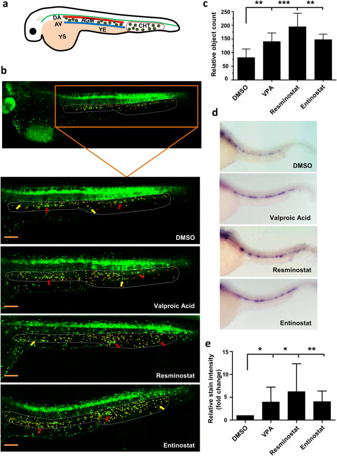
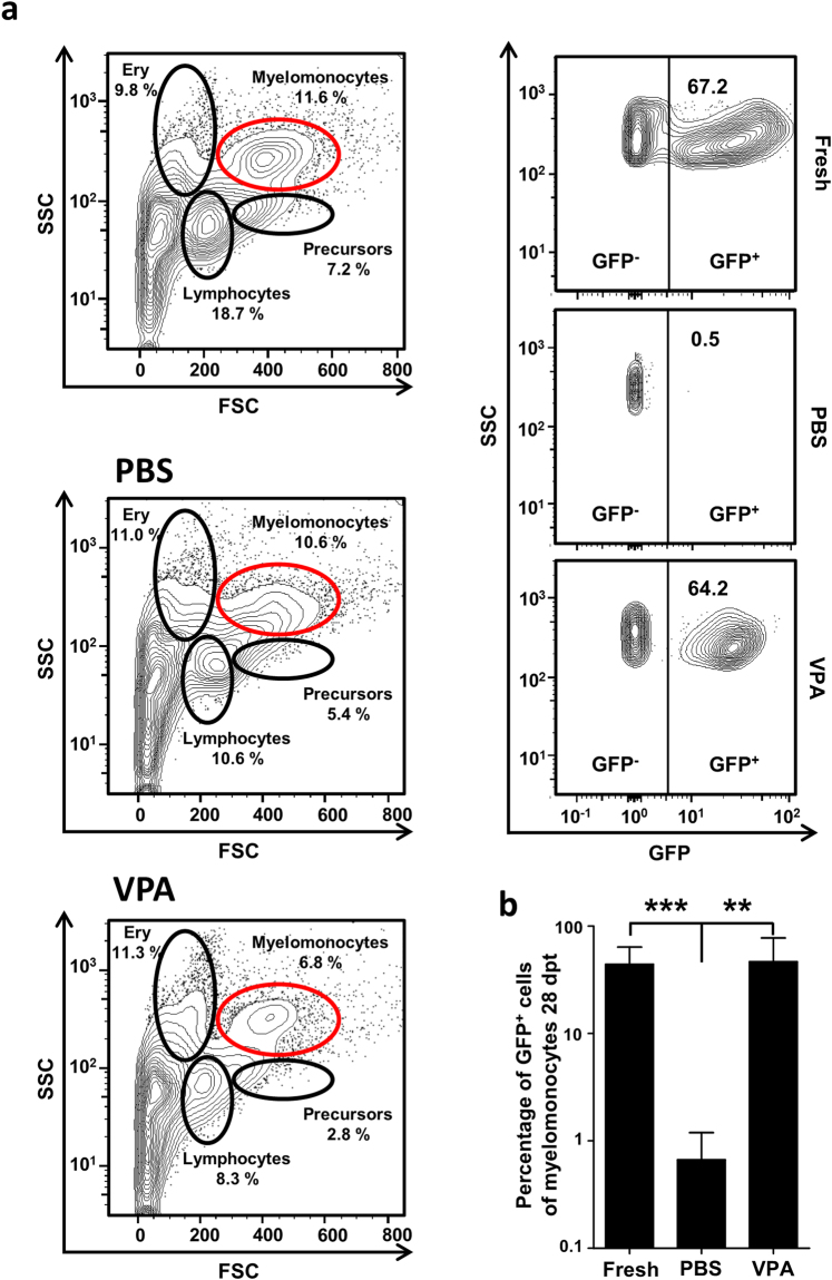
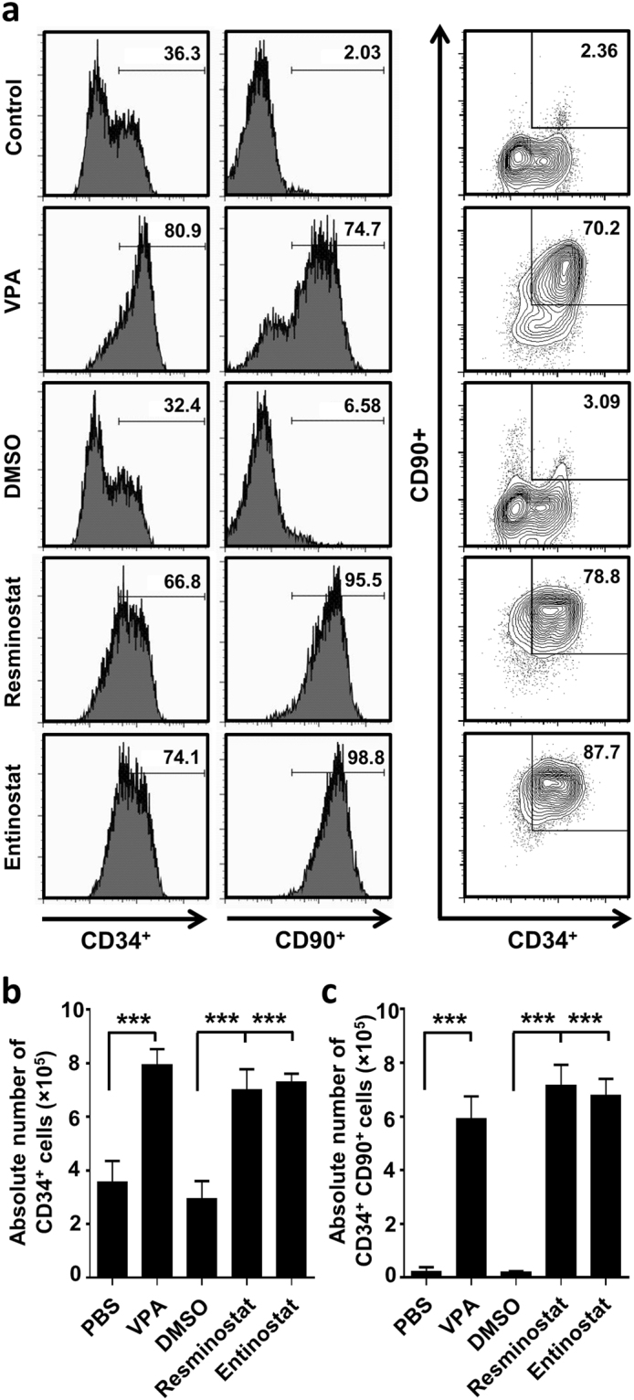
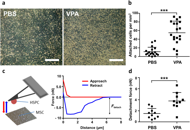
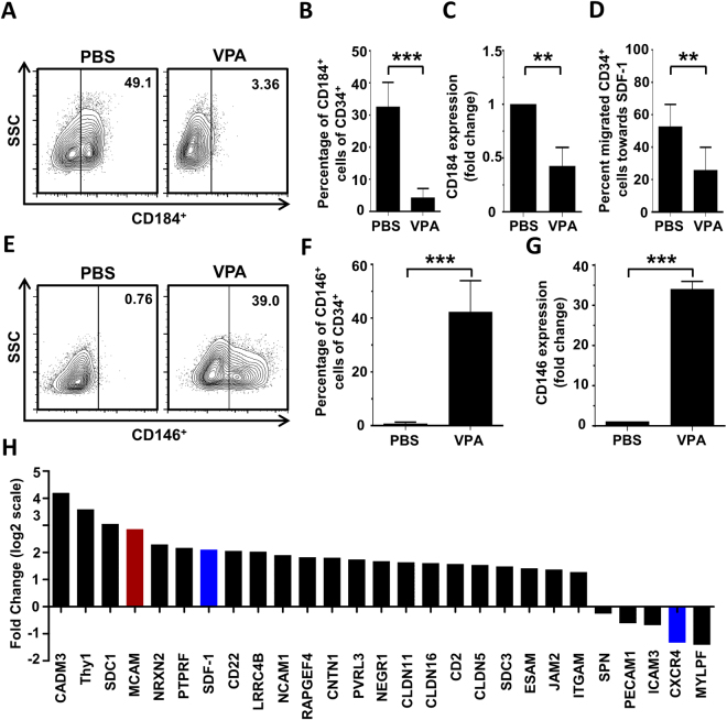
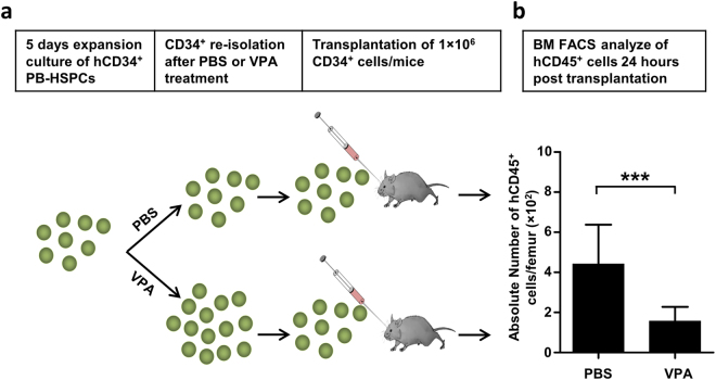
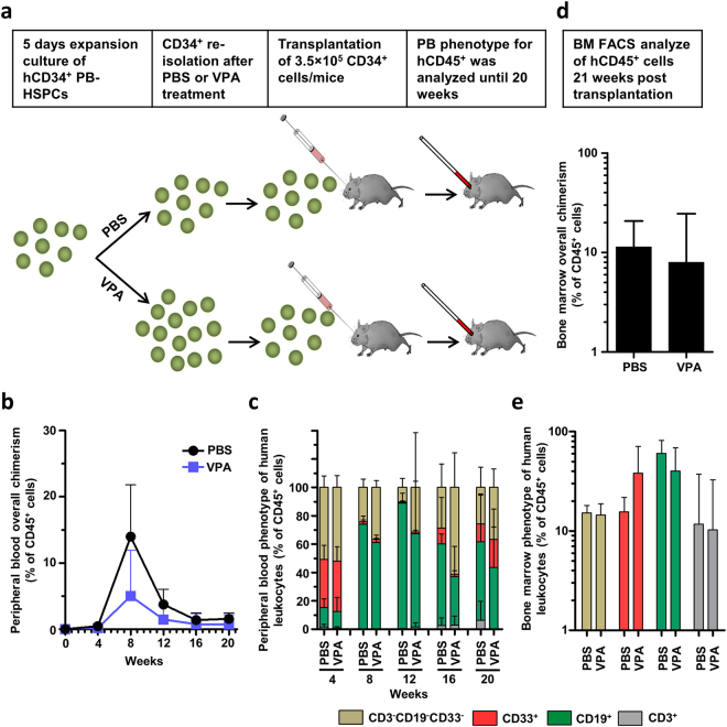
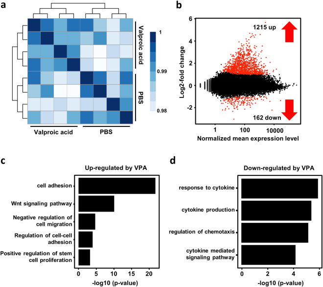
The identification of small molecules that either increase the number and/or enhance the activity of human hematopoietic stem and progenitor cells (hHSPCs) during ex vivo expansion remains challenging. We used an unbiased in vivo chemical screen in a transgenic (c-myb:EGFP) zebrafish embryo model and identified histone deacetylase inhibitors (HDACIs), particularly valproic acid (VPA), as significant enhancers of the number of phenotypic HSPCs, both in vivo and during ex vivo expansion. The long-term functionality of these expanded hHSPCs was verified in a xenotransplantation model with NSG mice. Interestingly, VPA increased CD34+ cell adhesion to primary mesenchymal stromal cells and reduced their in vitro chemokine-mediated migration capacity. In line with this, VPA-treated human CD34+ cells showed reduced homing and early engraftment in a xenograft transplant model, but retained their long-term engraftment potential in vivo, and maintained their differentiation ability both in vitro and in vivo. In summary, our data demonstrate that certain HDACIs lead to a net expansion of hHSPCs with retained long-term engraftment potential and could be further explored as candidate compounds to amplify ex-vivo engineered peripheral blood stem cells.


Conflict of interest statement

The authors declare that they have no competing interests.

Figures

Figure 1
Figure 1
HDACIs increase c-myb+ HSPC number and runx1 expression in zebrafish embryos. (a) Schematic representation of HSPC development in the AGM and CHT regions of a zebrafish embryo. YS – yolk sac; YE – yolk extension; DA – dorsal aorta; AV – axial vein; AGM – aorta-gonad-mesonephros; CHT – caudal hematopoietic tissue. Small green circles between the DA and AV and in CHT regions represent HSPCs. (b) Image based identification of c-myb+ cells in the AGM and CHT region identified HDACIs (valproic acid, resminostat, and entinostat) as enhancers of HSPC cell-count at 36 hpf (Bars = 200 µm). The region-of-interest (AGM and CHT) marked by white line and the c-myb+ cells are marked by red circle (indicated by red arrows) and the false positive objects that are excluded from the quantification are marked by yellow circle (indicated by yellow arrows). (c) Quantification of relative number of c-myb+ cells in the AGM and CHT region showing increased cell-count after 40 µM VPA, resminostat or entinostat treatment compared to DMSO (n = 5). (d) Validation of identified hits through whole-mount in situ hybridization for runx1 expression. (e) Quantification of runx1 relative stain intensity shows significantly higher runx1 expression in HDACI treated fish compared to DMSO controls. Intensity was calculated using area under curve analyses in ImageJ. Single images were split into 5 regions of interest (ROIs) and intensity was normalized to background signal. SD displays deviation among 5 ROIs in one image (n = 3). Data are shown as mean ± SD, *p < 0.05; **p < 0.01; *** p < 0.001.
Figure 2
Figure 2
Ex vivo VPA treatment preserves engraftment capacity of whole kidney marrow cells. (a) A representative plot showing gating strategy used to determine the degree of chimerism in the recipient and the FACS plot of PBS and VPA treated conditions. (b) PBS-treated control donor WKM cells failed to engraft the kidney of the recipient, but VPA treated cells demonstrated an engraftment capacity similar to that of freshly isolated cells (uncultured; n = 5 per group). Data are shown as mean ± SD, **p < 0.01, ***p < 0.001.
Figure 3
Figure 3
Ex vivo expansion of G-CSF mobilized CD34+ HSPCs treated for 5 days with HDACIs. (a) Representative flow-cytometry analysis of CD34 and CD90 expression after 5 d of ex vivo HDACI treatment. (b) Absolute cell numbers of CD34+ cells. (c) Absolute cell numbers of CD34+CD90+ cells. Both CD34+ and CD34+CD90+ cells were significantly increased after 5 days treatment with VPA (1 mM), resminostat (1.5 µM) or entinostat (1.5 µM) compared to controls (PBS and DMSO; n = 5). Data are shown as mean ± SD, ***p < 0.001.
Figure 4
Figure 4
Ex vivo VPA-expanded CD34+ cells exhibit increased adhesion to MSCs. (a) Freshly isolated CD34+ cells were seeded onto a confluent MSC layer. Representative images after 5 days of co-culture with MSCs showed an increase in the number of VPA-treated adherent cells (right) compared to control cells (left). Images were taken after sufficient washing with PBS. (Scale bar: 250 µm). (b) In the adhesion assay, a significantly higher number of VPA expanded cells were attached to MSCs compared to control (n = 4). (c) Schematic representation of atomic force microscopy-based single-cell force spectroscopy used to measure adhesive strength of VPA treated and control cells. (d) Plot showing detachment force measurement after 5 days of VPA or PBS (control) treatment. Adhesive strength of VPA treated cells was 2–3 fold higher than control (n = 3). Data are mean ± SD, ***p < 0.001.
Figure 5
Figure 5
Valproic acid affects adhesion of HSPCs and suppresses their migration toward SDF-1 in vitro. G-CSF mobilized CD34+ HSPCs were treated in vitro for 5 days with VPA or PBS and analyzed for the expression of molecules that are involved in cell adhesion and migration. The functional consequence of VPA-treatment on the migratory capacity toward SDF-1 was also evaluated. (a) Representative dot-plot of CD184 (CXCR4) expression on the cell surface of CD34+ cells as determined by flow cytometry. (b) VPA-treatment significantly reduced the expression of CXCR4 on the cell surface of CD34+ cells compared to control cells (n = 3), measured by flow cytometry. (c) Reduced CXCR4 expression was confirmed by quantitative PCR (n = 3). (d) Trans-well migration assay showed that VPA-treatment significantly reduced the migration capacity of CD34+ cells toward an SDF-1 gradient (100 ng/ml) compared to control cells (n = 4). (e) Representative plot of CD146 (MCAM) expression on the cell surface of CD34+ cells as determined by flow cytometry. (f) Flow cytometric analysis showed that VPA-treatment significantly increased surface expression of MCAM on CD34+ cells compared to controls (n = 4). (g) Significantly higher expression of MCAM in VPA expanded CD34+ cells was verified by quantitative PCR (n = 4). (h) RNA sequencing revealed that VPA-treatment substantially changed the expression of molecules involved in cell adhesion and migration in CD34+ cells compared to control cells, including CXCR4 and MCAM (n = 4). Data are mean ± SD, **p < 0.01, ***p < 0.001.
Figure 6
Figure 6
Valproic acid decreases bone marrow homing capacity of in vitro expanded CD34+ cells. (a) Schematic representation of the homing assay performed in NSG mice. CD34+ cells were re-isolated after 5 days ex vivo treatment with VPA or PBS. 1 × 106 CD34+ cells were transplanted by intravenous injection into the retro-orbital venous plexus of sub-lethally irradiated (100 cGy) NSG mice. Homing was quantified by flow cytometric analysis of human leucocytes (human CD45+) in the bone marrow of recipient mice 24 hours after injection. (b) The absolute number of human leukocytes homing to the femur of recipient mice was significantly reduced by ex vivo treatment of CD34+ with VPA compared to control cells (n = 8–14). Data are shown as mean ± SD, ***p < 0.001.
Figure 7
Figure 7
Valproic acid modifies short-term engraftment but does not influence long-term engraftment and differentiation capacity of VPA expanded CD34+ cells. (a) Schematic representation of the engraftment assay performed in NSG mice. Sub-lethally irradiated (100 cGy) mice were intravenously transplanted with 3.5 × 105 CD34+ VPA-treated or control cells. Engraftment of human CD34+ cells was monitored by analyzing chimerism and phenotype of circulating human leucocytes (human CD45+) in the peripheral blood of recipient mice every four weeks by flow cytometry. Long-term in vivo marrow repopulation capacity was determined at 20 weeks after transplantation by quantification and phenotyping of human leucocytes in the femur of NSG mice (n = 5 per group). (b) Overall peripheral blood chimerism increased by week 12 and subsequently declined. Mice transplanted with PBS-treated CD34+ cells showed elevated overall chimerism compared to mice that received VPA-treated CD34+ cells. Differences were most pronounced at week 8 but did not reach statistical significance (p = 0.913). Data represent mean + SD. (c) Lineage commitment of circulating human leucocytes was examined by analyzing CD3, CD19, and CD33 cell surface expression with no significant differences in the proportion of T-cells, B-cells or myeloid cells. (d) Absolute numbers of human leucocytes (human CD45+) per femur at week 20 after transplantation did not differ between groups (p = 0.61). (e) Lineage diversification of long-term marrow repopulating human leucocytes was similar in mice injected with VPA-treated or PBS-treated human CD34+ cells at 20 weeks after transplantation.
Figure 8
Figure 8
Gene expression profiling revealed induction of cell adhesion pathways and reduced expression of genes involved in chemotaxis. (a) Heat map of a sample-to-sample Pearson correlation and dendrogram showing sample-to-sample correlation. All biological replicates cluster well with each other, and all samples from different populations are clearly separated from each other (analysis by R). (b) The red dots represent differentially expressed genes (DEG) by VPA treatment (1% false discovery rate). (c,d) GO pathway analysis for differentially expressed genes. Plots show biological processes that are associated with genes that are up- or down-regulated in VPA treated CD34+ cells.

Similar articles

Cited by

References

    1. Niederwieser, D. et al. Hematopoietic stem cell transplantation activity worldwide in 2012 and a SWOT analysis of the Worldwide Network for Blood and Marrow Transplantation Group including the global survey. Bone Marrow Transplant. 1–8, 10.1038/bmt.2016.18 (2016). - PMC - PubMed
    1. Laughlin MJ, et al. Hematopoietic engraftment and survival in adult recipients of umbilical-cord blood from unrelated donors. N Engl J Med. 2001;344:1815–1822. doi: 10.1056/NEJM200106143442402. - DOI - PubMed
    1. Kasamon YL, et al. Outcomes of nonmyeloablative HLA-haploidentical blood or marrow transplantation with high-dose post-transplantation cyclophosphamide in older adults. in. Journal of Clinical Oncology. 2015;33:3152–3161. doi: 10.1200/JCO.2014.60.4777. - DOI - PMC - PubMed
    1. Pulsipher MA, et al. Donor, recipient, and transplant characteristics as risk factors after unrelated donor PBSC transplantation: Beneficial effects of higher CD34+ cell dose. Blood. 2009;114:2606–2616. doi: 10.1182/blood-2009-03-208355. - DOI - PMC - PubMed
    1. Dever DP, et al. CRISPR/Cas9 β-globin gene targeting in human haematopoietic stem cells. Nature. 2016;539:384–389. doi: 10.1038/nature20134. - DOI - PMC - PubMed

Publication types

MeSH terms