

Case Reports
doi: 10.4103/meajo.MEAJO_97_17.
A Case of Unilateral Retinitis Pigmentosa Associated with Full Thickness Macular Hole
Affiliations
- PMID: 28936059
- PMCID: PMC5598302
- DOI: 10.4103/meajo.MEAJO_97_17
Item in Clipboard
Case Reports
A Case of Unilateral Retinitis Pigmentosa Associated with Full Thickness Macular Hole
Middle East Afr J Ophthalmol.
2017 Apr-Jun.
Abstract
A 44-year-old Saudi female presented with poor right eye vision for 3 years. Visual acuity (VA) was 20/400 in the right eye and 20/20 in the left eye. Examination and imaging showed all the typical features of retinitis pigmentosa in the right eye associated with full thickness macular hole (FTMH) and an essentially normal left eye. The case underwent pars plana vitrectomy with internal limiting membrane peeling and gas tamponade that resulted in anatomical closure of the FTMH, but VA remained the same.
Keywords: Electroretinogram; fundus autofluorescence; macular hole; unilateral retinitis pigmentosa; vitrectomy.
Conflict of interest statement
There are no conflicts of interest.
Figures
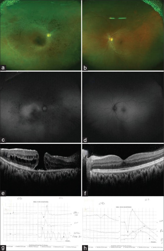
Baseline assessment of both eyes in a case of unilateral retinitis pigmentosa. (a) Color photograph of the right eye showing midperipheral intraretinal pigment migration, pale disc, and blood vessels attenuation. (b) Color photograph of the left eye with normal fundus appearance. (c) Fundus autofluorescence of the right eye showing decreased autofluorescence signal in the midperiphery associated with an abnormal parafoveal ring. (d) Autofluorescence of the left eye showing normal appearance. (e) Optical coherence tomography of the right eye showing full thickness macular hole with intraretinal fluid around it. (f) Optical coherence tomography of the left eye with normal foveal contour. (g) Photopic response of the full-field electroretinogram. It shows very diminished responses from the right eye and normal responses from the left eye. (h) Scotopic response full-field electroretinogram. It shows flat recordings from the right eye and normal responses from the left eye

Postoperative imaging of the right (affected) eye. (a) Color fundus photograph of the right eye showing the typical retinitis pigmentosa features. (b) Fundus autofluorescence of the right eye showing decreased autofluorescence signal in the midperiphery associated with an abnormal parafoveal ring. (c) Optical coherence tomography of the right eye showing closed hole with atrophic changes
Similar articles
-
Flap technique-assisted surgeries for advanced retinitis pigmentosa complicated with macular hole: a case report and literature review.BMC Ophthalmol. 2021 Sep 6;21(1):322. doi: 10.1186/s12886-021-02082-3. BMC Ophthalmol. 2021. PMID: 34488687 Free PMC article. Review.
-
UNILATERAL MACULAR HOLE IN A PATIENT WITH RETINITIS PIGMENTOSA TREATED WITH COVER FLAP TECHNIQUE WITH THE USE OF PLATELET-RICH PLASMA UNDER AIR TAMPONADE.Retin Cases Brief Rep. 2025 Jan 1;19(1):84-90. doi: 10.1097/ICB.0000000000001491. Epub 2024 Dec 13. Retin Cases Brief Rep. 2025. PMID: 37756670 Free PMC article.
-
Pre- and postoperative OCT features and surgical outcomes of advanced retinitis pigmentosa with macular hole: case series and literature review.BMC Ophthalmol. 2024 Aug 26;24(1):370. doi: 10.1186/s12886-024-03643-y. BMC Ophthalmol. 2024. PMID: 39187836 Free PMC article. Review.
-
Macular hole formation in patients with retinitis pigmentosa and prognosis of pars plana vitrectomy.Retina. 2008 Apr;28(4):610-4. doi: 10.1097/IAE.0b013e31815ec341. Retina. 2008. PMID: 18398364
-
BILATERAL LAMELLAR MACULAR HOLE SURGERY IN RETINITIS PIGMENTOSA.Retin Cases Brief Rep. 2016 Winter;10(1):83-5. doi: 10.1097/ICB.0000000000000166. Retin Cases Brief Rep. 2016. PMID: 26200384
Cited by
-
Flap technique-assisted surgeries for advanced retinitis pigmentosa complicated with macular hole: a case report and literature review.BMC Ophthalmol. 2021 Sep 6;21(1):322. doi: 10.1186/s12886-021-02082-3. BMC Ophthalmol. 2021. PMID: 34488687 Free PMC article. Review.
-
UNILATERAL MACULAR HOLE IN A PATIENT WITH RETINITIS PIGMENTOSA TREATED WITH COVER FLAP TECHNIQUE WITH THE USE OF PLATELET-RICH PLASMA UNDER AIR TAMPONADE.Retin Cases Brief Rep. 2025 Jan 1;19(1):84-90. doi: 10.1097/ICB.0000000000001491. Epub 2024 Dec 13. Retin Cases Brief Rep. 2025. PMID: 37756670 Free PMC article.
-
Pre- and postoperative OCT features and surgical outcomes of advanced retinitis pigmentosa with macular hole: case series and literature review.BMC Ophthalmol. 2024 Aug 26;24(1):370. doi: 10.1186/s12886-024-03643-y. BMC Ophthalmol. 2024. PMID: 39187836 Free PMC article. Review.
References
-
- Hagiwara A, Yamamoto S, Ogata K, Sugawara T, Hiramatsu A, Shibata M, et al. Macular abnormalities in patients with retinitis pigmentosa: Prevalence on OCT examination and outcomes of vitreoretinal surgery. Acta Ophthalmol. 2011;89:e122–5. - PubMed
-
- Fishman GA, Fishman M, Maggiano J. Macular lesions associated with retinitis pigmentosa. Arch Ophthalmol. 1977;95:798–803. - PubMed
Publication types
MeSH terms
LinkOut - more resources
Full Text Sources
Other Literature Sources
Miscellaneous
