

Combining SPECT and Quantitative EEG Analysis for the Automated Differential Diagnosis of Disorders with Amnestic Symptoms
- PMID: 28936173
- PMCID: PMC5594223
- DOI: 10.3389/fnagi.2017.00290
Combining SPECT and Quantitative EEG Analysis for the Automated Differential Diagnosis of Disorders with Amnestic Symptoms
Abstract
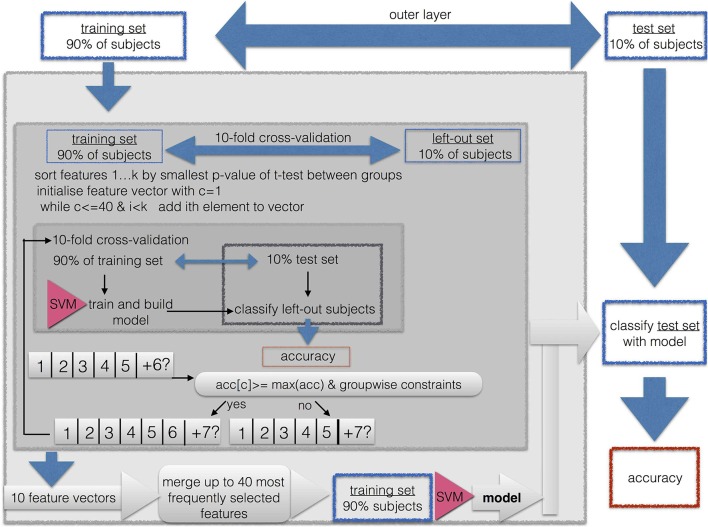
Single photon emission computed tomography (SPECT) and Electroencephalography (EEG) have become established tools in routine diagnostics of dementia. We aimed to increase the diagnostic power by combining quantitative markers from SPECT and EEG for differential diagnosis of disorders with amnestic symptoms. We hypothesize that the combination of SPECT with measures of interaction (connectivity) in the EEG yields higher diagnostic accuracy than the single modalities. We examined 39 patients with Alzheimer's dementia (AD), 69 patients with depressive cognitive impairment (DCI), 71 patients with amnestic mild cognitive impairment (aMCI), and 41 patients with amnestic subjective cognitive complaints (aSCC). We calculated 14 measures of interaction from a standard clinical EEG-recording and derived graph-theoretic network measures. From regional brain perfusion measured by 99mTc-hexamethyl-propylene-aminoxime (HMPAO)-SPECT in 46 regions, we calculated relative cerebral perfusion in these patients. Patient groups were classified pairwise with a linear support vector machine. Classification was conducted separately for each biomarker, and then again for each EEG- biomarker combined with SPECT. Combination of SPECT with EEG-biomarkers outperformed single use of SPECT or EEG when classifying aSCC vs. AD (90%), aMCI vs. AD (70%), and AD vs. DCI (100%), while a selection of EEG measures performed best when classifying aSCC vs. aMCI (82%) and aMCI vs. DCI (90%). Only the contrast between aSCC and DCI did not result in above-chance classification accuracy (60%). In general, accuracies were higher when measures of interaction (i.e., connectivity measures) were applied directly than when graph-theoretical measures were derived. We suggest that quantitative analysis of EEG and machine-learning techniques can support differentiating AD, aMCI, aSCC, and DCC, especially when being combined with imaging methods such as SPECT. Quantitative analysis of EEG connectivity could become an integral part for early differential diagnosis of cognitive impairment.
Keywords: EEG connectivity; SPECT; dementia; depression with cognitive impairment; mild cognitive impairment; subjective cognitive complaints.
Figures


Similar articles
-
Differentiating amnestic from non-amnestic mild cognitive impairment subtypes using graph theoretical measures of electroencephalography.Sci Rep. 2022 Apr 13;12(1):6219. doi: 10.1038/s41598-022-10322-9. Sci Rep. 2022. PMID: 35418202 Free PMC article.
-
A comparison of resting state EEG and structural MRI for classifying Alzheimer's disease and mild cognitive impairment.Neuroimage. 2020 Jul 15;215:116795. doi: 10.1016/j.neuroimage.2020.116795. Epub 2020 Apr 8. Neuroimage. 2020. PMID: 32278090
-
Cerebral perfusion (HMPAO-SPECT) in patients with depression with cognitive impairment versus those with mild cognitive impairment and dementia of Alzheimer's type: a semiquantitative and automated evaluation.Eur J Nucl Med Mol Imaging. 2009 May;36(5):801-10. doi: 10.1007/s00259-008-1028-2. Epub 2009 Jan 10. Eur J Nucl Med Mol Imaging. 2009. PMID: 19137294
-
[Molecular neuroimaging in the study of cognitive impairment: contribution of the cerebral blood flow SPECT with 99mTc-HMPAO and 18F-FDG PET/CT scan].Rev Esp Med Nucl. 2011 Sep-Oct;30(5):301-6. doi: 10.1016/j.remn.2011.03.010. Epub 2011 Jun 2. Rev Esp Med Nucl. 2011. PMID: 21640440 Spanish.
-
Dementia -- Caring, Ethics, Ethnical and Economical Aspects: A Systematic Review [Internet].Stockholm: Swedish Council on Health Technology Assessment (SBU); 2008 Jun. SBU Assessment No. 172. Stockholm: Swedish Council on Health Technology Assessment (SBU); 2008 Jun. SBU Assessment No. 172. PMID: 28876770 Free Books & Documents. Review.
Cited by
-
Neuro-Vulnerability in Energy Metabolism Regulation: A Comprehensive Narrative Review.Nutrients. 2023 Jul 11;15(14):3106. doi: 10.3390/nu15143106. Nutrients. 2023. PMID: 37513524 Free PMC article. Review.
-
Synaptic Molecular and Neurophysiological Markers Are Independent Predictors of Progression in Alzheimer's Disease.J Alzheimers Dis. 2021;83(1):355-366. doi: 10.3233/JAD-201234. J Alzheimers Dis. 2021. PMID: 34334389 Free PMC article.
-
Development and Validation of an Automatic System for Intracerebral Hemorrhage Medical Text Recognition and Treatment Plan Output.Front Aging Neurosci. 2022 Apr 8;14:798132. doi: 10.3389/fnagi.2022.798132. eCollection 2022. Front Aging Neurosci. 2022. PMID: 35462698 Free PMC article.
-
Long-Term Changes in Brain Connectivity Reflected in Quantitative Electrophysiology of Symptomatic Former National Football League Players.J Neurotrauma. 2023 Feb;40(3-4):309-317. doi: 10.1089/neu.2022.0029. Epub 2022 Nov 23. J Neurotrauma. 2023. PMID: 36324216 Free PMC article.
-
EEG-responses to mood induction interact with seasonality and age.Front Psychiatry. 2022 Aug 9;13:950328. doi: 10.3389/fpsyt.2022.950328. eCollection 2022. Front Psychiatry. 2022. PMID: 36016970 Free PMC article.
References
-
- Aertsen A., Preissl H. (1991). Dynamics of activity and connectivity in physiological neuronal networks, in Non Linear Dynamics and Neuronal Networks, ed Schuster H. (New York, NY: VCH; ), 281–302.
-
- Banzo I., Quirce R., Martnez-Rodrguez I., Jimnez-Bonilla J., Portilla-Quattrociocchi H., Medina-Quiroz P., et al. . (2011). Molecular neuroimaging in the study of cognitive impairment: Contribution of the cerebral blood flow SPECT with 99mtc-HMPAO and 18f-fdg pet/ct scan. Rev. Espanola de Med. Nucl. (English Edition) 30, 301–306. 10.1016/j.remngl.2011.03.011 - DOI - PubMed
Grants and funding
LinkOut - more resources
Full Text Sources
Other Literature Sources