

Bone Status in a Patient with Insulin-Like Growth Factor-1 Receptor Deletion Syndrome: Bone Quality and Structure Evaluation Using Dual-Energy X-Ray Absorptiometry, Peripheral Quantitative Computed Tomography, and Quantitative Ultrasonography
- PMID: 28936199
- PMCID: PMC5595156
- DOI: 10.3389/fendo.2017.00227
Bone Status in a Patient with Insulin-Like Growth Factor-1 Receptor Deletion Syndrome: Bone Quality and Structure Evaluation Using Dual-Energy X-Ray Absorptiometry, Peripheral Quantitative Computed Tomography, and Quantitative Ultrasonography
Abstract
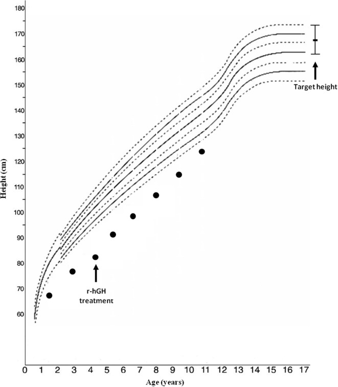
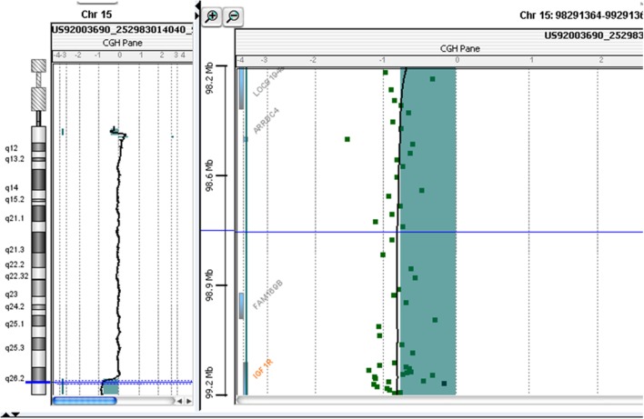
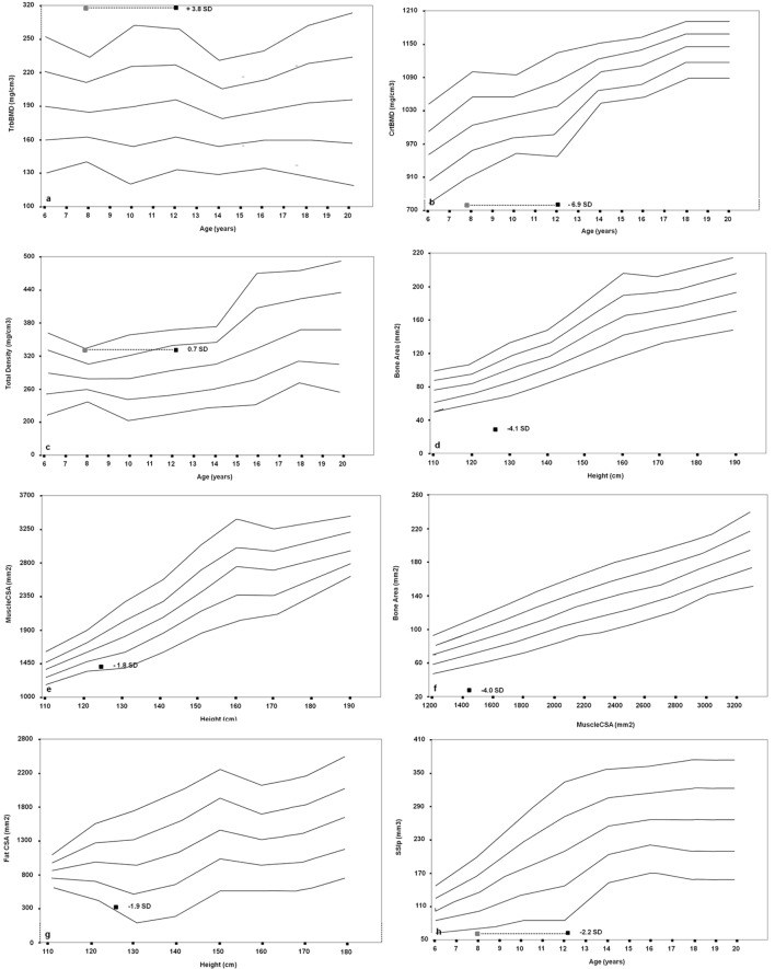
Haploinsufficiency of the insulin-like growth factor (IGF)-1 receptor (IGF1R) gene is a rare, probably under-diagnosed, cause of short stature. However, the effects of IGF1R haploinsufficiency on glucose metabolism, bone status, and metabolism have rarely been investigated. We report the case of a patient referred to our center at the age of 18 months for short stature, failure to thrive, and Silver-Russell-like phenotype. Genetic analysis did not show hypomethylation of the 11p15.5 region or uniparental disomy of chromosome 7. Growth hormone (GH) stimulation tests revealed GH deficiency, whereas IGF-1 was 248 ng/mL. r-hGH treatment showed only a slight improvement (from -4.4 to -3.5 SDS). At 10 years of age, the child was re-evaluated: CGH-array identified a heterozygous de novo 4.92 Mb deletion in 15q26.2, including the IGF1R gene. Dual-energy X-ray absorptiometry showed a normal bone mineral density z-score, while peripheral quantitative computed tomography revealed reduced cortical and increased trabecular elements. A phalangeal bone quantitative ultrasonography showed significantly reduced amplitude-dependent speed of sound and bone transmission time values. The changes in bone architecture, quality, and metabolism in heterozygous IGF1R deletion patients, support the hypothesis that IGF-1 can be a key factor in bone modeling and accrual.
Keywords: bone metabolism; insulin-like growth factor-I; insulin-like growth factor-I receptor; peripheral quantitative computed tomography; quantitative ultrasonography.
Figures
References
-
- Baylink D, Lau KH, Mohan S. The role of IGF system in the rise and fall in bone density with age. J Musculoskelet Neuronal Interact (2007) 7:304–5. - PubMed
Publication types
LinkOut - more resources
Full Text Sources
Other Literature Sources
Miscellaneous