

Modulatory Influence of Segmented Filamentous Bacteria on Transcriptomic Response of Gnotobiotic Mice Exposed to TCDD
- PMID: 28936204
- PMCID: PMC5594080
- DOI: 10.3389/fmicb.2017.01708
Modulatory Influence of Segmented Filamentous Bacteria on Transcriptomic Response of Gnotobiotic Mice Exposed to TCDD
Abstract
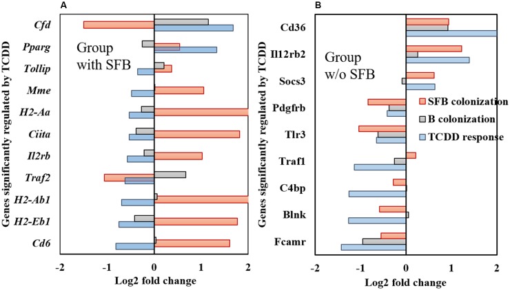
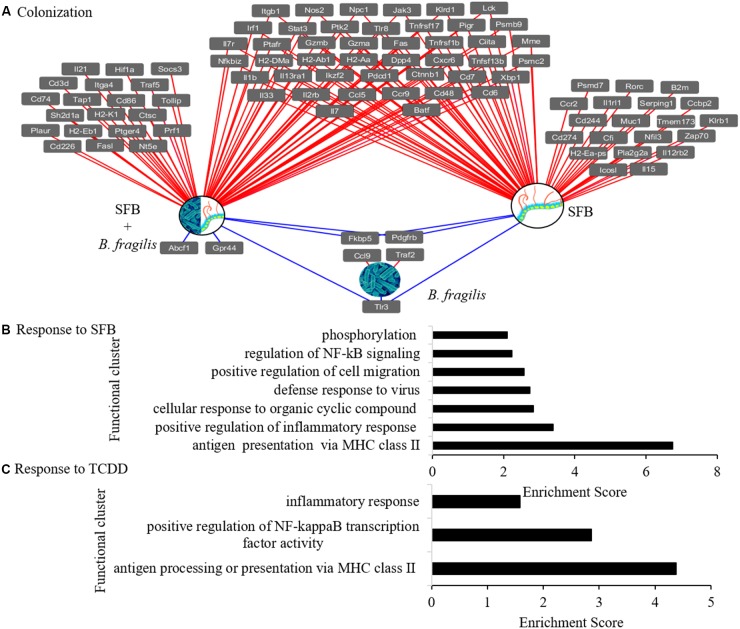
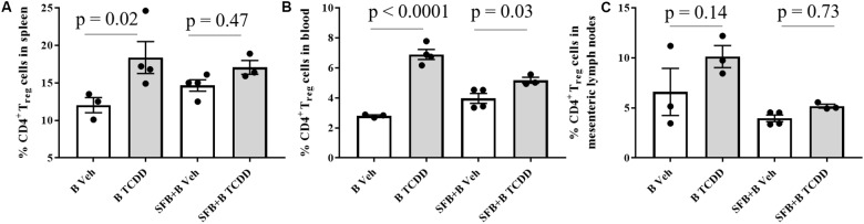
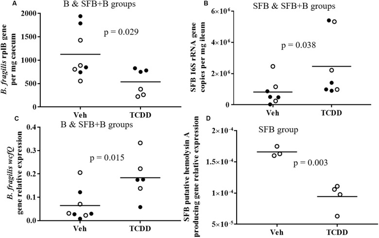
Environmental toxicants such as 2,3,7,8-tetrachlorodibenzo-p-dioxin (TCDD), an aryl hydrocarbon receptor (AhR), are known to induce host toxicity and structural shifts in the gut microbiota. Key bacterial populations with similar or opposing functional responses to AhR ligand exposure may potentially help regulate expression of genes associated with immune dysfunction. To examine this question and the mechanisms for AhR ligand-induced bacterial shifts, C57BL/6 gnotobiotic mice were colonized with and without segmented filamentous bacteria (SFB) - an immune activator. Mice were also colonized with polysaccharide A producing Bacteroides fragilis - an immune suppressor to serve as a commensal background. Following colonization, mice were administered TCDD (30 μg/kg) every 4 days for 28 days by oral gavage. Quantified with the nCounter® mouse immunology panel, opposing responses in ileal gene expression (e.g., genes associated with T-cell differentiation via the class II major histocompatibility complex) as a result of TCDD dosing and SFB colonization were observed. Genes that responded to TCDD in the presence of SFB did not show a significant response in the absence of SFB, and vice versa. Regulatory T-cells examined in the mesenteric lymph-nodes, spleen, and blood were also less impacted by TCDD in mice colonized with SFB. TCDD-induced shifts in abundance of SFB and B. fragilis compared with previous studies in mice with a traditional gut microbiome. With regard to the mouse model colonized with individual populations, results indicate that TCDD-induced host response was significantly modulated by the presence of SFB in the gut microbiome, providing insight into therapeutic potential between AhR ligands and key commensals.
Keywords: B. fragilis; TCDD; gnotobiotic mice; gut dysbiosis; host microbe response; regulatory T-cells; segmented filamentous bacteria.
Figures
Similar articles
-
MicroRNA-based host response to toxicant exposure is influenced by the presence of gut microbial populations.Sci Total Environ. 2021 Nov 25;797:149130. doi: 10.1016/j.scitotenv.2021.149130. Epub 2021 Jul 20. Sci Total Environ. 2021. PMID: 34311349 Free PMC article.
-
TCDD administered on activated carbon eliminates bioavailability and subsequent shifts to a key murine gut commensal.Appl Microbiol Biotechnol. 2017 Oct;101(19):7409-7415. doi: 10.1007/s00253-017-8460-9. Epub 2017 Aug 15. Appl Microbiol Biotechnol. 2017. PMID: 28812142 Free PMC article.
-
Hematopoietic MyD88 orchestrates the control of gut colonization by segmented filamentous bacteria.Mucosal Immunol. 2025 Jun;18(3):717-729. doi: 10.1016/j.mucimm.2025.03.002. Epub 2025 Mar 14. Mucosal Immunol. 2025. PMID: 40090466
-
Segmented Filamentous Bacteria - Metabolism Meets Immunity.Front Microbiol. 2018 Aug 24;9:1991. doi: 10.3389/fmicb.2018.01991. eCollection 2018. Front Microbiol. 2018. PMID: 30197636 Free PMC article. Review.
-
Segmented filamentous bacteria-induced immune responses: a balancing act between host protection and autoimmunity.Immunology. 2018 May 17;154(4):537-46. doi: 10.1111/imm.12950. Online ahead of print. Immunology. 2018. PMID: 29771448 Free PMC article. Review.
Cited by
-
Environmental toxicants in breast milk of Norwegian mothers and gut bacteria composition and metabolites in their infants at 1 month.Microbiome. 2019 Feb 27;7(1):34. doi: 10.1186/s40168-019-0645-2. Microbiome. 2019. PMID: 30813950 Free PMC article.
-
The Human Gut Microbiome - A Potential Controller of Wellness and Disease.Front Microbiol. 2018 Aug 14;9:1835. doi: 10.3389/fmicb.2018.01835. eCollection 2018. Front Microbiol. 2018. PMID: 30154767 Free PMC article. Review.
-
The central role of the gut in intensive care.Crit Care. 2022 Dec 7;26(1):379. doi: 10.1186/s13054-022-04259-8. Crit Care. 2022. PMID: 36476497 Free PMC article. Review.
-
The Dynamic Interplay between the Gut Microbiota and Autoimmune Diseases.J Immunol Res. 2019 Oct 27;2019:7546047. doi: 10.1155/2019/7546047. eCollection 2019. J Immunol Res. 2019. PMID: 31772949 Free PMC article. Review.
-
Aryl Hydrocarbon Receptor (AhR) Activation by 2,3,7,8-Tetrachlorodibenzo-p-Dioxin (TCDD) Dose-Dependently Shifts the Gut Microbiome Consistent with the Progression of Non-Alcoholic Fatty Liver Disease.Int J Mol Sci. 2021 Nov 18;22(22):12431. doi: 10.3390/ijms222212431. Int J Mol Sci. 2021. PMID: 34830313 Free PMC article.
References
Grants and funding
LinkOut - more resources
Full Text Sources
Other Literature Sources
Molecular Biology Databases