. 2015 May 5:4:3.
doi: 10.5772/60725. eCollection 2015 Jan-Dec.

Analytical Validation and Capabilities of the Epic CTC Platform: Enrichment-Free Circulating Tumour Cell Detection and Characterization

Affiliations

Analytical Validation and Capabilities of the Epic CTC Platform: Enrichment-Free Circulating Tumour Cell Detection and Characterization

Shannon L Werner et al. J Circ Biomark. .

Abstract

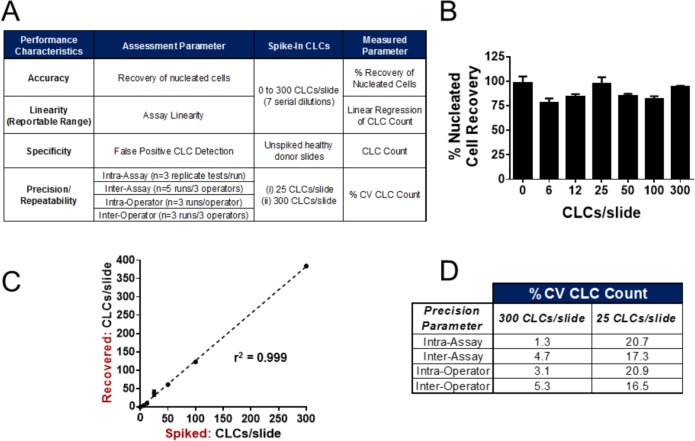
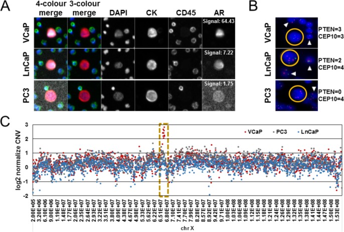
The Epic Platform was developed for the unbiased detection and molecular characterization of circulating tumour cells (CTCs). Here, we report assay performance data, including accuracy, linearity, specificity and intra/inter-assay precision of CTC enumeration in healthy donor (HD) blood samples spiked with varying concentrations of cancer cell line controls (CLCs). Additionally, we demonstrate clinical feasibility for CTC detection in a small cohort of metastatic castrate-resistant prostate cancer (mCRPC) patients. The Epic Platform demonstrated accuracy, linearity and sensitivity for the enumeration of all CLC concentrations tested. Furthermore, we established the precision between multiple operators and slide staining batches and assay specificity showing zero CTCs detected in 18 healthy donor samples. In a clinical feasibility study, at least one traditional CTC/mL (CK+, CD45-, and intact nuclei) was detected in 89 % of 44 mCRPC samples, whereas 100 % of samples had CTCs enumerated if additional CTC subpopulations (CK-/CD45- and CK+ apoptotic CTCs) were included in the analysis. In addition to presenting Epic Platform's performance with respect to CTC enumeration, we provide examples of its integrated downstream capabilities, including protein biomarker expression and downstream genomic analyses at single cell resolution.

Keywords: Analytical Validation; Biomarker; CTC; CTM; Circulating Tumour Cells; Clinical Feasibility; Epic CTC Platform; Fluid Biopsy; Liquid Biopsy; Metastasis.


Figures

Figure 1.
Figure 1.
Epic Platform workflow for sample preparation, CTC enumeration and biomarker analysis. Upon patient blood sample receipt at Epic Sciences, 1) whole blood is lysed and nucleated cells (3 × 10⁶ per slide) are deposited onto 10-12 microscope slides and are frozen at −80 °C until analysis. 2) Two slides per patient sample are thawed and stained with a cocktail of antibodies including cytokeratin, CD45, DAPI to perform CTC enumeration, and a fourth fluorescent channel is available for the evaluation of protein biomarker expression. 3) Stained slides are scanned and 4) the resulting images are analysed using a multi-parametric digital pathology algorithm to detect CTC candidates and quantitate biomarker expression levels. CTC classifications are displayed in a web-based report and are confirmed by trained technicians. 5) CTC enumeration and biomarker expression results are compiled and reported.
Figure 2.
Figure 2.
Representative CTC subtypes detected by the Epic Platform. CTCs from prostate cancer patient samples were enumerated using the Epic Platform. Representative 10X immunofluorescence images for the DAPI (blue), cytokeratin (red), CD45 (green) channels are shown for CTC subtypes and the surrounding white blood cells (WBCs), with the three-channel merge to the far left of each image. Classified CTC subtypes include A) Traditional CTCs (CK+, CD45-, DAPI+/intact), B) Small CTCs (CK+, CD45-, DAPI+/intact, with similar nuclear size to that of the surrounding WBCs), C) CTC clusters (two or more adjacent traditional CTCs that share cytoplasmic boundaries), D) CK- CTCs (CK-, CD45-, DAPI+/intact), and E) Apoptotic CTCs (CK+, CD45-, with DAPI staining pattern consistent with chromosomal condensation and/or nuclear fragmentation).
Figure 3.
Figure 3.
Analytical Validation of the Epic Platform. A) The analytical characteristics assessed to benchmark the performance of the Epic CTC platform. Varying concentrations of COLO-205 cell line cells (CLCs) were spiked into healthy donor blood, red blood cells lysed, and 3 × 10⁶ nucleated cells were deposited onto slides, ranging from 0-300 CLCs/slide. Slides were stained with a cocktail of CK, CD45 and DAPI antibodies, and assay accuracy, linearity, specificity and precision were determined as described in the methods. For each analysis, a “run” is comprised of three tests, with each test consisting of two replicate slides. B) The accuracy and repeatability of cell deposition was assessed calculating percent nucleated cell recovery (y-axis; Mean ± SEM) for one run each of six serial CLC dilutions (six, 12, 25, 50, 100 and 300 CLCs/slide), and for five slides of unspiked healthy donor (HD) blood (zero CLCs/slide). C) Assay linearity was characterized by plotting the actual CLCs/slide recovered (y-axis) versus the theoretical number of CLCs/slide (x-axis) for seven CLC concentrations (six slides tested/concentration), and the linear regression was calculated. Assay specificity was determined by measuring the number of CLCs detected on the unspiked healthy donor slides (zero CLCs/mL). D) Assay precision/repeatability was measured by calculating the percent coefficient of variation (%CV) for CLC counts from the 25 CLCs/slide and 300 CLCs/slide dilutions. Intra-assay variability was measured for one operator who performed one assay run, whereas inter-assay variability was measured across three operators who performed five assay runs total (one assay run per day). Intra-operator repeatability was measured for one operator who performed three assay runs on separate days, whereas inter-operator repeatability was measured for thee operators who performed one assay run each.
Figure 4.
Figure 4.
Clinical feasibility of CTC enumeration in metastatic castrate-resistant prostate cancer patient samples. Forty-four (44) mCRPC patient blood samples were tested for CTC enumeration using the Epic Platform, and the results were compared to those from 18 healthy donor (HD) blood samples. A) CTC incidence was calculated as CTC per millilitre (CTC/mL) of patient blood for traditional CTCs and CTC clusters (left, CTCs + clusters) and all CTC candidates (right; CTCs, CTC clusters, CK- CTCs, and apoptotic CTCs). Each dot on the graph is representative of the CTC/mL value for that patient sample. B) Summary of the range, median and mean CTC/mL values for 18 healthy donor and 44 mCRPC samples for traditional CTCs and CTC clusters (left) and all CTC candidates (right).
Figure 5.
Figure 5.
Epic Platform capabilities for the evaluation of protein and genetic biomarkers. Human prostate cancer cell line control cells (CLCs; VCaP, LnCaP or PC3) were spiked into healthy donor blood, processed onto slides and stained with CK, CD45, DAPI and N-terminal androgen receptor (AR) antibodies. Additional slides were processed for PTEN loss by FISH. Subsequently, individual CLCs were recovered and analysed for whole genome copy number variation by NGS. A) Representative images (10X) of individual CLCs detected, each with varying levels of AR expression (AR signal denoted in white). B) Representative images of PTEN gene deletion status in CLCs (yellow circles) and surrounding WBCs (white carrots), as determined by PTEN FISH analysis. Blue: DAPI, Red: CEP10 signals, Green: PTEN signals. The number of PTEN and CEP10 signals found in each CLC example are reported to the right of the image. C) Comparison of log2 CNV (y-axis) found within isolated VCaP (red), PC3 (grey), and LnCaP (blue) CLCs across the X chromosome (x-axis). Each data point represents the relative copy number within a 100,000 bp window normalized to healthy donor control WBC CNV. The highlighted window (yellow dotted line) contains the AR gene.

Similar articles

Cited by

References

    1. Gupta G P and Massague J (2006) Cancer metastasis: Building a framework. Cell. 127: 679–95 - PubMed
    1. Chaffer C L and Weinberg R A (2011) A perspective on cancer cell metastasis. Science. 331: 1559–64 - PubMed
    1. Hanahan D and Weinberg R A (2011) Hallmarks of cancer: The next generation. Cell. 144: 646–74 - PubMed
    1. Yu M Bardia A Wittner B S Stott S L Smas M E Ting D T Isakoff S J Ciciliano J C Wells M N Shah A M Concannon K F Donaldson M C Sequist L V Brachtel E Sgroi D Baselga J Ramaswamy S Toner M Haber D A and Maheswaran S (2013) Circulating breast tumor cells exhibit dynamic changes in epithelial and mesenchymal composition. Science. 339: 580–4 - PMC - PubMed
    1. Miller M C Doyle G V and Terstappen L W (2010) Significance of circulating tumor cells detected by the CellSearch system in patients with metastatic breast colorectal and prostate cancer. J Oncol. 2010: 617421. - PMC - PubMed