The relationship of mandibular radiomorphometric indices to skeletal age, chronological age and skeletal malocclusion type
- PMID: 28936286
- PMCID: PMC5601113
- DOI: 10.4317/jced.53819
The relationship of mandibular radiomorphometric indices to skeletal age, chronological age and skeletal malocclusion type
Abstract
Background: The present study was performed with the following aims: (1) to assess the relationship between skeletal age, measured using the cervical vertebral maturity (CVM) method, and chronological age; (2) to determine the correlation of skeletal and chronological age to the cortical thickness of the lower border of the mandible using the linear radiomorphometric; and (3) to explore the relationship between these indices and skeletal malocclusion type.
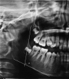
Material and methods: The data were collected from the records of 180 patients, including 57 males (31.7%) and 123 females (68.3%). The data were based on the panoramic and lateral cephalograms of each patient. The CVM stages were determined on the basis of the patients' lateral cephalograms. Three radiomorphometric indices were measured: AI, MI and GI. The patients were divided up into three groups of skeletal malocclusion: Class I, II, and III. For all the tests, statistical significance was set at P<0.05.
Results: The relationship between chronological age and skeletal age was 0.496. Furthermore, with an increase in chronological and skeletal age, the cortical thickness of the lower border of the mandible and consequently the radiomorphometric indices increase, except for the GI (P > 0.05). Lastly, the relationship between GI and skeletal malocclusion type proved significant.
Conclusions: AI and MI were found to increase significantly with increasing age, so the assessment of mandibular radiomorphometric indices could be clinically useful in estimating of the growth and maturation of the mandible. Key words:Orthodontics, Radiomorphometric indices, Skeletal age, Skeletal malocclusion.
Conflict of interest statement
Conflict of interest statement:The authors have declared that no conflict of interest exist.
Figures
Similar articles
-
Relationship between Skeletal Malocclusion and Radiomorphometric Indices of the Mandible in Long Face Patients.Diagnostics (Basel). 2024 Feb 20;14(5):459. doi: 10.3390/diagnostics14050459. Diagnostics (Basel). 2024. PMID: 38472932 Free PMC article.
-
Evaluation of Radiomorphometric Indices in Panoramic Radiograph - A Screening Tool.Open Dent J. 2015 Jul 31;9:303-10. doi: 10.2174/1874210601509010303. eCollection 2015. Open Dent J. 2015. PMID: 26464600 Free PMC article.
-
Correlation Between Dental Age, Chronological Age, and Cervical Vertebral Maturation in Patients with Class II Malocclusion: A Retrospective Study in a Romanian Population Group.Children (Basel). 2025 Mar 21;12(4):398. doi: 10.3390/children12040398. Children (Basel). 2025. PMID: 40310044 Free PMC article.
-
Radiomorphometric indices of the mandible in a British female population.Dentomaxillofac Radiol. 1999 May;28(3):173-81. doi: 10.1038/sj/dmfr/4600435. Dentomaxillofac Radiol. 1999. PMID: 10740473
-
Assessment of Growth Using Mandibular Canine Calcification Stages and Its Correlation with Modified MP3 Stages.Int J Clin Pediatr Dent. 2010 Jan-Apr;3(1):27-33. doi: 10.5005/jp-journals-10005-1050. Epub 2010 Apr 15. Int J Clin Pediatr Dent. 2010. PMID: 27625553 Free PMC article. Review.
Cited by
-
Relationship between Skeletal Malocclusion and Radiomorphometric Indices of the Mandible in Long Face Patients.Diagnostics (Basel). 2024 Feb 20;14(5):459. doi: 10.3390/diagnostics14050459. Diagnostics (Basel). 2024. PMID: 38472932 Free PMC article.
-
Evaluation of Fractal and Radiomorphometric Measurements of Mandibular Bone Structure in Pediatric Patients With Molar Incisor Hypomineralization.Int J Paediatr Dent. 2025 Sep;35(5):945-953. doi: 10.1111/ipd.13311. Epub 2025 Apr 1. Int J Paediatr Dent. 2025. PMID: 40166949 Free PMC article.
-
Oblique line contrast: A new radiomorphometric index for assessing bone quality in dental panoramic radiographs.Heliyon. 2022 Dec 14;8(12):e12266. doi: 10.1016/j.heliyon.2022.e12266. eCollection 2022 Dec. Heliyon. 2022. PMID: 36582704 Free PMC article.
References
-
- Baccetti T, Franchi L, Toth LR, McNamara JA Jr. Treatment timing for Twin-block therapy. American journal of orthodontics and dentofacial orthopedics : official publication of the American Association of Orthodontists, its constituent societies, and the American Board of Orthodontics. 2000;118:159–70. - PubMed
-
- Faltin KJ, Faltin RM, Baccetti T, Franchi L, Ghiozzi B, McNamara JA Jr. Long-term effectiveness and treatment timing for Bionator therapy. The Angle orthodontist. 2003;73:221–30. - PubMed
-
- Flores-Mir C, Burgess CA, Champney M, Jensen RJ, Pitcher MR, Major PW. Correlation of Skeletal Maturation Stages Determined by Cervical Vertebrae and Hand-wrist Evaluations. The Angle orthodontist. 2006;76:1–5. - PubMed
-
- Baccetti T, Franchi L, McNamara JA Jr. The cervical vertebral maturation method: some need for clarification. American journal of orthodontics and dentofacial orthopedics : official publication of the American Association of Orthodontists, its constituent societies, and the American Board of Orthodontics. 2003;123:19a–20a. - PubMed
-
- Bishara SE. Facial and dental changes in adolescents and their clinical implications. The Angle orthodontist. 2000;70:471–83. - PubMed
LinkOut - more resources
Full Text Sources
Other Literature Sources


