

Oral lymphoepithelial cyst: A clinicopathological study of 26 cases and review of the literature
- PMID: 28936296
- PMCID: PMC5601105
- DOI: 10.4317/jced.54072
Oral lymphoepithelial cyst: A clinicopathological study of 26 cases and review of the literature
Abstract
Introduction: Τo describe the clinicopathological features of 26 oral lymphoepithelial cysts (LECs) and review the literature.
Material and methods: Twenty-six cases of oral LECs diagnosed during a 37-year period were retrospectively collected. The patients' gender and age, as well as the main clinical features of the cysts were retrieved from the requisition forms. The main microscopic features were recorded after reevaluation of all cases. Pubmed and Google Scholar electronic databases were searched with the key word "oral LEC". Inclusion criteria were the microscopic confirmation of LEC diagnosis and the report at least two of three main clinical features (gender, age and cyst's location).
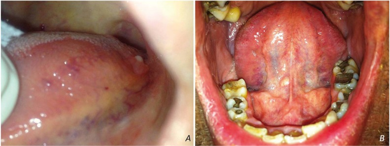
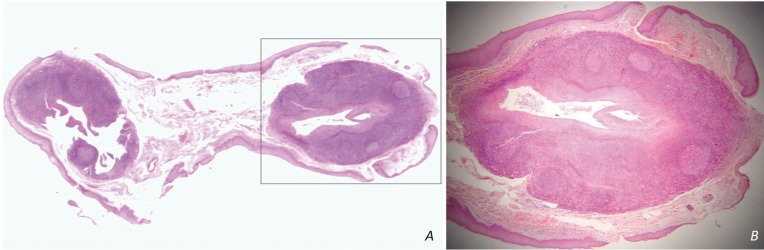
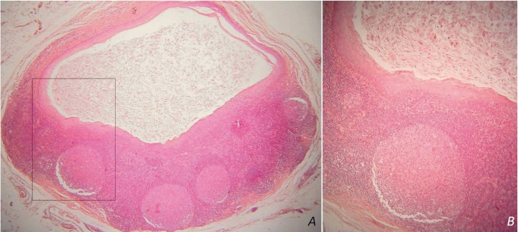
Results: The 26 oral LECs represented 0.08% of 31,564 biopsies accessioned during the study period. They affected 25 patients, 14 females and 11 males with a mean age of 33.04±9.81 years. They appeared as smooth (92%) nodules, with soft (24%) or firm (76%) consistency and normal (28%), yellow to normal (20%), yellow (32%) or white (20%) hue, in the tongue (69.23%) or the floor of mouth (30.77%). They were covered by parakeratinized squamous (92.31%) or non-keratinized (7.69%) epithelium and contained desquamated epithelial cells, amorphous eosinophilic material and/or inflammatory cells (100%). The lymphoid tissue surrounded the cystic cavity partially (34.62%) or completely (65.38%), often in a follicular pattern with prominent germinal centers (53.85%). Literature review yielded 316 cases of oral LECs derived from 25 case reports, 3 case studies/retrospective studies with detailed information for each case and 7 studies with summarized data.
Conclusions: Oral LEC is a pathologic entity with discrete clinical presentation that is, however, commonly misdiagnosed in clinical practice as other, mostly benign, entities. Its pathogenesis remains obscure, as its clinicopathologic features are consistent with both theories suggested up to date. Key words:Oral lymphoepithelial cyst; developmental cyst; non odontogenic cyst; lymphoid tissue; oral tonsil.
Conflict of interest statement
Conflict of interest statement:None declared.
Figures
Similar articles
-
Clinicopathological and immunohistochemical features of the oral lymphoepithelial cyst: A multicenter study.J Oral Pathol Med. 2020 Mar;49(3):219-226. doi: 10.1111/jop.12978. Epub 2019 Dec 16. J Oral Pathol Med. 2020. PMID: 31782199
-
Oral Lymphoepithelial Cyst: A Collaborative Clinicopathologic Study of 132 Cases from Brazil.Head Neck Pathol. 2022 Mar;16(1):268-277. doi: 10.1007/s12105-021-01352-2. Epub 2021 Jun 29. Head Neck Pathol. 2022. PMID: 34185247 Free PMC article.
-
Lymphoepithelial cysts of the pancreas: a report of 12 cases and a review of the literature.Mod Pathol. 2002 May;15(5):492-501. doi: 10.1038/modpathol.3880553. Mod Pathol. 2002. PMID: 12011254
-
Squamous-lined cysts of the pancreas: lymphoepithelial cysts, dermoid cysts (teratomas), and accessory-splenic epidermoid cysts.Semin Diagn Pathol. 2000 Feb;17(1):56-65. Semin Diagn Pathol. 2000. PMID: 10721807 Review.
-
Oral verruciform xanthoma: Report of 13 new cases and review of the literature.Med Oral Patol Oral Cir Bucal. 2018 Jul 1;23(4):e429-e435. doi: 10.4317/medoral.22342. Med Oral Patol Oral Cir Bucal. 2018. PMID: 29924759 Free PMC article. Review.
Cited by
-
Clinical features and management of lymphoepithelial cyst.Open Med (Wars). 2023 Dec 7;18(1):20230872. doi: 10.1515/med-2023-0872. eCollection 2023. Open Med (Wars). 2023. PMID: 38075029 Free PMC article.
-
Case of a Large Oropharyngeal Cyst.Cureus. 2019 Oct 5;11(10):e5843. doi: 10.7759/cureus.5843. Cureus. 2019. PMID: 31754578 Free PMC article.
-
Oral lymphoid lesions: a 47-year clinicopathological study in a Brazilian population.Med Mol Morphol. 2019 Sep;52(3):123-134. doi: 10.1007/s00795-018-0210-2. Epub 2018 Oct 31. Med Mol Morphol. 2019. PMID: 30382358 Review.
-
A Guide to Yellow Oral Mucosal Entities: Etiology and Pathology.Head Neck Pathol. 2019 Mar;13(1):33-46. doi: 10.1007/s12105-018-0977-4. Epub 2019 Jan 31. Head Neck Pathol. 2019. PMID: 30693453 Free PMC article. Review.
-
Oral lymphoepithelial cyst at the lateral border of the tongue.J Dent Sci. 2023 Oct;18(4):1941-1942. doi: 10.1016/j.jds.2023.06.013. Epub 2023 Jun 23. J Dent Sci. 2023. PMID: 37799928 Free PMC article. No abstract available.
References
-
- Nonaka CF, Henriques AC, de Matos FR, de Souza LB, Pinto LP. Nonodontogenic cysts of the oral and maxillofacial region: demographic profile in a Brazilian population over a 40-year period. Eur Arch Otorhinolaryngol. 2011;268:917–22. - PubMed
-
- Gold C. Branchial cleft cyst located in the floor of the mouth. Report of a case. Oral Surg Oral Med Oral Pathol. 1962;15:1118–20. - PubMed
-
- King ESJ. The Lateral Lymphoepithelial Cyst of the Neck (Branchial Cyst) Aust New Zeal J Surg. 1949;29:109–21. - PubMed
Publication types
LinkOut - more resources
Full Text Sources
Other Literature Sources