

Improved Calcium Scoring at Dual-Energy Computed Tomography Angiography Using a High-Z Contrast Element and Novel Material Separation Technique
- PMID: 28937491
- PMCID: PMC5860919
- DOI: 10.1097/RCT.0000000000000676
Improved Calcium Scoring at Dual-Energy Computed Tomography Angiography Using a High-Z Contrast Element and Novel Material Separation Technique
Abstract
Objectives: The aim of this study was to compare the accuracy of existing dual-energy computed tomography (CT) angiography coronary artery calcium scoring methods to those obtained using an experimental tungsten-based contrast material and a recently described contrast material extraction process (CMEP).
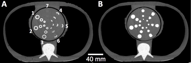
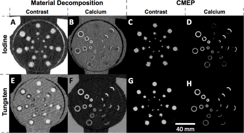
Methods: Phantom coronary arteries of varied diameters, with different densities and arcs of simulated calcified plaque, were sequentially filled with water, iodine, and tungsten contrast materials and scanned within a thorax phantom at rapid-kVp-switching dual-energy CT. Calcium and contrast density images were obtained by material decomposition (MD) and CMEP. Relative calcium scoring errors among the 4 reconstructed datasets were compared with a ground truth, 120-kVp dataset.
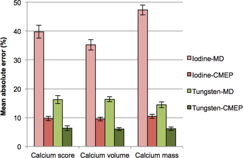
Results: Compared with the 120-kVp dataset, tungsten CMEP showed a significantly lower mean absolute error in calcium score (6.2%, P < 0.001) than iodine CMEP, tungsten MD, and iodine MD (9.9%, 15.7%, and 40.8%, respectively).
Conclusions: Novel contrast elements and material separation techniques offer improved coronary artery calcium scoring accuracy and show potential to improve the use of dual-energy CT angiography in a clinical setting.
Figures
Similar articles
-
An Image-Domain Contrast Material Extraction Method for Dual-Energy Computed Tomography.Invest Radiol. 2017 Apr;52(4):245-254. doi: 10.1097/RLI.0000000000000335. Invest Radiol. 2017. PMID: 27875338 Free PMC article.
-
Feasibility of coronary artery calcium scoring on virtual unenhanced images derived from single-source fast kVp-switching dual-energy coronary CT angiography.J Cardiovasc Comput Tomogr. 2014 Sep-Oct;8(5):391-400. doi: 10.1016/j.jcct.2014.08.005. Epub 2014 Aug 28. J Cardiovasc Comput Tomogr. 2014. PMID: 25301045 Clinical Trial.
-
Modified Dual-Energy Algorithm for Calcified Plaque Removal: Evaluation in Carotid Computed Tomography Angiography and Comparison With Digital Subtraction Angiography.Invest Radiol. 2017 Nov;52(11):680-685. doi: 10.1097/RLI.0000000000000391. Invest Radiol. 2017. PMID: 28542096
-
Dual-energy CT revisited with multidetector CT: review of principles and clinical applications.Diagn Interv Radiol. 2011 Sep;17(3):181-94. doi: 10.4261/1305-3825.DIR.3860-10.0. Epub 2010 Nov 14. Diagn Interv Radiol. 2011. PMID: 20945292 Review.
-
Dual-Energy Spectral CT: Various Clinical Vascular Applications.Radiographics. 2016 Jul-Aug;36(4):1215-32. doi: 10.1148/rg.2016150185. Radiographics. 2016. PMID: 27399244 Review.
Cited by
-
Multi-energy computed tomography and material quantification: Current barriers and opportunities for advancement.Med Phys. 2020 Aug;47(8):3752-3771. doi: 10.1002/mp.14241. Epub 2020 Jun 23. Med Phys. 2020. PMID: 32453879 Free PMC article. Review.
References
-
- Montalescot G, Sechtem U, Achenbach S, et al. 2013 ESC guidelines on the management of stable coronary artery disease. Eur Heart J. 2013;34(38):2949–3003. - PubMed
-
- Staniak HL, Bittencourt MS, Pickett C, et al. Coronary CT angiography for acute chest pain in the emergency department. J Cardiovasc Comput Tomogr. 2014;8(5):359–67. - PubMed
-
- Goldstein JA, Chinnaiyan KM, Abidov A, et al. The CT-STAT (Coronary Computed Tomographic Angiography for Systematic Triage of Acute Chest Pain Patients to Treatment) trial. J Am Coll Cardiol. 2011;58(14):1414–22. - PubMed
-
- Leschka S, Scheffel H, Desbiolles L, et al. Combining dual-source computed tomography coronary angiography and calcium scoring: added value for the assessment of coronary artery disease. Heart. 2008;94(9):1154–61. - PubMed
MeSH terms
Substances
Grants and funding
LinkOut - more resources
Full Text Sources
Other Literature Sources
