. 2017 Sep 22;22(10):1589.
doi: 10.3390/molecules22101589.

Aspalathin Reverts Doxorubicin-Induced Cardiotoxicity through Increased Autophagy and Decreased Expression of p53/mTOR/p62 Signaling

Affiliations

Aspalathin Reverts Doxorubicin-Induced Cardiotoxicity through Increased Autophagy and Decreased Expression of p53/mTOR/p62 Signaling

Rabia Johnson et al. Molecules. .

Abstract

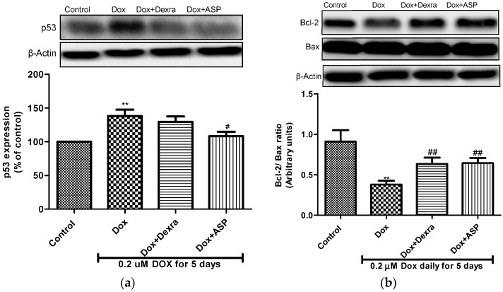
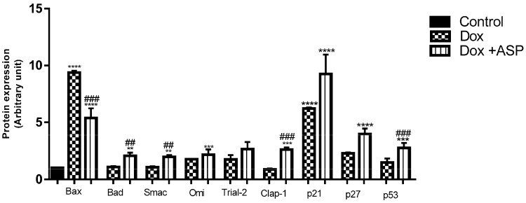
Doxorubicin (Dox) is an effective chemotherapeutic agent used in the treatment of various cancers. Its clinical use is often limited due to its potentially fatal cardiotoxic side effect. Increasing evidence indicates that tumour protein p53 (p53), adenosine monophosphate-activated protein kinase (AMPK), nucleoporin p62 (p62), and the mammalian target of rapamycin (mTOR) are critical mediators of Dox-induced apoptosis, and subsequent dysregulation of autophagy. Aspalathin, a polyphenolic dihydrochalcone C-glucoside has been shown to activate AMPK while decreasing the expression of p53. However, the role that aspalathin could play in the inhibition of Dox-induced cardiotoxicity through increased autophagy flux remained unexplored. H9c2 cardiomyocytes and Caov-3 ovarian cancer cells were cultured in Dulbecco's Modified Eagle's medium and treated with or without Dox for five days. Thereafter, cells exposed to 0.2 µM Dox were co-treated with either 20 µM Dexrazozane (Dexra) or 0.2 µM aspalathin (ASP) daily for 5 days. Results obtained showed that ASP mediates its cytoprotective effect in a p53-dependent manner, by increasing the Bcl-2/Bax ratio and decreasing apoptosis. The latter effect was diminished through ASP-induced activation of autophagy-related genes (Atgs) with an associated decrease in p62 through induction of AMPK and Fox01. Furthermore, we showed that ASP was able to potentiate this effect without decreasing the anti-cancer efficacy of Dox, as could be observed in Caov-3 ovarian cancer cells. Taken together, the data presented in this study provides a credible mechanism by which ASP co-treatment could protect the myocardium from Dox-induced cardiotoxicity.

Keywords: apoptosis; aspalathin; autophagy; cardiomyopathy; cardiotoxicity; doxorubicin; oxidative stress.


Conflict of interest statement

The authors declare no conflict of interest.

Figures

Figure 1
Figure 1
The effect of aspalathin (ASP) co-treatment on metabolic activity of H9c2 cells exposed to Doxorubicin. H9c2 cells were treated with 0.2 µM doxorubicin (Dox) alone (positive control) or co-treated in combination with either 20 µM dexrazoxane (Dexra) or 0.2 µM ASP daily for 5 days. Control samples were either exposed to media only or 0.2 µM ASP for 5 days. Results are expressed as the mean ± SEM of three independent biological experiments, each with three technical replicates (n = 9). *** p ≤ 0.001, **** p ≤ 0.0001 versus control and ### p ≤ 0.001, #### p ≤ 0.0001 versus Dox.
Figure 2
Figure 2
The effect of aspalathin on autophagy proteins. (A) Represents the expression of phosphor (p)adenosine monophosphate-activated protein kinase (AMPK); (B) the expression of mammalian target of rapamycin (mTOR); (C) the expression of LC3-II; and (D) the expression of p62 in doxorubicin (Dox)-induced cardiotoxicity. H9c2 cells were treated with 0.2 µM Dox alone or in combination with either 20 µM dexrazoxane (Dexra) or 0.2 µM ASP daily for 5 days. Control samples were exposed to media only. All blots were normalized to beta actin. Results are expressed as the mean ± SEM of three independent biological experiments, each with three technical replicates (n = 9). ** p *** p ≤ 0.01 versus control and # p ≤ 0.05, ## p ≤ 0.01 and ### p ≤ 0.01 versus Dox.
Figure 3
Figure 3
(a) The effect of aspalathin (ASP) on apoptosis proteins. (a) Represents the expression of p53 and (b) the expression of the Bcl-2/Bax ratio. H9c2 cells were treated with 0.2 µM Dox alone or in combination with either 20 µM dexrazoxane (Dexra) or 0.2 µM ASP daily for 5 days. Control samples were exposed to media only. Results are expressed as the mean ± SEM of three independent biological experiments, each with three technical replicates (n = 9). ** p ≤ 0.01 versus control and # p ≤ 0.05, ## p ≤ 0.01 versus Dox.
Figure 4
Figure 4
The effect of aspalathin (ASP) on caspase 3/7 activity on H9c2 cells treated with 0.2 µM Dox for 5 days. H9c2 cells were treated with 0.2 µM Dox alone or in combination with either 20 µM dexrazoxane (Dexra) or 0.2 µM ASP daily for 5 days. Control samples were exposed to media only. Results are expressed as the mean ± SEM of three independent biological experiments, each with three technical replicates (n = 9). ** p < 0.01 versus control and ## p < 0.05 and ### p < 0.01 versus Dox.
Figure 5
Figure 5
The effect of aspalathin (ASP) co-treatment on metabolic activity of Caov-3 cells exposed to Doxorubicin. Caov-3 cells were treated with either 0.2 µM Dox alone or co-treated in combination with 0.2 µM ASP, daily for 5 days. Control samples were exposed to either media only or 0.2 µM ASP for 5 days. Results are expressed as the mean ± SEM of three independent biological experiments, each with three technical replicates (n = 9). *** p ≤ 0.0001 versus control and ### p ≤ 0.001 versus Dox.
Figure 6
Figure 6
The effect of aspalathin (ASP) on pro-apoptotic protein expression in Caov-3 cells. Caov-3 ovarian cancer cells were treated with 0.2 µM Dox alone or in combination with 0.2 µM ASP daily for 5 days. Control samples were exposed to media only or ASP. Results are expressed as the mean ± SEM of three independent biological experiments, each with three technical replicates (n = 9). ** p ≤ 0.01, *** p ≤ 0.001 and **** p ≤ 0.0001 versus control and ## p ≤ 0.01, and ### p ≤ 0.001 versus Dox.
Figure 7
Figure 7
Aspalathin inhibits apoptosis and increase autophagy in H9c2 cells exposed to Doxorubicin (Dox). AMPK is a master regulator known to play an important role in energy homeostasis. Activation of AMPK attenuates Dox-induced activation of mTOR, restoring autophagy flux in H9c2 cells. Chronic exposure to Dox induces apoptosis through increased p53 and caspase-3/7 activity, a process known to be associated with cardiomyocyte loss. Activation of AMPK was able to inhibit p53 expression and enhances cardiomyocyte survival. AMPK, adenosine monophosphate-activated protein kinase ; Bax, Bcl-2-like protein 4; Bcl-2, B-cell lymphoma-2; ATG, autophagy-related genes; Cyt c, cytochrome C; mTOR, Mammalian target of rapamycin; LC3, Microtubule-associated protein 1A/1B-light chain 3 (LC3); P62, Nucleoporin p62.

References

    1. Sui X., Han W., Pan H. P53-induced autophagy and senescence. Oncotarget. 2015;6:11723–11724. doi: 10.18632/oncotarget.4170. - DOI - PMC - PubMed
    1. Maiuri M.C., Zalckvar E., Kimchi A., Kroemer G. Self-eating and self-killing: Crosstalk between autophagy and apoptosis. Nat. Rev. Mol. Cell Biol. 2007;8:741–752. doi: 10.1038/nrm2239. - DOI - PubMed
    1. Marino G., Niso-Santano M., Baehrecke E.H., Kroemer G. Self-consumption: The interplay of autophagy and apoptosis. Nat. Rev. Mol. Cell Biol. 2014;15:81–94. doi: 10.1038/nrm3735. - DOI - PMC - PubMed
    1. Thorburn A. Apoptosis and autophagy: Regulatory connections between two supposedly different processes. Apop. Int. J. Progr. Cell Death. 2008;13:1–9. doi: 10.1007/s10495-007-0154-9. - DOI - PMC - PubMed
    1. Bjorkoy G., Lamark T., Pankiv S., Overvatn A., Brech A., Johansen T. Monitoring autophagic degradation of p62/sqstm1. Methods Enzymol. 2009;452:181–197. - PubMed

LinkOut - more resources