. 2018 Feb;25(2):268-281.
doi: 10.1038/cdd.2017.153. Epub 2017 Sep 22.

A vitronectin-derived peptide reverses ovariectomy-induced bone loss via regulation of osteoblast and osteoclast differentiation

Affiliations

A vitronectin-derived peptide reverses ovariectomy-induced bone loss via regulation of osteoblast and osteoclast differentiation

Seung-Ki Min et al. Cell Death Differ. 2018 Feb.

Abstract

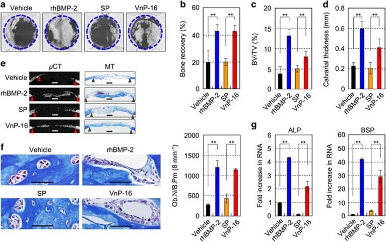
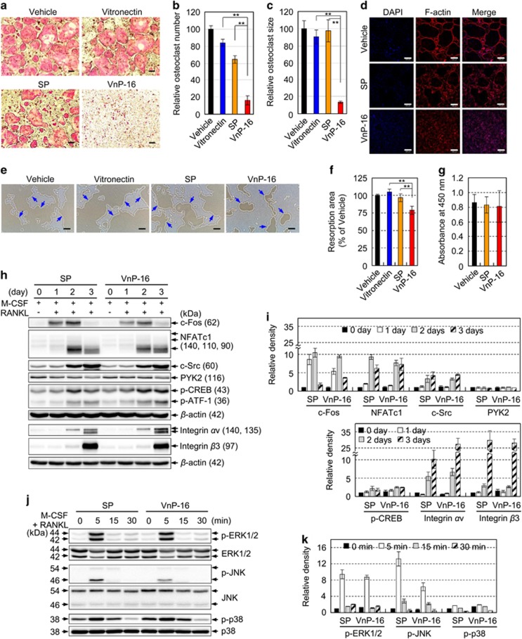
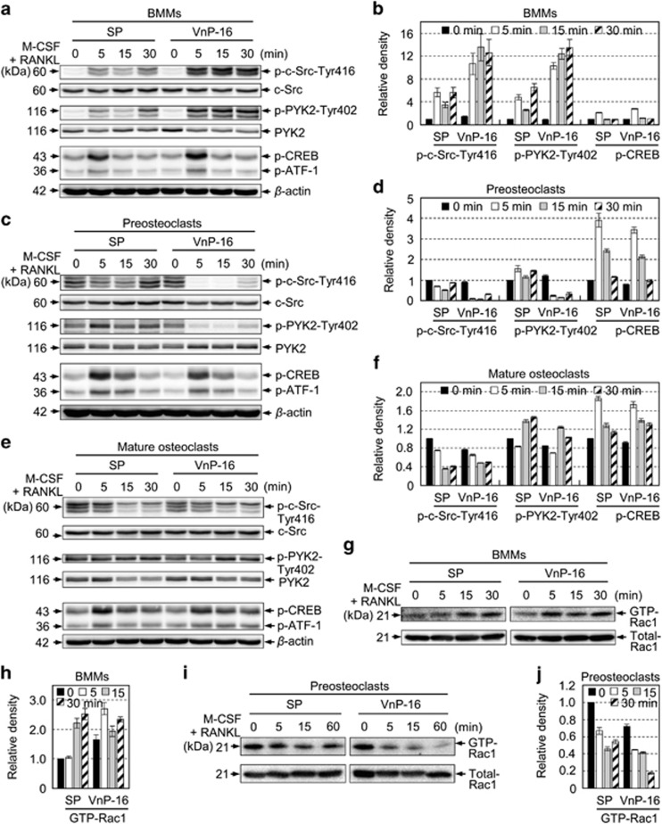
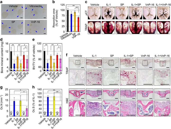
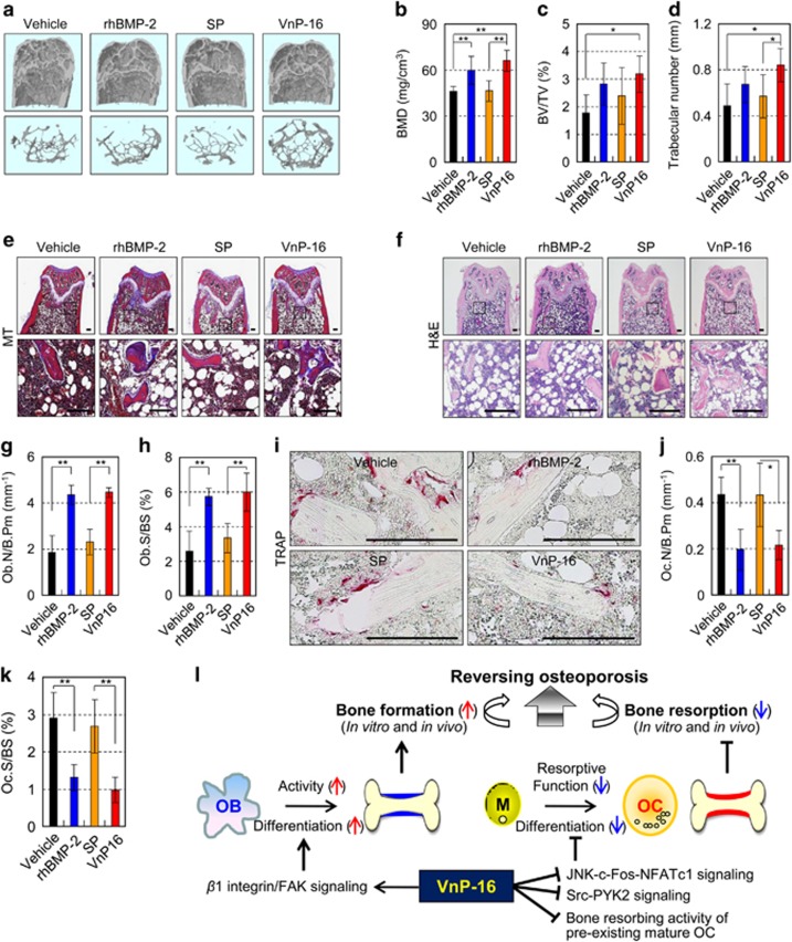
Osteoporosis affects millions of people worldwide by promoting bone resorption and impairing bone formation. Bisphosphonates, commonly used agents to treat osteoporosis, cannot reverse the substantial bone loss that has already occurred by the time of diagnosis. Moreover, their undesirable side-effects, including osteonecrosis of the jaw, have been reported. Here, we demonstrated that a new bioactive core vitronectin-derived peptide (VnP-16) promoted bone formation by accelerating osteoblast differentiation and activity through direct interaction with β1 integrin followed by FAK activation. Concomitantly, VnP-16 inhibited bone resorption by restraining JNK-c-Fos-NFATc1-induced osteoclast differentiation and αvβ3 integrin-c-Src-PYK2-mediated resorptive function. Moreover, VnP-16 decreased the bone resorbing activity of pre-existing mature osteoclasts without changing their survival rate. Furthermore, VnP-16 had a strong anabolic effect on bone regeneration by stimulating osteoblast differentiation and increasing osteoblast number, and significantly alleviated proinflammatory cytokine-induced bone resorption by restraining osteoclast differentiation and function in murine models. Moreover, VnP-16 could reverse ovariectomy-induced bone loss by both inhibiting bone resorption and promoting bone formation. Given its dual role in promoting bone formation and inhibiting bone resorption, our results suggest that VnP-16 could be an attractive therapeutic agent for treating osteoporosis.


Conflict of interest statement

The authors declare no conflict of interest.

Figures

Figure 1
Figure 1
Analyses of purified rVn truncations by SDS-PAGE and circular dichroism spectroscopy and their effects on cell behavior. (a) Schematic diagram of the rVn truncations. The amino acid (aa) positions and the domain structure of full-length vitronectin are indicated. The black box and colored bars represent the signal peptide and the positions of the recombinant proteins, respectively. (b) Schematic diagram and SDS-PAGE analyses (10% polyacrylamide gels under reducing conditions) of the purified rVn proteins, expressed as His6-tagged fusions. The bands were visualized by Coomassie staining. (c) Gel mobilities (12.5% SDS-PAGE) of the purified rVn proteins treated with or without dithiothreitol (DTT). (d) Circular dichroism analysis of the rVn fragments in PBS (pH 3.0) at 23 °C. (e) The dose-dependent effects of the truncated rVn proteins on the attachment of human osteogenic cells. The cells were seeded onto rVn-treated plates for 1 h in serum-free medium. (f and g) Cell attachment (f) and spreading (g) of osteogenic cells induced by BSA (1%), vitronectin (0.23 μg/cm2), and the truncated rVn proteins (5.7 μg/cm2) for 1 h (f) or 3 h (g) in serum-free medium. (h) Migration of osteogenic cells induced by vitronectin and the truncated rVn proteins. Osteogenic cells were seeded into the upper chambers of Transwell filters coated with vitronectin or rVn proteins and were incubated for 24 h. ND, not detected. Data in (eh) represent the mean±SD (n=4). **P<0.01
Figure 2
Figure 2
VnP-16 promotes cell behavior in fibroblast lineages. (a) The locations of the synthetic peptides (VnP-11 to VnP-21) in the amino acid sequence of the central region of human vitronectin (residues 230–322). (b) Dose-dependent cell attachment to immobilized synthetic peptides. Human osteogenic cells were allowed to adhere to peptide-treated plates for 1 h in serum-free medium. (c and d) Cell attachment (c) and spreading (d) of osteogenic cells induced by treatment with BSA (1%), vitronectin (0.23 μg/cm2), rVn-FII (5.7 μg/cm2), or the synthetic peptides (9.1 μg/cm2) for 1 h (c) or 3 h (d) in serum-free medium. (e) The viabilities of osteogenic cells treated with VnP-16 for 24 or 48 h. (f) The amino acid sequence of N- and C-terminal truncated peptides for VnP-16. (g) Attachment of osteogenic cells induced by vitronectin, rVn-FII, and peptides. (h) Attachment of NHEKs, NHOKs, NHDFs, NHOFs, PC-12 cells, MC3T3-E1 cells, CV-1 cells, and NIH/3T3 cells to VnP-16 (9.1 μg/cm2). The cells were allowed to adhere to VnP-16-treated plates for 1 h in serum-free medium. (i) The predicted structure of the VnP-16 dodecapeptide computed using the PSIPRED protein structure prediction server. The positions of the two β-strands (yellow arrows) on the coil (black line) are indicated. Conf, confidence of prediction; Pred, predicted secondary structure; AA, target sequence. Data in (be,g,h) represent the mean±SD (n=4). **P<0.01
Figure 3
Figure 3
VnP-16 promotes osteogenic cell attachment through direct interaction with β1 integrin. (a) Attachment of human osteogenic cells pretreated with EDTA (5 mM), MnCl2 (500 μM), or heparin (100 μg/ml) to VnP-16. The cells were seeded onto plates that were precoated with VnP-16 (9.1 μg/cm2) for 1 h. (b) The effects of various integrin-blocking antibodies on cell attachment to VnP-16. (c–e) Immunoblotting (c) and densitometric analysis (d) of β1 integrin, and cell attachment to VnP-16 (e) in osteogenic cells that were transfected with a control (Con) or β1 integrin-specific siRNA (10 nM; β1 integrin). (f) Streptavidin-bead pulldown assay with the biotinylated SP or biotinylated VnP-16 peptides from extracts of osteogenic cells that were cultured on biotinylated SP- or biotinylated VnP-16-coated dishes for 30 min. Data in (a,b,e) (n=4), and (d) (n=2) represent the mean±SD. **P<0.01
Figure 4
Figure 4
VnP-16 promotes osteogenic differentiation through β1 integrin/FAK signaling. (a,b) Immunoblotting (a) and densitometric analyses (b) of phospho-FAK, phospho-Akt Ser473, phospho-PKCδ Thr505, and phospho-c-Src Tyr416 in osteogenic cells that were cultured for 3 h on plates coated with vitronectin (0.23 μg/cm2), SP, or VnP-16 (9.1 μg/cm2). (c,d) Immunoblotting (c) and densitometric analyses (d) of total FAK (t-FAK) and phospho-FAK Tyr397 in osteogenic cells that were pretreated with PF-573228 for 1 h. (e) Attachment of cells that were treated with PF-573228 for 1 h in serum-free medium to plates precoated with VnP-16 (9.1 μg/cm2). (f) The effects of VnP-16 on alkaline phosphatase activity and calcium deposition in SKP-derived mesenchymal precursors (MPs), mouse calvarial osteoblast precursors (MC3T3-E1), and human osteogenic cells (Osteogenic). The cells were cultured in osteogenic differentiation medium containing VnP-16 or SP (50 μg/0.5 ml) for 2 weeks. (g) The effects of PF-573228 on alkaline phosphatase activity and calcium deposition in SKP-derived mesenchymal precursors, MC3T3-E1, and human osteogenic cells. The cells were cultured on VnP-16-treated (9.1 μg/cm2) plates in osteogenic differentiation medium with or without 1 μM PF-573228 for 2 weeks. (h–j) Immunoblotting (h) and densitometric analyses (i) of t-FAK, and dose-dependent attachment (j) of control or FAK-specific siRNA-treated (100 nM) osteogenic cells to VnP-16. (k) Determination of apoptotic cells in osteogenic cells that were cultured for 24, 48, and 96 h on plates coated with SP or VnP-16 (9.1 μg/cm2) by TUNEL assay. Data in (b, d and i) (n=3), and (e, j and k) (n=4) represent the mean±SD. *P<0.05 or **P<0.01 compared to vehicle or control siRNA
Figure 5
Figure 5
The effects of VnP-16 on bone regeneration in vivo. (a) μCT images of the bone defects in each group. Critical-sized rat calvarial defects were implanted with absorbable collagen sponges treated with vehicle (DMSO), rhBMP-2 (2 μg/scaffold), SP (1 mg/scaffold), or VnP-16 (1 mg/scaffold). (bd) Quantitative bone morphometric analyses of the bone recovery rate (b), BV/TV (c), and calvarial thickness (d) in the region of the defects. (e) μCT images of the bone defects and Masson’s trichrome staining (MT) of rat calvarial sections 2 weeks after transplantation. The triangles indicate the wound edges. For histomorphometric analysis, the specimens were decalcified with 12% EDTA for 4 weeks and embedded in paraffin. The paraffin-embedded samples were sectioned at a thickness of 4 μm and then stained with Masson’s trichrome. Scale bars, 1 mm. (f) The number of osteoblasts per bone perimeter from rat calvarial sections 2 weeks after transplantation. Ob.N, osteoblast number; B.Pm, bone perimeter. (Scale bars, 50 μm.) (g) qPCR analysis of the expression levels of osteogenic markers in each group. ALP, alkaline phosphatase; BSP, bone sialoprotein. Data in (bd, f and g) represent the mean±SD (n=5 per group). **P<0.01
Figure 6
Figure 6
The effects of VnP-16 on M-CSF- and RANKL-induced osteoclast formation, F-actin-mediated cytoskeletal organization, resorptive activity, and the expression levels of osteoclastogenesis-related genes. (ad) BMMs were cultured for 6 days on plates that were precoated with vehicle (DMSO), vitronectin (0.23 μg/cm2), SP (9.1 μg/cm2), or VnP-16 (9.1 μg/cm2), in the presence of 30 ng/ml M-CSF and 100 ng/ml RANKL. The induced cells were stained for TRAP (a) and immunostained with DAPI (blue) and rhodamine-phalloidin (F-actin, red) (d). TRAP-positive multinucleated cells containing three or more nuclei were counted as osteoclasts (b). The sizes of the osteoclasts were obtained by measuring the diameters of multinucleated TRAP-positive cells on × 40 photomicrographs (c). Scale bars, 200 μm. (e and f) The effects of VnP-16 on the bone resorbing activity of osteoclasts. BMMs were cultured for 6 days on Osteo Assay Surface plates that were coated with vehicle (DMSO), vitronectin (0.23 μg/cm2), or synthetic peptide (9.1 μg/cm2), in the presence of 30 ng/ml M-CSF and 100 ng/ml RANKL. The cells were removed and the resorbed pits were photographed (e). The blue arrows indicate the resorption pits formed by osteoclasts. Bone resorption was assessed by pit area measurements (f). Scale bars, 200 μm. (g) The effect of the concentration of VnP-16 (9.1 μg/cm2) that blocked osteoclast formation on the proliferation and viability of BMMs. (h and i) Immunoblotting (h) and densitometric analyses (i) of the osteoclastogenesis-related proteins in BMMs that were cultured for 1–3 days on plates precoated with SP or VnP-16 (9.1 μg/cm2), in the presence of 30 ng/ml M-CSF and 100 ng/ml RANKL. CREB, cAMP-response element-binding protein; ATF-1, activating transcription factor 1. (j,k) Immunoblotting (j) and densitometric analyses (k) of MAPKs in BMMs that were cultured for 3 days on plates precoated with SP or VnP-16 (9.1 μg/cm2), in the presence of 30 ng/ml M-CSF and 100 ng/ml RANKL, serum-starved for 3 h, and stimulated with M-CSF (30 ng/ml) and RANKL (100 ng/ml) for the indicated times. ERK, extracellular signal-regulated kinase. Data in (b,c,f and g) (n=4), and (i and k) (n=3) represent the mean±SD. **P<0.01
Figure 7
Figure 7
The effects of VnP-16 on M-CSF- and RANKL-induced activation of cytoskeletal organizers during osteoclastogenesis. (af) Immunoblotting (a,c,e) and densitometric analyses (b, d and f) of c-Src, PYK2, and CREB in BMMs (a and b), preosteoclasts (c and d), and mature osteoclasts (e and f). The cells were cultured for 1 day (a and e) or 3 days (c) on plates that were precoated with SP or VnP-16 (9.1 μg/cm2), in the presence of 30 ng/ml M-CSF and 100 ng/ml RANKL, serum-starved for 3 h, and then stimulated with M-CSF (30 ng/ml) and RANKL (100 ng/ml) for the indicated times. (gj) Immunoblotting (g and i) and densitometric analyses (h and j) of active (GTP-Rac1) and total Rac1 levels in BMMs (g and h) and preosteoclasts (i and j). The assay conditions were the same as those described for (a and c). The cells were lysed and incubated with PAK1 PBD Agarose beads for 1 h at 4 °C. Active Rac1 proteins were detected by immunoblotting using an anti-Rac1 antibody. Data in (b, d, f, h and j) represent the mean±SD (n=3)
Figure 8
Figure 8
The effects of VnP-16 on bone resorbing activity in vitro and IL-1-induced bone destruction in vivo. (a and b) The effects of VnP-16 on the bone resorbing activity of mature osteoclasts. The assay conditions were the same as in Figure 6e, except that mature osteoclasts cultured for 12 h on Osteo Assay Surface plates were used (a). Bone resorption was assessed by pit area measurements (b). Scale bars, 200 μm. (ce) The effects of VnP-16 on IL-1-induced bone destruction in vivo. A collagen sponge treated with vehicle (DMSO), IL-1 (2 μg), synthetic peptides (125 μg), or synthetic peptides (125 μg) plus IL-1 (2 μg) was implanted over the calvarial bone of 5-week-old ICR mice. TRAP staining and μCT imaging of whole calvariae were performed (c); the black spots indicate eroded surfaces. Bone mineral content (d) and BV/TV (e) were measured by quantitative bone morphometric analysis. (fh) Histological sections of calvarial bones were stained with H&E (f, bottom) and histochemically for TRAP (f, top). The osteoclast number (g) and surface area (h) were determined by histomorphometric analysis. Oc.N, osteoclast number; Oc.S, osteoclast surface. Scale bars, 200 μm. Data in (b, d, e, g and h) represent the mean±SD (n=5 per group). *P<0.05, **P<0.01
Figure 9
Figure 9
VnP-16 substantially alleviates osteoporosis by reversing OVX-induced bone loss. (ad) μCT reconstruction of metaphyses of distal femurs (a) as well as BMD (b), BV/TV (c), and trabecular number (d) in OVX mice at 1 week after administration of vehicle, rhBMP-2, SP, or VnP-16. The images represent femurs (a, upper panel) and its trabecular bones (a, lower panel) of vehicle-, rhBMP-2-, or peptide-treated OVX mice. (e and f) Masson’s trichrome (e) and H&E (f) staining of femur sections from OVX mice at 1 week after administration of vehicle, rhBMP-2, SP, or VnP-16. Scale bars, 200 μm. (g and h) Morphometric analysis of the osteoblast number (g) and osteoblast surface (h) in OVX mice at 1 week after administration of vehicle, rhBMP-2, SP, or VnP-16. (i) TRAP staining of osteoclasts surrounding trabecular bones in OVX mice at 1 week after administration of vehicle, rhBMP-2, SP, or VnP-16. Scale bars, 200 μm. (j and k) Morphometric analysis of the osteoclast number (j) and osteoclast surface (k) in OVX mice at 1 week after administration of vehicle, rhBMP-2, SP, or VnP-16. (l) Proposed pathway for reversing estrogen deficiency-induced bone loss by a vitronectin-derived peptide VnP-16. OB, osteoblasts; OC, osteoclasts; M, bone marrow-derived macrophages. Data in (bd, g, h, j and k) represent the mean±SD (n=7 per group). *P<0.05, **P<0.01

References

    1. Schieker M, Mutschler W. Bridging posttraumatic bony defects. Established and new methods. Unfallchirurg 2006; 109: 715–732. - PubMed
    1. Yaszemski MJ, Payne RG, Hayes WC, Langer R, Mikos AG. Evolution of bone transplantation: molecular, cellular and tissue strategies to engineer human bone. Biomaterials 1996; 17: 175–185. - PubMed
    1. Wiesmann HP, Joos U, Meyer U. Biological and biophysical principles in extracorporal bone tissue engineering. Part II. Int J Oral Maxillofac Surg 2004; 33: 523–530. - PubMed
    1. Tanaka S, Nakamura K, Takahasi N, Suda T. Role of RANKL in physiological and pathological bone resorption and therapeutics targeting the RANKL-RANK signaling system. Immunol Rev 2005; 208: 30–49. - PubMed
    1. Broadhead ML, Clark JC, Dass CR, Choong PF, Myers DE. Therapeutic targeting of osteoclast function and pathways. Expert Opin Ther Targets 2011; 15: 169–181. - PubMed

Publication types