. 2017 Sep 22;13(9):e1007022.
doi: 10.1371/journal.pgen.1007022. eCollection 2017 Sep.

Interference with plastome gene expression and Clp protease activity in Arabidopsis triggers a chloroplast unfolded protein response to restore protein homeostasis

Affiliations

Interference with plastome gene expression and Clp protease activity in Arabidopsis triggers a chloroplast unfolded protein response to restore protein homeostasis

Ernesto Llamas et al. PLoS Genet. .

Abstract

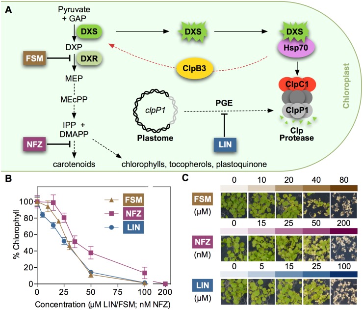
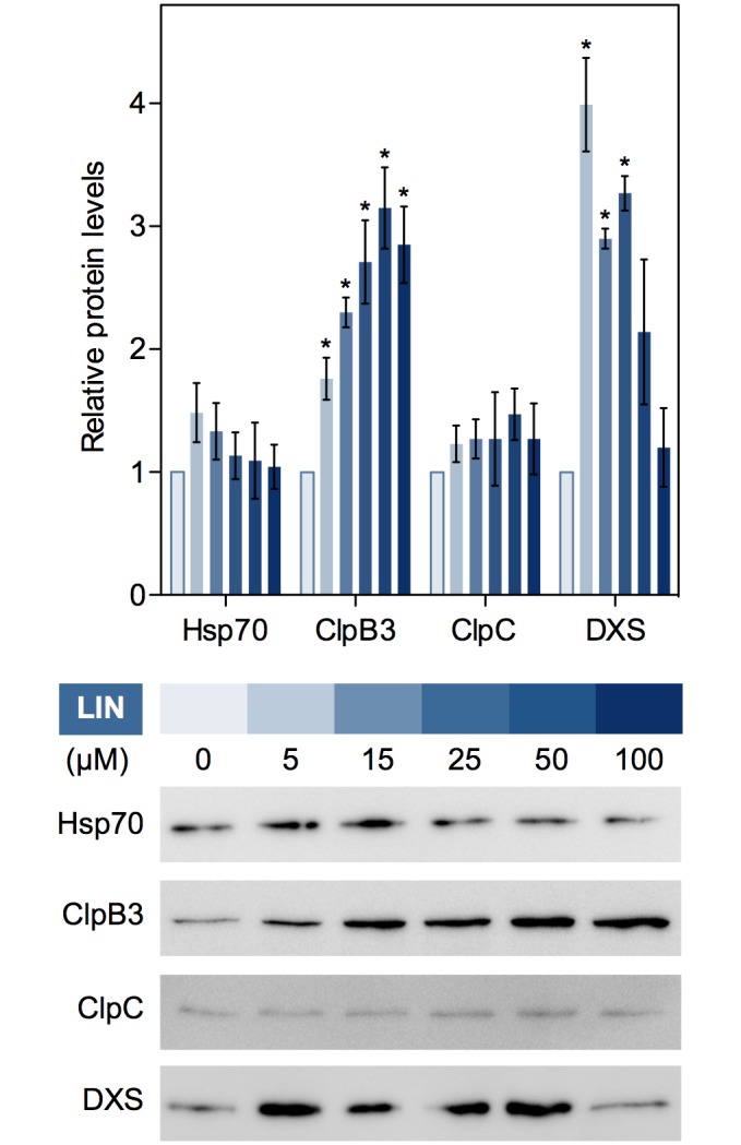
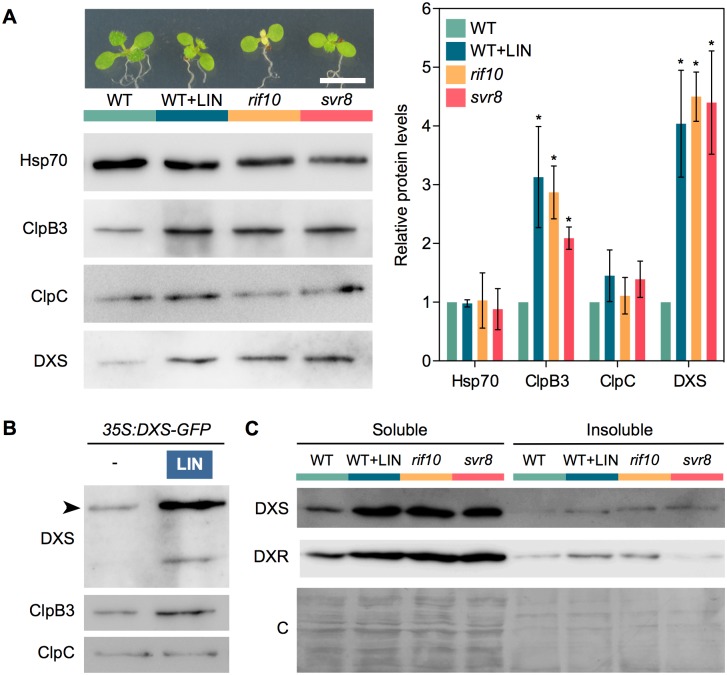
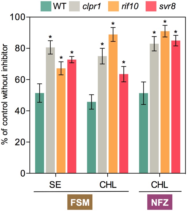
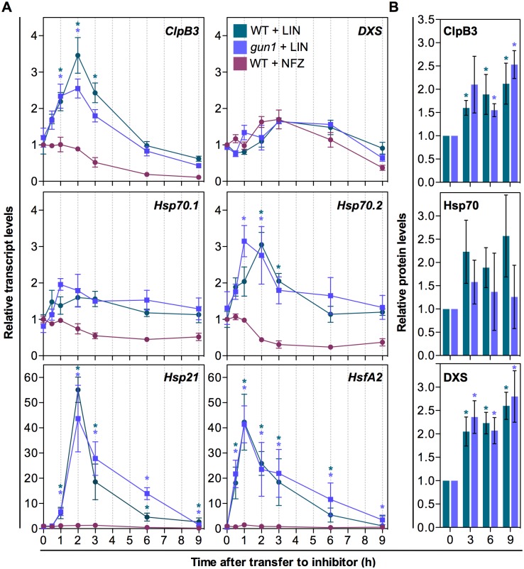
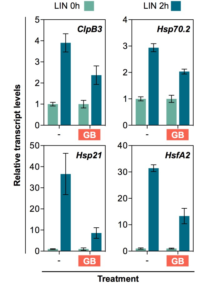
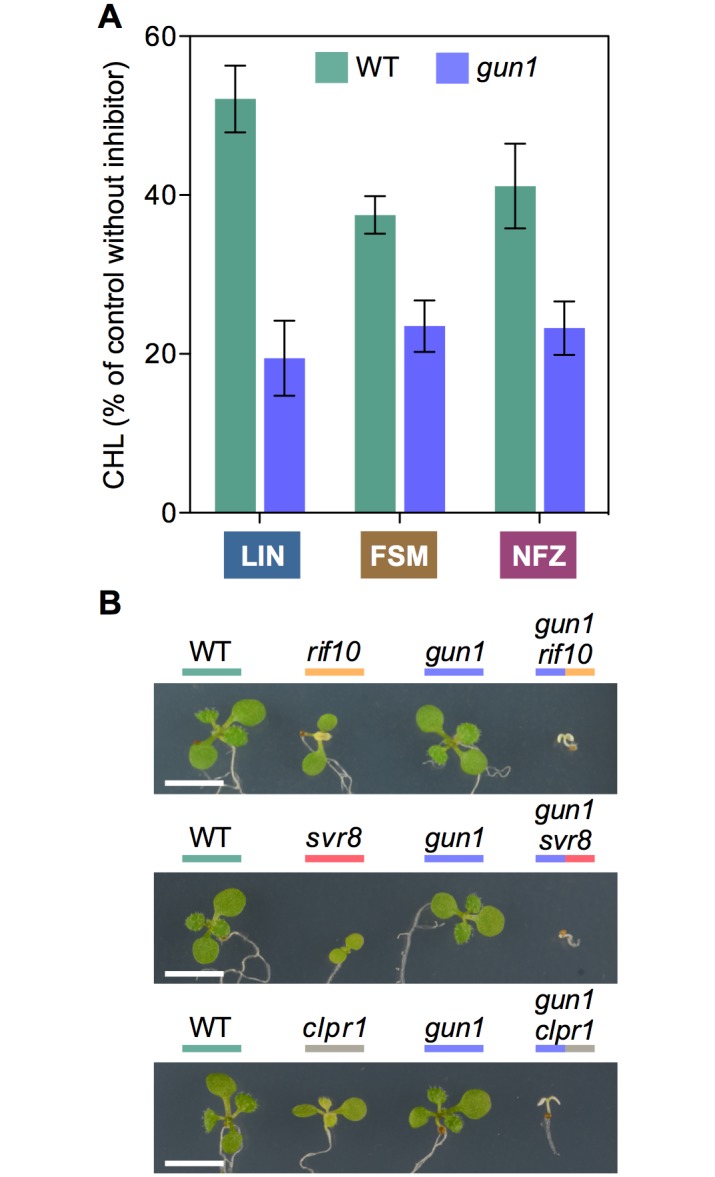
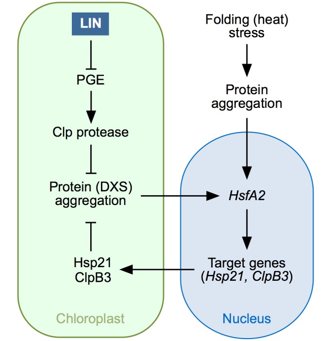
Disruption of protein homeostasis in chloroplasts impairs the correct functioning of essential metabolic pathways, including the methylerythritol 4-phosphate (MEP) pathway for the production of plastidial isoprenoids involved in photosynthesis and growth. We previously found that misfolded and aggregated forms of the first enzyme of the MEP pathway are degraded by the Clp protease with the involvement of Hsp70 and Hsp100/ClpC1 chaperones in Arabidopsis thaliana. By contrast, the combined unfolding and disaggregating actions of Hsp70 and Hsp100/ClpB3 chaperones allow solubilization and hence reactivation of the enzyme. The repair pathway is promoted when the levels of ClpB3 proteins increase upon reduction of Clp protease activity in mutants or wild-type plants treated with the chloroplast protein synthesis inhibitor lincomycin (LIN). Here we show that LIN treatment rapidly increases the levels of aggregated proteins in the chloroplast, unleashing a specific retrograde signaling pathway that up-regulates expression of ClpB3 and other nuclear genes encoding plastidial chaperones. As a consequence, folding capacity is increased to restore protein homeostasis. This sort of chloroplast unfolded protein response (cpUPR) mechanism appears to be mediated by the heat shock transcription factor HsfA2. Expression of HsfA2 and cpUPR-related target genes is independent of GUN1, a central integrator of retrograde signaling pathways. However, double mutants defective in both GUN1 and plastome gene expression (or Clp protease activity) are seedling lethal, confirming that the GUN1 protein is essential for protein homeostasis in chloroplasts.


Conflict of interest statement

The authors have declared that no competing interests exist.

Figures

Fig 1
Fig 1. Inhibitors and mechanisms modulating metabolic flux to isoprenoids in chloroplasts.
(A) Schematic representation of: (1) MEP pathway and derived products, with the position of enzymes (DXS, DXR) and inhibitor (FSM, NFZ) targets; (2) Hsp70-dependent pathways for misfolded and aggregated forms of DXS to be degraded (via ClpC1 and the Clp protease) or, alternatively, reactivated (via ClpB3); and (3) proposed mechanism by which interference with PGE (e.g. with LIN) impacts the activity of the Clp protease complex, based on the production of the plastome-encoded ClpP1 subunit. Red arrow represents the stress-induced refolding pathway. Dashed arrows represent multiple steps. GAP, glyceraldehyde 3-phosphate; DXP, deoxyxylulose 5-phosphate. See text for other acronyms. (B) Quantification of the inhibitor resistance phenotype estimated from chlorophyll levels in the absence (100%) or presence of inhibitors. (C) Representative images of Arabidopsis WT (Columbia) seedlings germinated and grown for 10 days under LD in the presence of the indicated concentrations of inhibitors.
Fig 2
Fig 2. LIN treatment boosts accumulation of ClpB3 and DXS proteins.
Chart represents Hsp70, ClpB3, ClpC and DXS protein levels detected by immunoblot analysis in 10-day-old WT plants grown at the concentrations of LIN indicated below. The mean and SEM values of n≥3 independent experiments are shown. Asterisks mark statistically significant differences (t test: p<0.05) relative to untreated controls. Representative immunoblots are also shown.
Fig 3
Fig 3. Interference with PGE promotes the accumulation of soluble DXS protein.
(A) Immunoblot analysis of Hsp70, ClpB3, ClpC and DXS levels in WT plants and PGE-defective mutants rif10-2 and svr8-2. PGE was also blocked in WT plants germinated and grown in the presence of 15 μM LIN (WT+LIN sample). Representative images of the plants used for immunoblot analysis are shown on top (bar, 5 mm). Graph shows the quantification of immunoblot data from n≥3 experiments represented as mean and SEM values relative to untreated WT plants. Asterisks mark statistically significant differences (t test: p<0.05) relative to WT samples. (B) Immunoblot analysis with the indicated antibodies of 35S:DXS-GFP plants grown on media with (+) or without (-) 15 μM LIN. Arrowhead marks the position of the DXS-GFP protein. (C) DXS and DXR protein distribution in soluble and insoluble fractions isolated from the indicated samples. A Coomassie-Blue (C) staining of the blots is shown for reference.
Fig 4
Fig 4. Mutants defective in PGE and Clp protease activity are resistant to plastidial isoprenoid inhibitors.
Resistance to FSM was estimated by quantifying seedling establishment (SE, number of plants producing true leaves) and chlorophyll levels (CHL) in plants germinated and grown in the presence of 30 μM FSM relative to those obtained with no inhibitor (100%). Similarly, NFZ resistance was calculated based on chlorophyll levels in media with 35 nM NFZ. Data correspond to the mean and SEM values of n≥3 independent experiments and asterisks mark statistically significant differences (t test: p<0.05) relative to WT samples.
Fig 5
Fig 5. Resistance to FSM and NFZ is improved by disrupting PGE with LIN.
(A) Resistance of WT plants and the indicated mutants was estimated by quantifying SE after germination and growth on media supplemented with 15 μM LIN (L), 30 μM FSM (F), or both (F+L) relative to non-supplemented medium. (B) Resistance of WT plants quantified as CHL levels in media supplemented with 15 μM LIN (L), 30 μM FSM (F), 35 nM NFZ (N) or the indicated combinations relative to non-supplemented medium. Data correspond to the mean and SEM values of n≥3 independent experiments.
Fig 6
Fig 6. Blockage of PGE results in protein aggregation in chloroplasts.
(A) TGX Stain-Free gel showing protein fractions from isolated Arabidopsis chloroplasts treated with LIN for the indicated times. An untreated control is also shown. Following ultracentrifugation of chloroplast lysates, supernatant and pellet fractions were collected (corresponding to non-aggregated and aggregated proteins, respectively) and separated by SDS-PAGE. Bands of aggregated proteins whose intensity increased after LIN treatment are marked with black arrowheads; the white arrowhead marks a major band whose intensity did not increase. (B) Quantification of total protein levels from TGX Stain-Free gel runs corresponding to chloroplast lysates before ultracentrifugation (labeled as “total”) and after separation of non-aggregated and aggregated protein fractions. Protein levels are represented relative to those in untreated controls and correspond to the mean and SEM values of n = 3 independent experiments. Asterisks marks statistically significant difference (t test: p<0.05) relative to the untreated sample. (C) Confocal microscopy detection of GFP (green) and chlorophyll (red) fluorescence in chloroplasts of siblings harboring the same T-DNA insertion with the 35S:DXS-GFP construct in WT or svr8-2 mutant backgrounds. The images were obtained with the same confocal parameters and are to the same scale. They correspond to chloroplasts from the cotyledons of 10-day-old seedlings grown in the presence of absence of 15 μM LIN. (D) Quantification of DXS protein levels detected by immunoblot analysis of insoluble protein fractions isolated from leaves infiltrated with the indicated inhibitors. Results correspond to the mean and SEM values of n = 3 independent experiments are represented relative to those in NFZ-treated samples.
Fig 7
Fig 7. Interference with PGE triggers a rapid but transient expression of specific genes.
(A) Quantitative RT-PCR (qPCR) analysis of transcript levels of the indicated genes in 7-day-old WT and gun1-101 plants after transferring to medium with 400 μM LIN or 400 nM NFZ. (B) Levels of ClpB3, Hsp70 and DXS proteins detected by immunoblot analysis in LIN-treated samples. Data correspond to the mean and SEM values of n≥3 independent experiments, and asterisks mark statistically significant differences (t test: p<0.05) relative to untreated (0h) samples.
Fig 8
Fig 8. Pre-treatment with a chemical chaperone desensitizes the molecular response to LIN.
Quantitative (qPCR) analysis of transcript levels of the indicated genes in 7-day-old WT plants grown in the presence or absence of glycine betaine (GB) and then transferred for 2h to fresh medium supplemented with 400 μM LIN. Data correspond to the mean and SEM values of n = 3 independent experiments and they are represented relative to the levels before the LIN treatment. For all the genes tested, statistically significant differences (t test: p<0.05) were found between the LIN-triggered induction of control and GB-grown samples.
Fig 9
Fig 9. GUN1 contributes to chloroplast protein homeostasis and is required for survival of PGE-defective mutants.
(A) Resistance of WT and gun1-101 plants to the indicated inhibitors estimated as CHL level of plants germinated and grown in the presence of 15 μM LIN, 30 μM FSM or 35 nM NFZ relative to no-inhibitor controls. Data correspond to the mean and SEM values of n≥3 independent experiments. (B) Phenotype of 10-day-old WT and mutant lines of the indicated genotypes grown in the same plate. Bar, 5 mm.
Fig 10
Fig 10. Model for the cpUPR mechanism in Arabidopsis.
LIN treatment represses PGE and this eventually causes a reduced activity of the Clp protease. Normal Clp protease activity removes misfolded proteins and hence prevents protein aggregation. When Clp protease activity is compromised, however, the misfolded proteins that fail to be degraded (including DXS) aggregate. Build-up of protein aggregates somehow sends an unknown retrograde signal to upregulate the expression of HsfA2, a gene that can also be induced (at much higher levels) by heat stress episodes causing protein aggregation in other cell compartments. HsfA2 encodes a transcription factor that in turn induces the expression of target genes encoding chloroplast chaperones such as Hsp21 and ClpB3. As a result, more of these chaperones are synthesized and imported into plastids, eventually contributing to alleviate protein folding stress in this organelle.

Similar articles

Cited by

References

    1. Grimm B, Dehesh K, Zhang L, Leister D (2014) Intracellular communication. Mol Plant 7: 1071–1074. doi: 10.1093/mp/ssu073 - DOI - PubMed
    1. Kleine T, Leister D (2016) Retrograde signaling: Organelles go networking. Biochim Biophys Acta 1857: 1313–1325. doi: 10.1016/j.bbabio.2016.03.017 - DOI - PubMed
    1. Chan KX, Phua SY, Crisp P, McQuinn R, Pogson BJ (2016) Learning the Languages of the Chloroplast: Retrograde Signaling and Beyond. Annu Rev Plant Biol 67: 25–53. doi: 10.1146/annurev-arplant-043015-111854 - DOI - PubMed
    1. Sun AZ, Guo FQ (2016) Chloroplast Retrograde Regulation of Heat Stress Responses in Plants. Front Plant Sci 7: 398 doi: 10.3389/fpls.2016.00398 - DOI - PMC - PubMed
    1. Lin YF, Haynes CM (2016) Metabolism and the UPR(mt). Mol Cell 61: 677–682. doi: 10.1016/j.molcel.2016.02.004 - DOI - PMC - PubMed

MeSH terms