. 2017 Sep 22;11(9):e0005927.
doi: 10.1371/journal.pntd.0005927. eCollection 2017 Sep.

Recombinant vaccines of a CD4+ T-cell epitope promote efficient control of Paracoccidioides brasiliensis burden by restraining primary organ infection

Affiliations

Recombinant vaccines of a CD4+ T-cell epitope promote efficient control of Paracoccidioides brasiliensis burden by restraining primary organ infection

Rodrigo Assunção Holanda et al. PLoS Negl Trop Dis. .

Abstract

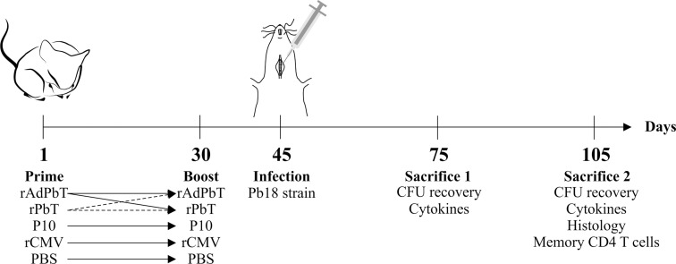
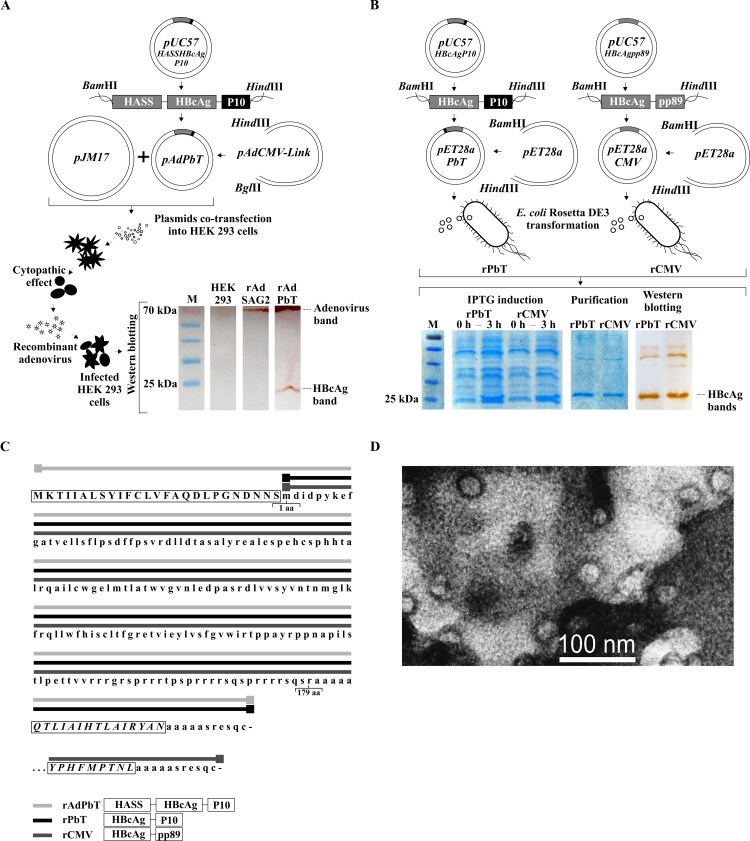
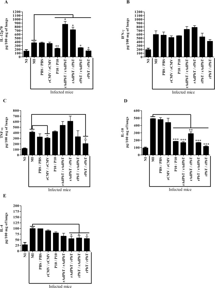
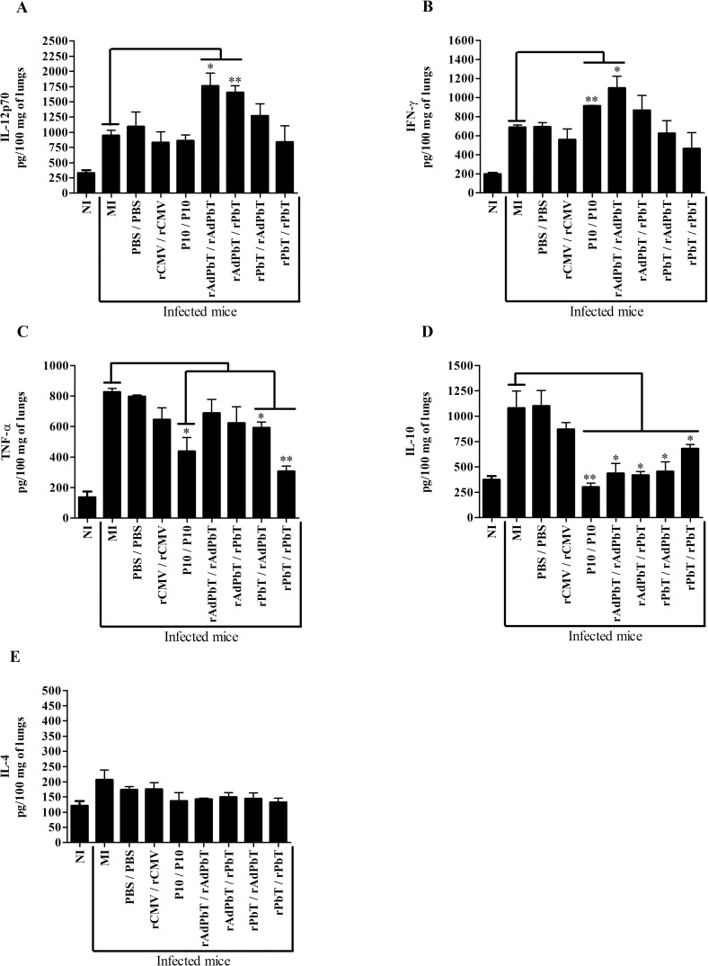
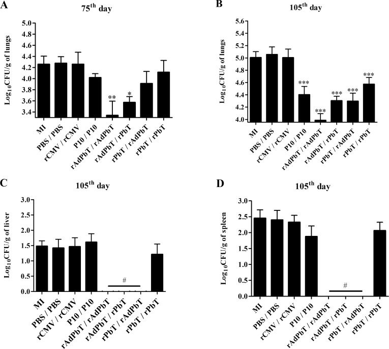
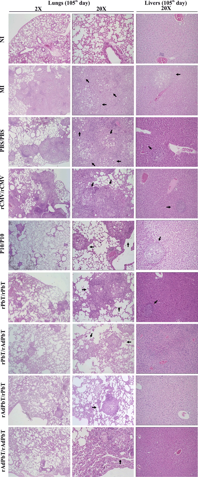
Paracoccidioidomycosis (PCM) is an infectious disease endemic to South America, caused by the thermally dimorphic fungi Paracoccidioides. Currently, there is no effective human vaccine that can be used in prophylactic or therapeutic regimes. We tested the hypothesis that the immunogenicity of the immunodominant CD4+ T-cell epitope (P10) of Paracoccidioides brasiliensis gp43 antigen might be significantly enhanced by using a hepatitis B virus-derived particle (VLP) as an antigen carrier. This chimera was administered to mice as a (His)6-purified protein (rPbT) or a replication-deficient human type 5 adenoviral vector (rAdPbT) in an immunoprophylaxis assay. The highly virulent Pb18 yeast strain was used to challenge our vaccine candidates. Fungal challenge evoked robust P10-specific memory CD4+ T cells secreting protective Th-1 cytokines in most groups of immunized mice. Furthermore, the highest level of fungal burden control was achieved when rAdPbT was inoculated in a homologous prime-boost regimen, with 10-fold less CFU recovering than in non-vaccinated mice. Systemic Pb18 spreading was only prevented when rAdPbT was previously inoculated. In summary, we present here VLP/P10 formulations as vaccine candidates against PCM, some of which have demonstrated for the first time their ability to prevent progression of this pernicious fungal disease, which represents a significant social burden in developing countries.


Conflict of interest statement

I have read the journal's policy and the authors of this manuscript have the following competing interests: RAH, DAS, CPT and OBR are inventors on patent application [BR1020150311150] of the recombinant vaccines that was assigned by National Institute of Industrial Property, Brazil.

Figures

Fig 1
Fig 1. Scheme of murine immunization protocols used to test paracoccidioidomycosis prophylaxis.
Mice were immunized subcutaneously with proteins or viruses as indicated (homologous or heterologous prime-boost protocols), and further challenged by intratracheal inoculation of Pb18 yeast cells. At the times indicated mice were sacrificed to evaluate tissue injuries, fungal burden, inflammatory responses, and humoral and/or cellular immune responses. rAdPbT is a recombinant adenovirus expressing a VLP/P10 chimera; rPbT is a purified recombinant VLP/P10 protein chimera; P10 is a synthetic peptide; rCMV is a purified recombinant VLP/pp89 protein chimera used as control; mock-immunized mice were challenged with Pb18 strain but previously non-immunized; PBS indicates phosphate buffered saline emulsified in adjuvant as control (non-immunized).
Fig 2
Fig 2. Recombinant VLP chimeras.
(A) Cloning and co-transfection steps for generating rAdPbT adenoviral vector. Extracts of non-infected and adenovirus-infected HEK293 cells were incubated with dual anti-HBcAg/anti-adenoviral polyclonal sera. HBcAg and adenoviral proteins displayed 25 and 70 kDa bands, respectively. rAdSAG2 is a control recombinant adenovirus that encodes a surface antigen of Toxoplasma gondii. (B) Cloning, expression, purification and western blot reactivity tests of rPbT and rCMV (control) proteins from E. coli Rosetta DE3 bacteria. All HBcAg-derived bands display molecular weights of around 25 kDa. M = protein molecular weight ladder; 0h, 3h = times after IPTG induction. (C) Amino acid sequences of chimeric proteins, indicating the presence of signal peptide (HASS, uppercase letters within initial rectangle), hepatitis B virus core sequences (HBcAg, lowercase letters), P. brasiliensis epitope P10 (lower set of uppercase letters within a rectangle) or MCMV pp89 epitope (lowest set of uppercase letters within a rectangle). Amino acid residues comprising HASS-HBcAg-P10 (continuous light grey line following the letters), HBcAg-P10 (continuous black line following the letters) and HBcAg-pp89 (continuous dark grey line following the letters) are displayed. Amino-acids 1 and 179 of HBcAg are also indicated. (D) Transmission electron micrographs of the rPbT virus-like particles obtained from E. coli bacteria.
Fig 3
Fig 3. Cytokine quantification in lungs at the 75th day.
IL12p70 (A), IFN-γ (B), TNF-α (C), IL-10 (D) and IL-4 (E) were quantified in 100 mg of lung homogenates from the different groups of mice. Mock-immunized (MI), PBS/PBS, rCMV/rCMV, P10/P10, rAdPbT/rAdPbT, rAdPbT/rPbT, rPbT/rAdPbT and rPbT/rPbT mice were all intratracheally inoculated with virulent Pb18 yeast cells, while non-infected mice (NI) were intratracheally inoculated with sterile PBS. Data represent the mean of two independent experiments (three animals per experiment). ***(p<0,001), **(p<0,01) and *(p<0,05) were considered to be significant.
Fig 4
Fig 4. Cytokine quantification in lungs at the 105th day.
IL12p70 (A), IFN-γ (B), TNF-α (C), IL-10 (D) and IL-4 (E) were quantified from 100 mg of lung homogenate. Mock-immunized (MI), PBS/PBS, rCMV/rCMV, P10/P10, rAdPbT/rAdPbT, rAdPbT/rPbT, rPbT/rAdPbT and rPbT/rPbT were groups intratracheally inoculated with virulent Pb18 yeast cells, while non-infected mice (NI) were intratracheally inoculated with sterile PBS. Data represent the mean of two independent experiments (three animals per experiment). ***(p<0,001), **(p<0,01) and *(p<0,05) were considered to be significant.
Fig 5
Fig 5. Specific proliferation of CFSE-labeled CD4+ T lymphocytes.
Cellular immunity recalled by Pb18 infection in immunized (rAdPbT/rAdPbT, rAdPbT/rPbT, rPbT/rAdPbT, rPbT/rPbT, P10/P10, rCMV/rCMV and PBS/PBS) and mock-immunized (MI) mice at the 105th experimentation day (75 days after the last immunization). (A) Lymphocyte population was delimited based on its size (FSC) and granularity (SSC) and CD4+ T lymphocytes were searched for by anti-CD3 and anti-CD4 surface marking. (B) Controls of non-stimulated proliferative CD4+ T cells were monitored in CD3+CD4+CFSEHIGH histogram peaks (non-specific proliferation; dotted line) and ConA-stimulated CD4+ T cells were monitored in CD3+CD4+CFSELOW histogram peaks (continuous line) using splenocytes from non-infected and non-immunized mice. Proliferative CD4+ T cells of rAdPbT/rAdPbT-immunized mice (red line), rAdPbT/rPbT (blue line), rPbT/rAdPbT (purple line), P10/P10 (green line) and rPbT/rPbT-immunized mice (black line) recalled by intratracheal Pb18-challenge are shown as overlaid histograms (C) and the corresponding percentage of cells that experimented proliferation (D). Data represent the mean of two independent experiments (three animals per experiment). ***(p<0,001), **(p<0,01) and *(p<0,05) were considered to be significant.
Fig 6
Fig 6. Memory CD4+ T lymphocytes recalled by P. brasiliensis infection.
Central (TCM) and effector (TEM) phenotyping were evaluated at the 105th experimentation day (75 days after the last immunization) in CD4+ T lymphocytes region (CD3+CD4+) using anti-CD44 and anti-CD62L surface marking, according to the staining controls for negative and positive populations (A). TCM and TEM phenotypes were evidenced in CD3+CD4+CD44HIGHCD62LHIGH (filled bars) and in CD3+CD4+CD44HIGHCD62LLOW (open bars) regions, respectively (B), as well as the percentages of these cells were calculated (C). Mock-immunized (MI), PBS/PBS, rCMV/rCMV, P10/P10, rAdPbT/rAdPbT, rAdPbT/rPbT, rPbT/rAdPbT and rPbT/rPbT were groups intratracheally inoculated by virulent Pb18 yeast. Data represent the mean of two independent experiments (three animals per experiment). *** or ### (p<0,001), ** or ## (p<0,01) and * or # (p<0,05) were considered to be significant.
Fig 7
Fig 7. Fungal burden measurement.
Plots representing the numbers of colony-forming units detected per gram of tissue at the 75th and the 105th days, as indicated, in different organs. (A and B) lungs; (C) livers; (D) spleens. All groups were intratracheally inoculated with virulent Pb18 yeast. Data represent the mean of two independent experiments (six animals per experiment). ***(p<0,001), **(p<0,01), *(p<0,05) and # (p<0,0001) were considered to be significant.
Fig 8
Fig 8. Histopathology.
Lungs and livers were stained with Hematoxilin-Eosin at day 105 (2-20X magnification). Organs of normal uninfected mice intratracheally inoculated with sterile PBS (NI) and of those challenged with virulent Pb18 yeast after immunization (MI, PBS/PBS, rCMV/rCMV, P10/P10, rAdPbT/rAdPbT, rAdPbT/rPbT, rPbT/rAdPbT and rPbT/rPbT) were evaluated. Arrows indicate the presence of granulomas containing yeast cells.

Similar articles

Cited by

References

    1. Munoz JF, Gallo JE, Misas E, Priest M, Imamovic A, Young S, et al. Genome update of the dimorphic human pathogenic fungi causing paracoccidioidomycosis. Plos Neglect Trop D 2014; 8:e3348. - PMC - PubMed
    1. Franco M, Bagagli E, Scapolio S, da Silva Lacaz C. A critical analysis of isolation of Paracoccidioides brasiliensis from soil. Med Mycol 2000; 38:185–91. - PubMed
    1. Bocca AL, Amaral AC, Teixeira MM, Sato PK, Shikanai-Yasuda MA, Soares Felipe MS. Paracoccidioidomycosis: eco-epidemiology, taxonomy and clinical and therapeutic issues. Future Microbiol 2013; 8:1177–91. doi: 10.2217/fmb.13.68 - DOI - PubMed
    1. Stevens DA, Brummer E, Clemons KV. Interferon- gamma as an antifungal. The J Infect Dis 2006; 194 Suppl 1:S33–7. - PubMed
    1. Iwai LK, Yoshida M, Sadahiro A, da Silva WR, Marin ML, Goldberg AC, et al. T-cell recognition of Paracoccidioides brasiliensis gp43-derived peptides in patients with paracoccidioidomycosis and healthy individuals. Clin Vaccine Immunol 2007; 14:474–6. doi: 10.1128/CVI.00458-06 - DOI - PMC - PubMed

MeSH terms

LinkOut - more resources