. 2017 Sep 22;13(9):e1006639.
doi: 10.1371/journal.ppat.1006639. eCollection 2017 Sep.

The Kaposi's sarcoma-associated herpesvirus (KSHV) non-structural membrane protein K15 is required for viral lytic replication and may represent a therapeutic target

Affiliations

The Kaposi's sarcoma-associated herpesvirus (KSHV) non-structural membrane protein K15 is required for viral lytic replication and may represent a therapeutic target

Bizunesh Abere et al. PLoS Pathog. .

Abstract

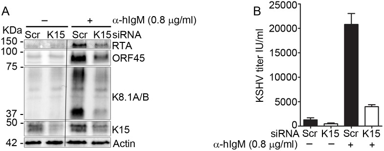
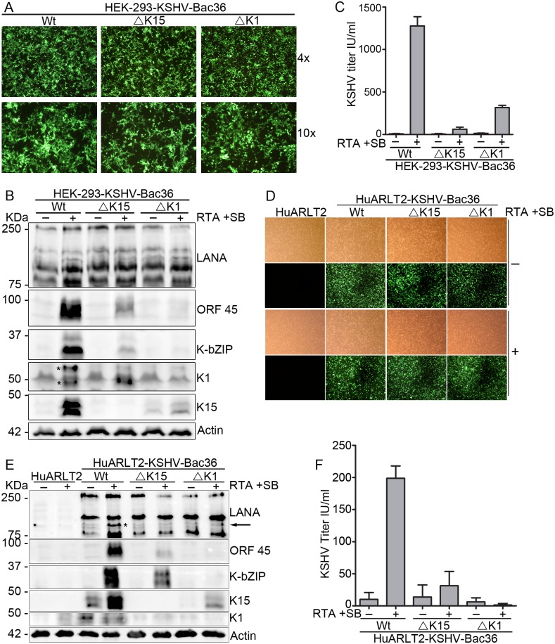
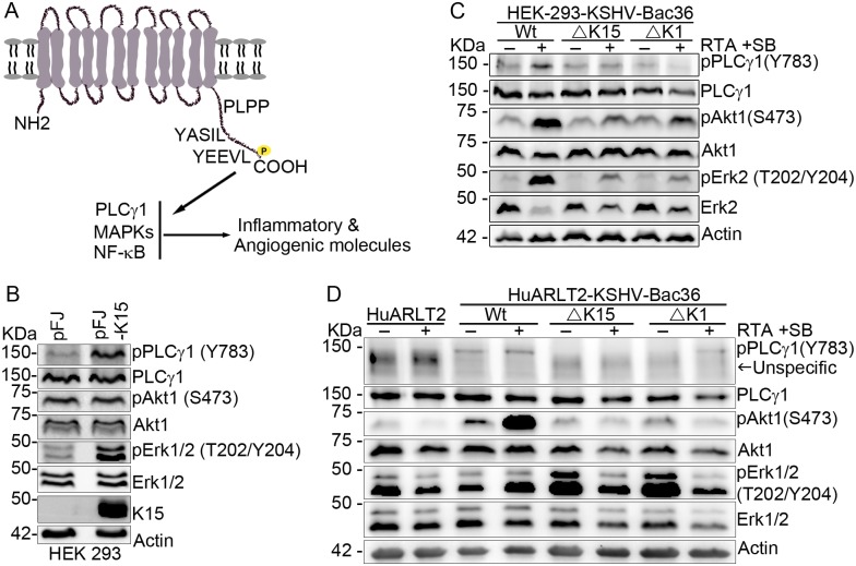
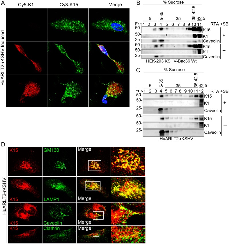
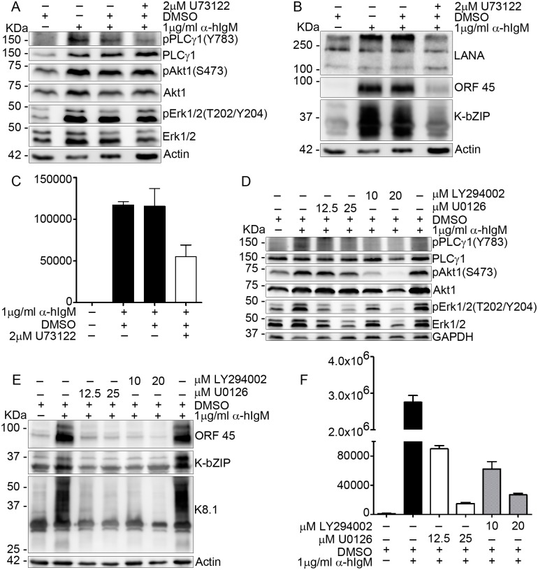
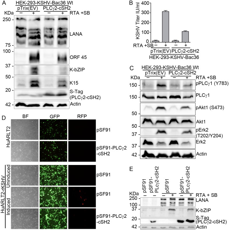
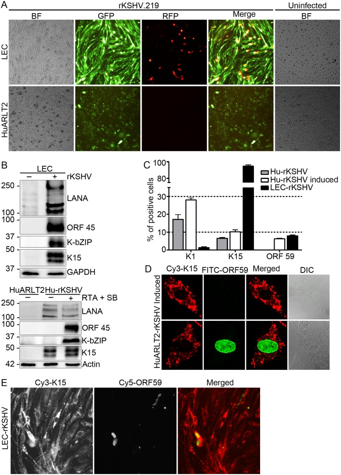
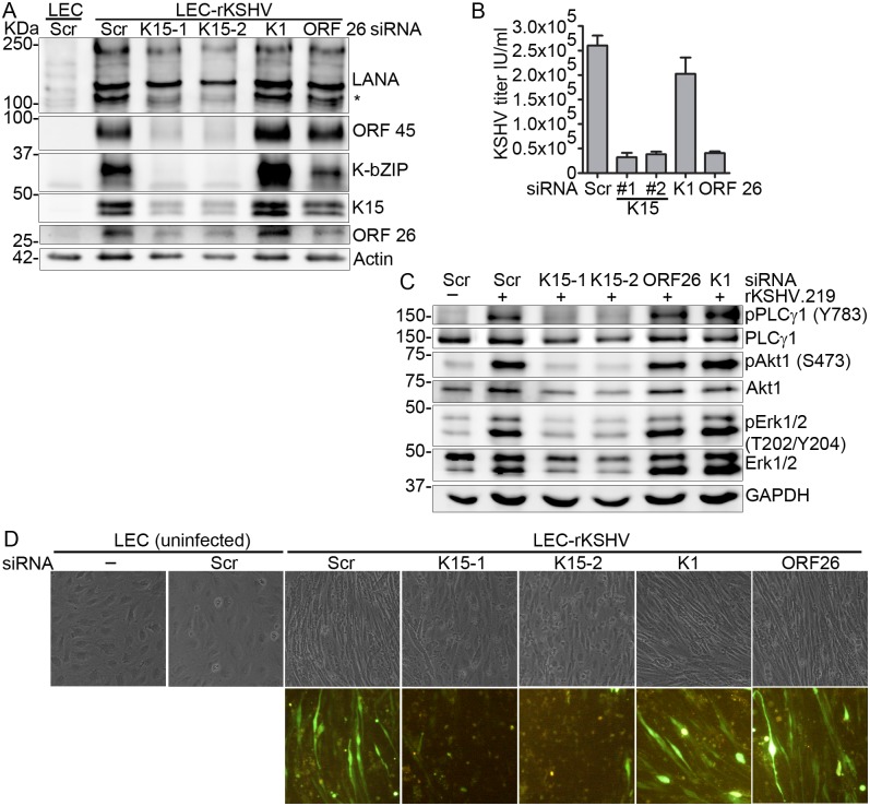
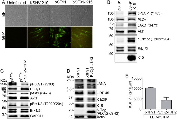
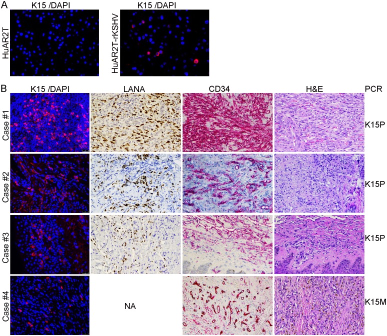
Kaposi's sarcoma-associated herpesvirus (KSHV) is the infectious cause of the highly vascularized tumor Kaposi's sarcoma (KS), which is characterized by proliferating spindle cells of endothelial origin, extensive neo-angiogenesis and inflammatory infiltrates. The KSHV K15 protein contributes to the angiogenic and invasive properties of KSHV-infected endothelial cells. Here, we asked whether K15 could also play a role in KSHV lytic replication. Deletion of the K15 gene from the viral genome or its depletion by siRNA lead to reduced virus reactivation, as evidenced by the decreased expression levels of KSHV lytic proteins RTA, K-bZIP, ORF 45 and K8.1 as well as reduced release of infectious virus. Similar results were found for a K1 deletion virus. Deleting either K15 or K1 from the viral genome also compromised the ability of KSHV to activate PLCγ1, Erk1/2 and Akt1. In infected primary lymphatic endothelial (LEC-rKSHV) cells, which have previously been shown to spontaneously display a viral lytic transcription pattern, transfection of siRNA against K15, but not K1, abolished viral lytic replication as well as KSHV-induced spindle cell formation. Using a newly generated monoclonal antibody to K15, we found an abundant K15 protein expression in KS tumor biopsies obtained from HIV positive patients, emphasizing the physiological relevance of our findings. Finally, we used a dominant negative inhibitor of the K15-PLCγ1 interaction to establish proof of principle that pharmacological intervention with K15-dependent pathways may represent a novel approach to block KSHV reactivation and thereby its pathogenesis.


Conflict of interest statement

The authors have declared that no competing interests exist.

Figures

Fig 1
Fig 1. Depleting the K15 mRNA from KSHV infected cells compromises its ability to undergo lytic reactivation.
BJAB-rKSHV.219 cells were microporated with either a control siRNA (Scr) or siRNA against the KSHV K15 gene and the lytic cycle was induced 24 hours later with 0.8 μg/ml of anti-hIgM antibody. Forty-eight hours after lytic induction, (A) cells were lysed and the expression level of the indicated lytic viral proteins was assessed by western blot and (B) infectious virus titer released into the supernatant was determined by infecting HEK-293 cells. Results are representative of two or more independent experiments. Bar graphs (B) represent the means ± SD of three replicates.
Fig 2
Fig 2. Absence of either the K1 or K15 gene from the viral genome impairs KSHV lytic reactivation.
(A) Stably transfected polyclonal HEK-293-KSHV-Bac36 Wt/ΔK15/ΔK1 cells. In these cells, the KSHV lytic cycle was induced with 1 mM Sodium Butyrate (SB) and SF-9 cell supernatant containing KSHV-RTA expressing baculovirus. After 48 hours, cells and culture supernatant were collected; (B) expression level of the indicated lytic viral proteins and (C) infectious virus titer was analyzed as described in materials and methods. (D)–(F) In HuARLT2 cells stably infected with the indicated KSHV-Bac36 viruses, the KSHV lytic cycle was induced as before by using a cocktail of SB and RTA. (D) Stably infected cells after 48 hours with or without induction of the lytic cycle. (E) The expression level of the indicated lytic proteins and (F) infectious virus titer analyzed as described in materials and methods. Results are representative of at least three independent experiments. Bar graphs represent the means ± SD of (C) two or (F) four replications. * denotes K1 bands (panel B)or low molecular weight isoforms of LANA (panel E; see text).
Fig 3
Fig 3. Deleting either K1 or K15 from the viral genome compromises PLCγ1, Akt1 and Erk1/2 signaling activation during KSHV reactivation.
(A) Schematic representation of K15 activated signaling. HEK-293 cells were transfected with a plasmid DNA expressing K15 or an empty vector control; cells were collected 48 hours after transfection and (B) the phosphorylation level of the indicated signaling components was analyzed by western blot. The KSHV lytic cycle was induced in either (C) HEK-293 or (D) HuARLT2 cells stably infected with the indicated KSHV Bac36 viruses; forty eight hours after lytic induction, cells were collected and western blot was performed as in B. Results are representative of at least two independent experiments.
Fig 4
Fig 4. Sub-cellular localization of the K1 and K15 proteins in KSHV infected cells.
(A) HuARLT2-rKSHV cells after lytic induction were co-stained with a rat anti-K15 and a mouse anti-K1 monoclonal antibodies followed by Cy3-conjugated anti-rat IgG (Green) and a Cy5-conjugated anti-Mouse IgG (Red) secondary antibodies; representative cells expressing only K15 (upper panel) or together with K1(lower two panels) are shown. (B and C) Cellular lysate from either HEK-293-KSHV-Bac36 Wt or HuARLT2-rKSHV cells with or without induction of the lytic cycle was subjected to membrane flotation assay and fractions of the sucrose gradient were analyzed for the presence of the indicated proteins by western blot. (D) HuARLT2-rKSHV cells without induction of the KSHV lytic cycle were co-stained with a rat anti-K15 and a rabbit antibody to the indicated cellular organelle markers followed by Cy3-conjugated anti-rat IgG (red) and a FITC-conjugated anti-rabbit IgG (green) secondary antibodies. Images (A and D) were acquired using a Leica confocal microscope. Experiments were performed two or more times.
Fig 5
Fig 5. Inhibiting the activation of the PLCγ, MAPK and PI3K/Akt signaling pathways blocks KSHV reactivation.
BJAB-rKSHV.219 cells were treated with the indicated inhibitors or DMSO as a control and the KSHV lytic cycle was induced with 1 μg/ml of anti-human IgM antibody applied together with the inhibitors. Seventy two hours after lytic induction, cells were lysed and the activation level of the indicated signaling components (A and D) as well as the expression level of lytic viral proteins (B and E) was assessed by western blot and (C and F) infectious virus titer was determined from the culture supernatant. Results are representative of three independent experiments. Bar graphs (C and F) represent the means ± SD of three replications.
Fig 6
Fig 6. The isolated PLCγ2-cSH2 domain blocks KSHV reactivation.
HEK-293-KSHV-Bac36 Wt cells were transfected with a plasmid expressing the PLCγ2-cSH2 domain or its empty vector control and the lytic cycle was induced 24 hours later. Cells and cell culture supernatant were collected 48 hours after induction of the lytic cycle; (A) expression level of KSHV lytic proteins, (B) virus titer in the supernatant and (C) phosphorylation level of the indicated signaling components was analyzed as described before. HuARLT2 or HuARLT2-rKSHV cells were transduced with a retrovirus vector expressing either the PLCγ2-cSH2 or the empty vector control and the KSHV lytic cycle was induced 24 hours later. Forty eight hours after induction of the lytic cycle, images were taken for GFP and RFP expression (D), cells were lysed and expression level of the indicated viral proteins was analyzed by western blot (E). Experiments were performed at least two times. Bar graphs in (B) represent the means ± SD of 2 replications.
Fig 7
Fig 7. K15 protein expression in KSHV stably infected LEC and HuARLT2 cells.
LEC and HuARLT2 cells were infected with rKSHV.219 virus at MOI 1 and (A) images were taken 7 days after infection. Two weeks post infection, cells were collected and (B) the expression level of the indicated viral lytic proteins in the stably infected LECs (upper panel) or HuARLT2 cells (lower panel) before and after lytic induction was analyzed by western blot. (C) HuARLT2-rKSHV cells with or without induction of the KSHV lytic cycle or stably infected LEC-rKSHV cells were stained with a mouse anti-K1 mAb, a rat anti-K15 mAb or a mouse anti-ORF59 mAb followed by a Cy3-conjugated anti-mouse or anti-rat IgG; the number of cells expressing K1, K15 or ORF 59 protein was manually counted. Bar graphs represent the means ± SD of 3 replications. (D) HuARLT2-rKSHV cells after lytic reactivation were co-stained with a rat anti-K15 mAb and a mouse anti-ORF 59 mAb followed by a Cy3-conjugated anti-rat (red) and FITC-conjugated anti-mouse (green) secondary antibodies and the representative images were acquired using a Leica confocal microscope. (E) LEC-rKSHV cells were co-stained with a rat anti-K15 mAb and a mouse anti-ORF 59 mAb followed by a Cy3-conjugated anti-rat (red) and Cy5-conjugated anti-mouse (green) secondary antibodies. Images for (C) and (E) were acquired using a Zeiss Observer.Z1 epi-fluorescent microscope.
Fig 8
Fig 8. Depletion of K15 from stably infected LECs impairs KSHV lytic replication and reverses endothelial cell spindling.
LEC or LEC-rKSHV cells were transfected with the indicated siRNAs. Seventy-two hours after siRNA mediated knock-down, cells and cell culture supernatant were collected; (A) western blot of KSHV lytic proteins, (B) KSHV infectious virus titer, (C) western blot analysis of signaling components and (D) representative images of siRNA transfected cells taken before cell lysis. Results are representative of two or more independent experiments. Bar graphs in (B) represent the means ± SD of 3 replications. * Low molecular weight isoforms of LANA (see text).
Fig 9
Fig 9. K15 expression in LECs induces signaling activation and endothelial cell spindling similar to KSHV infection.
LECs were transduced with a retrovirus vector expressing K15 or an empty vector control, or in parallel infected with rKSHV.219 virus or left uninfected. After 48 hours (A) representative images were taken and (B) the activation level of the indicated signaling components was assessed by western blot. (C)(E) stably infected LEC-rKSHV cells were transduced with a retrovirus vector expressing the PLCγ2-cSH2 or an empty vector control. Seventy two hours later, cells and cell culture supernatant were collected; (C) activation of signaling components and (D) expression level of lytic viral proteins were analyzed by western blot, and (E) KSHV infectious titer was analyzed as described before. Bar graphs in (E) represent the means ± SD of 2 replications.
Fig 10
Fig 10. K15 is abundantly expressed in KS tissue biopsies.
(A) IF staining, using antibody clone number 18E5, of K15 (red) in sections prepared from paraffin embedded blocks of HuARLT2 or HuARLT2-rKSHV cells. (B) KS tissue biopsies obtained from HIV positive individuals were stained for K15/DAPI by IF (first column), LANA (dark brown nuclei), CD34 (red) and Hematoxylin and Eosin (last column); the respective KSHV genotype (K15P or M) was determined by PCR and is shown on the right. NA—staining not available because of insufficient biopsy material.

Similar articles

Cited by

References

    1. Chang Y, Cesarman E, Pessin MS, Lee F, Culpepper J, Knowles DM, et al. Identification of herpesvirus-like DNA sequences in AIDS-associated Kaposi's sarcoma. Science. 1994;266(5192):1865–9. . - PubMed
    1. Cesarman E, Chang Y, Moore PS, Said JW, Knowles DM. Kaposi's sarcoma-associated herpesvirus-like DNA sequences in AIDS-related body-cavity-based lymphomas. The New England Journal of Medicine. 1995;332(18):1186–91. doi: 10.1056/NEJM199505043321802 . - DOI - PubMed
    1. Soulier J, Grollet L, Oksenhendler E, Cacoub P, Cazals-Hatem D, Babinet P, et al. Kaposi's sarcoma-associated herpesvirus-like DNA sequences in multicentric Castleman's disease. Blood. 1995;86(4):1276–80. . - PubMed
    1. Friedman-Kien AE. Disseminated Kaposi's sarcoma syndrome in young homosexual men. Journal of the American Academy of Dermatology. 1981;5(4):468–71. . - PubMed
    1. Ganem D. KSHV and the pathogenesis of Kaposi sarcoma: listening to human biology and medicine. The Journal of Clinical Investigation. 2010;120(4):939–49. doi: 10.1172/JCI40567 . - DOI - PMC - PubMed

Substances