Review
. 2017 Sep 21;24(9):1101-1119.
doi: 10.1016/j.chembiol.2017.08.027.

Small-Molecule Inhibitors Targeting DNA Repair and DNA Repair Deficiency in Research and Cancer Therapy

Affiliations
Review

Small-Molecule Inhibitors Targeting DNA Repair and DNA Repair Deficiency in Research and Cancer Therapy

Sarah R Hengel et al. Cell Chem Biol. .

Abstract

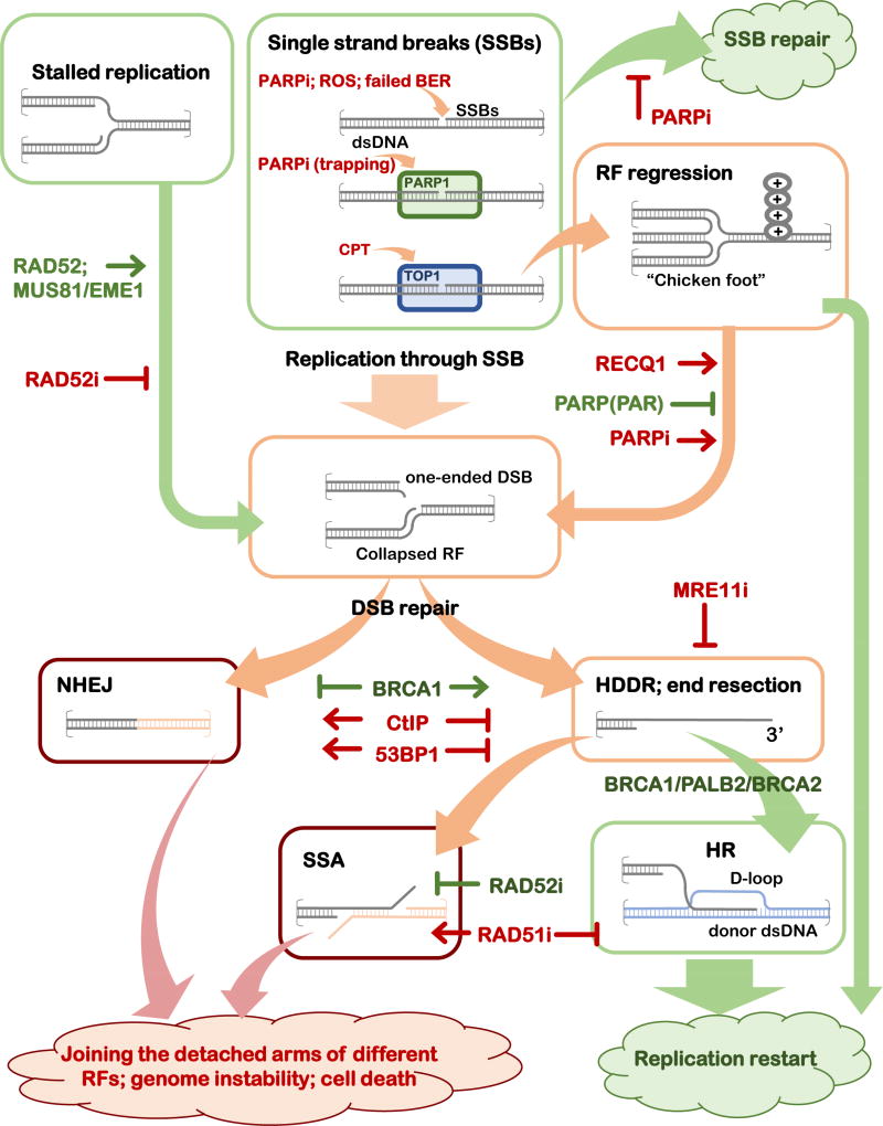
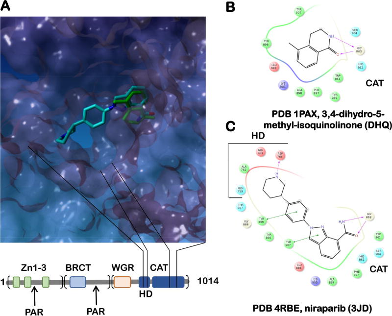
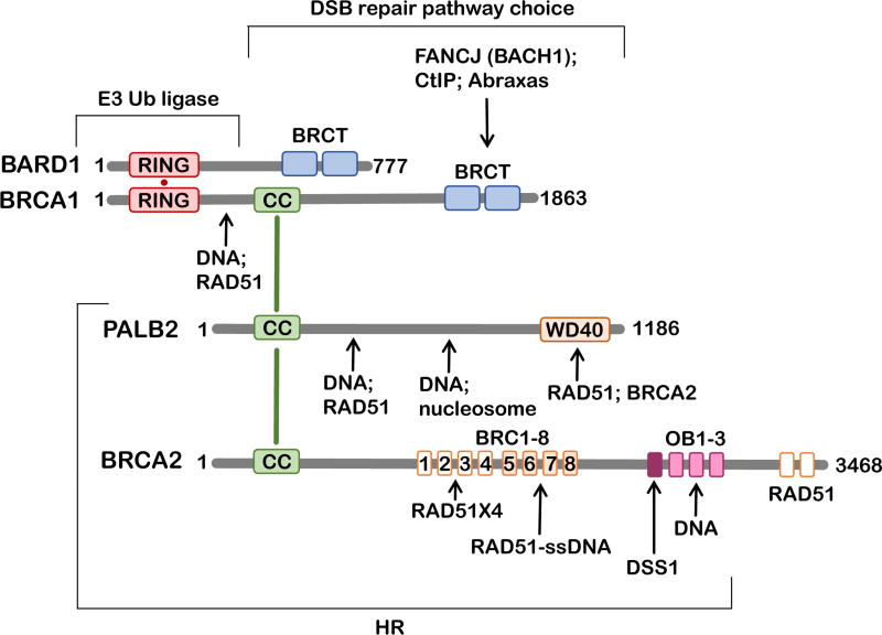
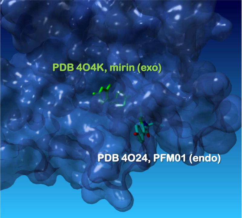
To maintain stable genomes and to avoid cancer and aging, cells need to repair a multitude of deleterious DNA lesions, which arise constantly in every cell. Processes that support genome integrity in normal cells, however, allow cancer cells to develop resistance to radiation and DNA-damaging chemotherapeutics. Chemical inhibition of the key DNA repair proteins and pharmacologically induced synthetic lethality have become instrumental in both dissecting the complex DNA repair networks and as promising anticancer agents. The difficulty in capitalizing on synthetically lethal interactions in cancer cells is that many potential targets do not possess well-defined small-molecule binding determinates. In this review, we discuss several successful campaigns to identify and leverage small-molecule inhibitors of the DNA repair proteins, from PARP1, a paradigm case for clinically successful small-molecule inhibitors, to coveted new targets, such as RAD51 recombinase, RAD52 DNA repair protein, MRE11 nuclease, and WRN DNA helicase.

Keywords: BLM DNA helicase; BRCA1; BRCA2; DNA repair; MRE11; PARP inhibitors; PARP1 (poly(ADP-ribose)polymerase1); RAD51; RAD52; RECQ1 DNA helicase; TOP1; WRN DNA helicase; homologous recombination.


Figures

Figure 1
Figure 1. Roles of the DNA repair inhibitors in altering the outcomes of the DNA repair events in the presence of DNA replication
Schematic representation of the progression of a DNA lesion (a single-strand break, SSB, or stalled replication fork, RF) through various DNA repair mechanisms leading to DNA repair and restoration of replication fork, or to genome destabilizing events. The intermediates and mechanisms that are likely to lead to accurate repair if processed through an appropriate mechanism are depicted in green boxes, the intermediates that can lead to either positive (replication fork restoration) or negative (genome instability and cell death) outcomes are in orange boxes, and the intermediates and mechanisms that funnel the DNA repair intermediates into the genome destabilizing outcomes are shown in red boxes. The key regulators (proteins and inhibitors) that affect the outcome of each event are shown.
Figure 2
Figure 2. BRCA-interacting network
Schematic representation of the primary structures of BRCA1, BRCA2 and their interacting partners BARD1 and PALB2. Structural domains and regions of significance to this review are shown as rectangles: RING domains (red), CC – coiled coil domains (green), WD40 domain (orange), BRCT domains (blue), BRC motifs 1 – 8 (orange), OB folds 1 – 3 and DSS1-interacting region (purple), BRCA2 C-terminal RAD51-interacting domains (orange). Arrows point to the respective interaction sites. Brackets highlight regions important for the stated cellular function.
Figure 3
Figure 3. PARP inhibitors
A. The overlap of a nicotineamide analog DHQ (green; PDB 1PAX) bound to the active site of PARP1 and the second generation PARP1 inhibitor niraparib (light blue; PDB 1PAX), which occupies both the nicotinamide-ribose binding pocket and the adenosine-ribose binding pocket of ADP-ribosyl transferase catalytic site, making additional contacts with E763 and D766 of the regulatory α-helical sub-domain (HD) (Thorsell et al., 2017). PARP1 ADP-ribosyl transferase catalytic domain (CAT; PDB 1PAX) is shown as a surface representation. The primary structure of PARP1 is shown below the active site image. The bead-on-a-string architecture of PARP1 (Langelier and Pascal, 2013) includes the N-terminal DNA binding region (the three Zn-finger domains are shown as green rectangles), the middle region containing a BRCT domain and the main auto-PARylation site (Altmeyer et al., 2009), and the C-terminal region that contains the WGR domain responsible for the interdomain contacts and the cross-talk between the DNA-binding/damage recognition domains, the regulatory HD domain as well as the CAT domain. B. and C. The ligand maps for DHQ and niraparib, respectively.
Figure 4
Figure 4. MRE11 separation of function inhibitors
Surface representation of the MRE11 nuclease active site with bound mirin (green, PDB: KO4K), an exonuclease inhibitor and PFM01 (elemental colors, from PDB: 4O24), an endonuclease inhibitor (Shibata et al., 2014). See text for the details of the inhibition mechanisms.

References

    1. Aggarwal M, Banerjee T, Sommers JA, Brosh RM., Jr Targeting an Achilles’ heel of cancer with a WRN helicase inhibitor. Cell cycle (Georgetown, Tex) 2013a;12:3329–3335. - PMC - PubMed
    1. Aggarwal M, Banerjee T, Sommers JA, Iannascoli C, Pichierri P, Shoemaker RH, Brosh RM., Jr Werner syndrome helicase has a critical role in DNA damage responses in the absence of a functional fanconi anemia pathway. Cancer research. 2013b;73:5497–5507. - PMC - PubMed
    1. Aggarwal M, Sommers JA, Shoemaker RH, Brosh RM., Jr Inhibition of helicase activity by a small molecule impairs Werner syndrome helicase (WRN) function in the cellular response to DNA damage or replication stress. Proceedings of the National Academy of Sciences of the United States of America. 2011;108:1525–1530. - PMC - PubMed
    1. Alagpulinsa DA, Ayyadevara S, Shmookler Reis RJ. A Small-Molecule Inhibitor of RAD51 Reduces Homologous Recombination and Sensitizes Multiple Myeloma Cells to Doxorubicin. Frontiers in oncology. 2014;4:289. - PMC - PubMed
    1. Altmeyer M, Messner S, Hassa PO, Fey M, Hottiger MO. Molecular mechanism of poly(ADP-ribosyl)ation by PARP1 and identification of lysine residues as ADP-ribose acceptor sites. Nucleic acids research. 2009;37:3723–3738. - PMC - PubMed

MeSH terms