Review
. 2017 Sep 21;24(9):1161-1180.
doi: 10.1016/j.chembiol.2017.08.028.

Targeting Metabolism for Cancer Therapy

Affiliations
Review

Targeting Metabolism for Cancer Therapy

Alba Luengo et al. Cell Chem Biol. .

Abstract

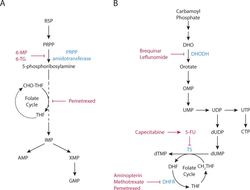
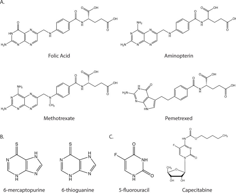
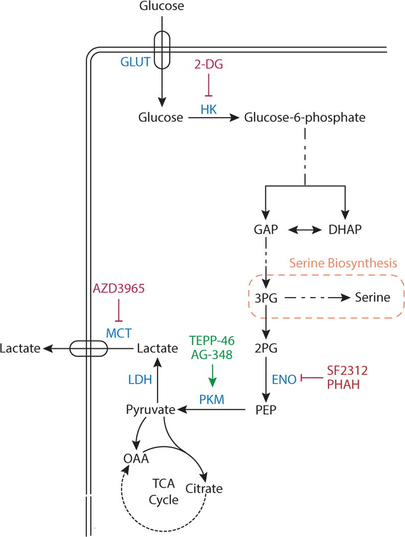
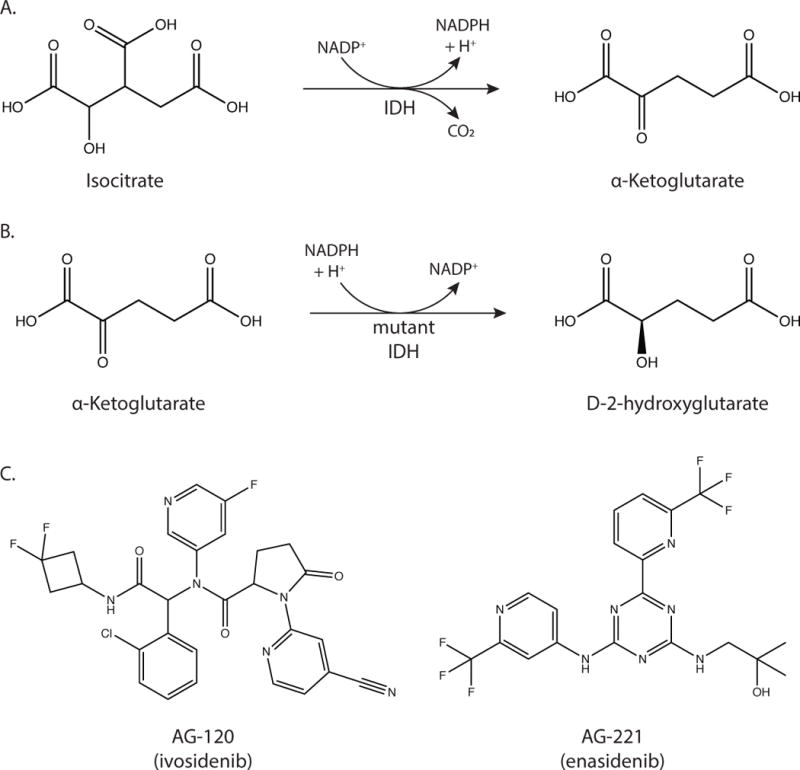
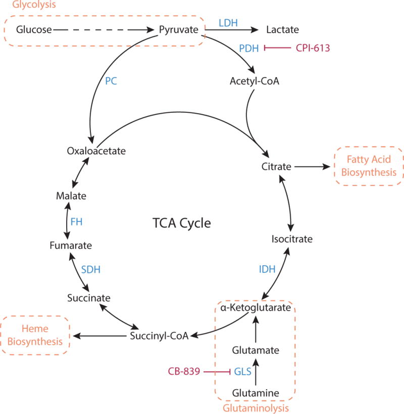
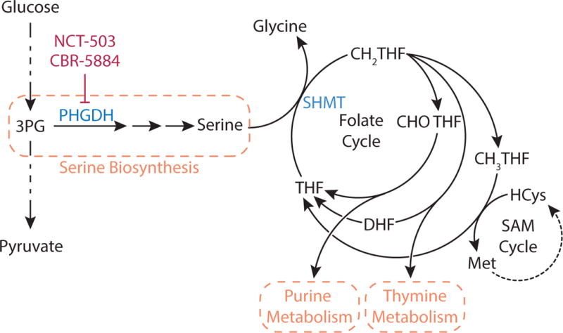
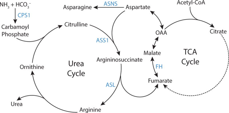
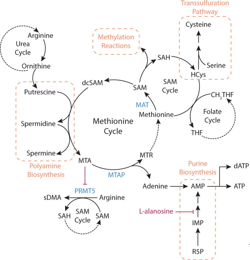
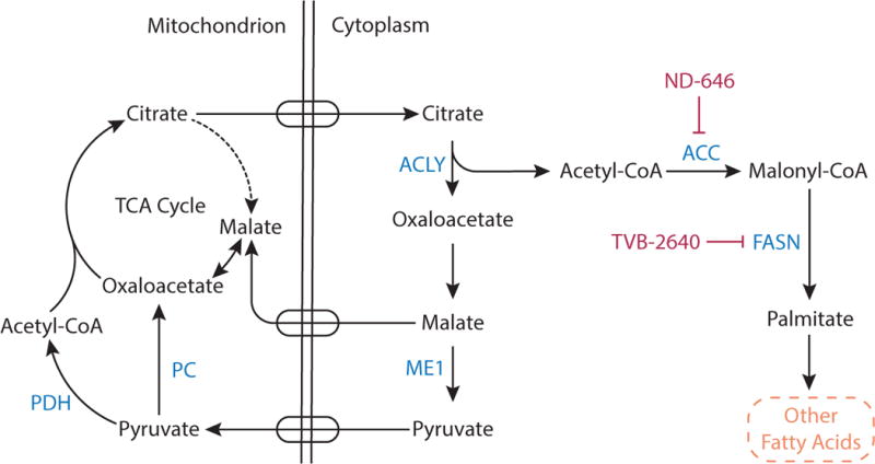
Metabolic reprogramming contributes to tumor development and introduces metabolic liabilities that can be exploited to treat cancer. Chemotherapies targeting metabolism have been effective cancer treatments for decades, and the success of these therapies demonstrates that a therapeutic window exists to target malignant metabolism. New insights into the differential metabolic dependencies of tumors have provided novel therapeutic strategies to exploit altered metabolism, some of which are being evaluated in preclinical models or clinical trials. Here, we review our current understanding of cancer metabolism and discuss how this might guide treatments targeting the metabolic requirements of tumor cells.

Keywords: cancer metabolism; cell metabolism; chemotherapy.


Figures

Figure 1
Figure 1. Nucleotide Biosynthesis
(A) Purine nucleotide synthesis. The first reaction in purine production generates 5-phosphoribosyl-1-pyrophosphatae (PRPP) from ribose 5-phosphate (R5P). The second step is catalyzed by PRPP amidotransferase, and commits PRPP to purine synthesis. This step can be inhibited by the antimetabolites, 6-mercaptopurine (6-MP) and 6-thioguanine (6-TG). Subsequent steps in the pathway assemble the purine ring and result in the formation of inosine monophosphate (IMP), which in turn can be converted to either adenosine monophosphate (AMP) or guanosine monophosphate (GMP) by distinct reactions. The synthesis of the purine ring requires N10-formyl-tetrahydrofolate (CHO-THF) via a reaction that can be inhibited by pemetrexed. (B) Pyrimidine nucleotide synthesis. Pyrimidine nucleotide synthesis begins with the conversion of carbamoyl phosphate to the pyrimidine base orotate. One of the steps in pathway is catalyzed by dihydroorotate dehydrogenase (DHODH), which can be inhibited by brequinar sodium and leflunomide. Next, orotate is combined with PRPP to generate orotate monophosphate (OMP), which is subsequently converted to uridine monophosphate (UMP). UMP can be phosphorylated to form UDP and UTP, and the latter can be further converted to citidine triphosphate (CTP). Uridine nucleotides can also be used for de novo thymine nucleotide synthesis. UDP is converted to deoxy-UMP (dUMP), and the enzyme thymidylate synthase (TS) generates dTMP by catalyzing the methylation of dUMP using N5,N10-methylene-tetrahydrofolate (CH2-THF) as the methyl donor. TS activity is inhibited by the antipyrimidine 5-fluorouracil (5-FU) and the 5-FU pro-drug capecitabine. Thymidine synthesis can also be inhibited by the antifolates aminopterin, methotrexate, and pemetrexed, as these drugs inhibit the enzyme dihydrofolate reductase (DHFR), limiting the availability of CH2-THF.
Figure 2
Figure 2. Antimetabolites
(A) Structures of folic acid and the antifolate compounds aminopterin, methotrexate and pemetrexed. (B) Structures f the purine analogues 6-mercaptopurine and 6-thioguanine (C) Structures of pyrimidine analogue 5-fluorouracil and the 5-fluorouracil prodrug capecitabine.
Figure 3
Figure 3. Glycolysis
Glucose is imported in cells by one of several glucose transporters (GLUT). Glucose is phosphorylated by hexokinase (HK), a step that can be competitively inhibited by the compound 2-deoxyglucose (2-DG). In a later step of glycolysis, glucose carbon is cleaved into two interconvertible three-carbon units, dihydroxyacetone phosphate (DHAP) and glyceraldehyde 3-phosphate (GAP). Downstream of GAP, 3-phosphoglycerate (3-PG) is converted to 2-phosphoglycerate (2-PG), and the enzyme enolase (ENO) generates phosphoenolpyruvate (PEP) from 2-PG. ENO activity can be inhibited by the compounds phosphonoacetohydroxamate (PHAH) and SF-2312. PEP is converted to pyruvate by pyruvate kinase (PKM), which can be activated by the drugs TEPP-46 and AG-348. Pyruvate can be oxidized in the TCA cycle, or it can produce lactate via lactate dehydrogenase (LDH), with lactate excreted by monocarboxylate transporters (MCT). MCT isoform 1 can be inhibited by the compound AZD3965.
Figure 4
Figure 4. Mutant IDH
(A) The reaction catalyzed by wild-type IDH is the decarboxylation of isocitrate to α-ketoglutarate. (B) The reaction catalyzed by mutant IDH1 and IDH2 is the reduction of α-ketoglutarate to D-2-hydroxyglutarate. (C) Structures of AG-120 (ivosidenib) and AG-221 (enasidenib), which inhibit mutant IDH1 and IDH2 respectively.
Figure 5
Figure 5. TCA Cycle
Pyruvate produced by glycolysis can be metabolized by the TCA cycle. Pyruvate is oxidized via pyruvate dehydrogenase (PDH) to the two-carbon unit acetyl-CoA, and subsequently combined with the four-carbon oxaloacetate to generate citrate. The cycle regenerates oxaloacetate while generating precursors that are important for biosynthetic processes including heme and fatty acid synthesis. When molecules are removed from the TCA cycle to feed these pathways, TCA intermediates must be replenished in a process termed anaplerosis. Pyruvate can serve as an anaplerotic substrate when converted to oxaloacetate by the enzyme pyruvate carboxylase (PC). Glutaminolysis, the conversion of glutamine to glutamate via glutaminase (GLS), can also support anaplerosis via production of α-ketoglutarate. PDH activity is inhibited by the lipoic acid derivative CPI-613 and GLS activity is inhibited by CB-839.
Figure 6
Figure 6. Serine Biosynthesis and Folate Cycle
The glycolytic intermediate 3-phophoglycerate (3-PG) is metabolized to serine in a three-step pathway where the first step is catalyzed by the enzyme phosphoglycerate dehydrogenase (PHGDH), which can be inhibited by NCT-503 and CBR-5884. Serine metabolism to glycine by serine hydroxymethyltransferase (SHMT) transfers a one carbon unit to tetrahydrofolate (THF) to form N5,N10-methylene-tetrahydrofolate (CH2-THF). CH2-THF is the methyl donor for thymidine nucleotides, or it can be converted to N10-formyl-tetrahydrofolate (CHO-THF) for purine biosynthesis or to N5-methyl-THF (CH3-THF) to support methionine production and numerous methylation reactions via the S-adenosyl methionine (SAM) cycle.
Figure 7
Figure 7. Urea Cycle
The urea cycle permits excretion of excess nitrogen as urea. Carbamoyl phosphate is synthesized from ammonia and bicarbonate by the enzyme carbamoyl phosphate synthetase 1 (CPS1), and subsequently carbamoyl phosphate and ornithine are used to produce citrulline. The enzyme argininosuccinate synthase (ASS1) condenses citrulline with aspartate to form argininosuccinate, which is then cleaved to arginine and fumarate by argininosuccinate lyase (ASL). Arginine is then hydrolyzed to produce urea and regenerate ornithine, completing the cycle. Fumarate produced by the urea cycle can regenerate aspartate via enzymes involved in the tricarboxylic acid (TCA) cycle as shown. Asparagine synthetase (ASNS) catalyzes production of asparagine from aspartate.
Figure 8
Figure 8. Methionine Cycle
Methionine is an essential amino acid that can be used for methylation reactions, cysteine synthesis, and polyamine generation. Methionine is converted to S-adenosylmethionine (SAM) by methionine adenosyltransferase (MAT). Methyl transferases utilize SAM as the methyl donor for many methylation reactions in cells. Demethylation of SAM generates S-adenosylhomocysteine (SAH), which is hydrolyzed to homocysteine (HCys) and adenosine. Methionine is regenerated from homocysteine by transfer of the methyl group from N5-methyl-THF (CH3-THF). Homocysteine is also an intermediate of cysteine synthesis, where serine and homocysteine condense and form cysteine via the transsulfuration pathway. SAM can also support polyamine synthesis when SAM is decarboxylated to form S-adenosylmethioninamine, also known as decarboxylated S-adenosylmethionine (dcSAM). Together with the urea cycle metabolite ornithine, this compound generates putrescine and subsequently spermine and spermidine. In these reactions, dcSAM is converted to 5′-methylthioadenosine (MTA). Accumulation of MTA inhibits the enzyme protein arginine N-methyltrasferase 5 (PRMT5), which uses SAM as a methyl donor to synthesize symmetrical dimethylarginine (sDMA) from arginine. In the methionine cycle, MTA is cleaved to 5-methylthioribose-1-phosphate (MTR) and adenine by the enzyme methylthioadenosine phosphorylase (MTAP). Adenine can be converted to AMP, and the adenosine analogue L-alanosine can limit AMP production via the purine synthesis pathway. MTR can be converted to methionine, completing the methionine cycle.
Figure 9
Figure 9. Fatty Acid Synthesis
Citrate is a carrier of acetyl groups from the mitochondria to the cytoplasm to support fatty acid synthesis. Mitochondrial citrate is transported into the cytosol where ATP citrate lyase (ACLY) cleaves citrate to acetyl-CoA and oxaloacetate. Oxaloacetate is converted to malate, and malate can be transported back into the mitochondrial matrix or can be converted into pyruvate in the cytoplasm by malic enzyme 1 (ME1). Cytosolic acetyl-CoA contributes to fatty acid synthesis. First, the enzyme acetyl-CoA carboxylase (ACC) catalyzes carboxylation of acetyl CoA to malonyl CoA. Fatty acid synthase (FASN) then uses malonyl-CoA to sequentially add two carbon units to a growing acyl chain and synthesize the saturated 16-carbon fatty acid palmitate, which serves as a precursor to other fatty acids. The compound ND-464 and TVB-2640 are inhibitors of ACC and FASN, respectively.

Similar articles

Cited by

References

    1. Adam J, Yang M, Bauerschmidt C, Kitagawa M, O’Flaherty L, Maheswaran P, Ozkan G, Sahgal N, Baban D, Kato K, et al. A role for cytosolic fumarate hydratase in urea cycle metabolism and renal neoplasia. Cell Rep. 2013;3:1440–1448. - PMC - PubMed
    1. Altman BJ, Stine ZE, Dang CV. From Krebs to clinic: glutamine metabolism to cancer therapy. Nat Rev Cancer. 2016;16:619–634. - PMC - PubMed
    1. Anastasiou D, Yu Y, Israelsen WJ, Jiang JK, Boxer MB, Hong BS, Tempel W, Dimov S, Shen M, Jha A, et al. Pyruvate kinase M2 activators promote tetramer formation and suppress tumorigenesis. Nat Chem Biol. 2012;8:839–847. - PMC - PubMed
    1. Appel IM, den Boer ML, Meijerink JP, Veerman AJ, Reniers NC, Pieters R. Up-regulation of asparagine synthetase expression is not linked to the clinical response L-asparaginase in pediatric acute lymphoblastic leukemia. Blood. 2006;107:4244–4249. - PubMed
    1. Appleyard MV, Murray KE, Coates PJ, Wullschleger S, Bray SE, Kernohan NM, Fleming S, Alessi DR, Thompson AM. Phenformin as prophylaxis and therapy in breast cancer xenografts. Br J Cancer. 2012;106:1117–1122. - PMC - PubMed

MeSH terms