. 2017 Dec 1;158(12):4270-4285.
doi: 10.1210/en.2017-00495.

IL-1β Inhibits Connexin 43 and Disrupts Decidualization of Human Endometrial Stromal Cells Through ERK1/2 and p38 MAP Kinase

Affiliations

IL-1β Inhibits Connexin 43 and Disrupts Decidualization of Human Endometrial Stromal Cells Through ERK1/2 and p38 MAP Kinase

Jie Yu et al. Endocrinology. .

Abstract

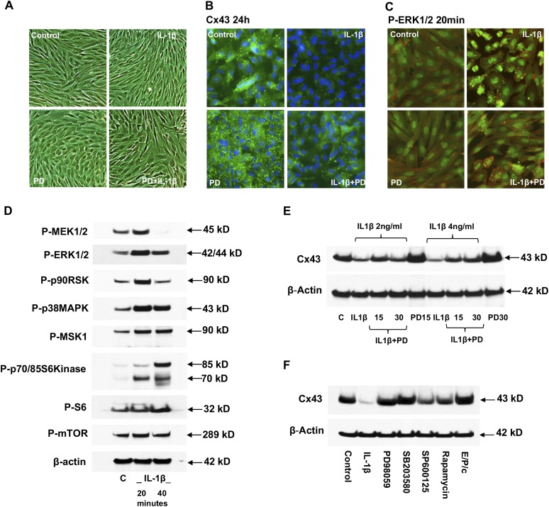
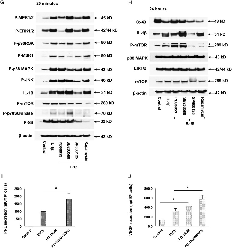
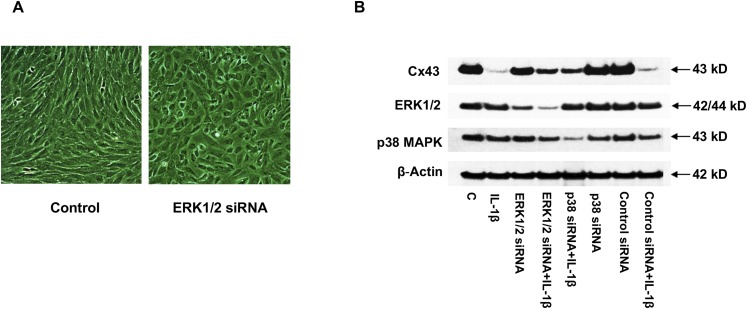
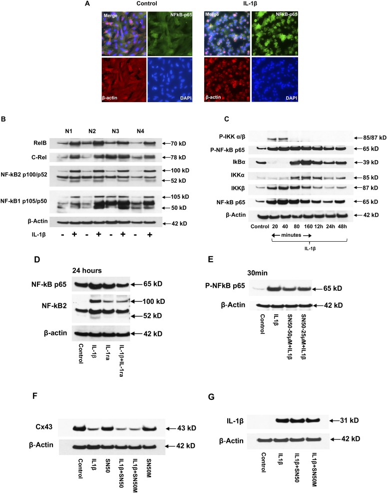
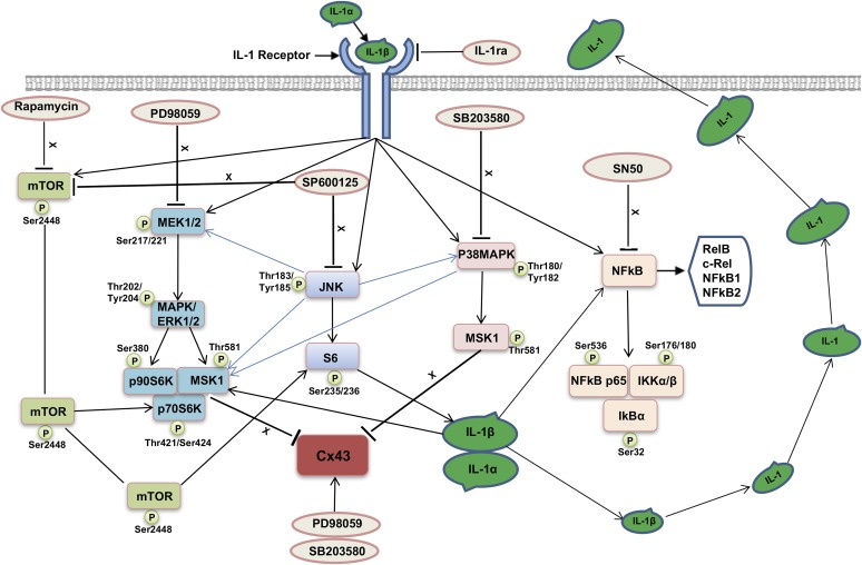
Inflammation can interfere with endometrial receptivity. We examined how interleukin 1β (IL-1β) affects expression of the uterine gap junction protein connexin 43 (Cx43), which is known to be critical for embryonic implantation. We used an in vitro model of human endometrial stromal cells (ESCs), Western blotting, and a combination of validated, selective kinase inhibitors to evaluate five canonical IL-1β signaling pathways. Cx43 and two other markers of ESC differentiation (prolactin and VEGF) were inhibited predominantly via IL-1β-activated ERK1/2 and p38 MAP kinase cascades. The findings were corroborated using small interfering RNA to silence critical genes in either pathway. By contrast, upregulation of endogenous pro-IL-1α and pro-IL-1β following recombinant IL-1β treatment was mediated via the Jun N-terminal kinase pathway. The clinicopharmacological significance of our findings is that multiple signaling cascades may need to be neutralized to reverse deleterious effects of IL-1β on human endometrial function.


Figures

Figure 1.
Figure 1.
Endometrial morphology in vivo and in vitro. (A) CD68+ tissue macrophages (green) infiltrate the mesenchyme; the epithelium and stroma are IL-1β+ (red). The merged image (yellow) shows IL-1β highly concentrated in extravasating macrophages. DAPI (blue) highlights basal nuclei with subnuclear epithelial vacuoles and perivascular stromal edema typical of secretory phase endometrium. (B) Infiltrating CD68+ macrophages (green) have typical amoeboid shape and are surrounded by Cx43+ (red) stroma; the epithelium is negative. The merged image shows coexpression of CD68 and Cx43 in stromal macrophages (yellow). DAPI (blue) reveals the glandular architecture. (C) ESCs exposed to decidualizing hormone cocktail (E/P/c, bottom-left panel) for 3 days undergo “rounded” differentiation. By contrast, IL-1β induced a pronounced bipolar, fibroblastic phenotype in with parallel alignment (top-right panel). The combination of E/P/c and IL-1β suppressed the roundness noted with E/P/c (bottom-right panel).
Figure 2.
Figure 2.
Biochemical responses of ESCs to E/P/c and IL-1β. (A) Proliferative-phase ESCs show a dramatic increase in prolactin (PRL) production over control 72 hours after E/P/c treatment; however, addition of recombinant IL-1β reduced prolactin by 62% ± 3% (*P < 0.01, Student t test). (B) VEGF was blunted 48% ± 1% by IL-1β (*P < 0.01). (C) IL-1β dose-dependently suppressed VEGF (IC50 ∼0.2 ng/mL) and stimulated IL-6 secretion from (D) ESCs with comparable pharmacokinetics. Similar results were noted in three independent experiments.
Figure 3.
Figure 3.
Western blot and RT-qPCR analyses of of IL-1β on ESCs. (A) Cx43 expression was upregulated 72 hours after E/P/c incubation but dramatically reduced with IL-1β (top panel). Five nanograms per milliliter of recombinant IL-1β (+) auto-upregulated endogenous pro-IL-1β (31-kD bands) and mature IL-1β (17-kD bands) (middle panel). (B) Cx43 was suppressed by IL-1β (+) in four out of four independent ESC preparations (top panel, 43-kD bands), whereas pro-IL-1β (top panel, 31-kD bands) increased in three out of four cases. No effect on β-actin was noted (bottom panel). (C) Incubation with IL-1β reduced Cx43, whereas IL-1α and -β were upregulated (panels 2 and 3). Active cleaved caspase 3 was only observed 48 hours after IL-1β (panel 4), corresponding to near total depletion of Cx43. (D) IL-1ra (500 ng/mL) neutralized the inhibitory effect of IL-1β on Cx43 and returned the gap junction protein to basal levels (lane 4, top panel). IL-1ra also blocked auto-upregulation of pro-IL-1β (middle panel) but had no effect on β-actin (bottom panel). (E) Suppression of Cx43 by IL-1β was mediated, in part, at the mRNA level (IC50 ∼2 ng/mL). Insets show parallel amplicon amplification and sharp melting characteristics.
Figure 4.
Figure 4.
Effects of kinase inhibitors. (A) IL-1β induced a fibroblastic phenotype (top right). PD reversed this effect (bottom right) and alone promoted cell “rounding” (bottom left). (B) Punctate green Cx43 foci were reduced by IL-1β (top right) but were restored or increased by PD (bottom panels). DAPI (blue) = nuclei. (C) Phospho(P)-ERK1/2 (green) nuclear accumulation after IL-1β (top right) was blocked by PD (bottom right). β-Actin (red) stained background. (D) ESC lysates after 20 to 40 minutes IL-1β showed increased P-MEK1/2, P-ERK1/2, P-p90RSK, P-p38MAPK, P-MSK1, P-p70S6Kinase (70- and 85-kD isoforms), and P-S6. P-mTOR and β-actin levels were unchanged. (E) Two and four nanograms per milliliter IL-1β for 24 hours reduced Cx43, whereas coincubation with 15 or 30 μM PD partially reversed effects (top panel). PD alone (15 and 30 μM) stimulated Cx43 80% above baseline (top panel). (F) IL-1β inhibited Cx43. PD and SB203580 increased Cx43 to levels approaching E/P/c-treated ESCs. SP600125 and rapamycin suppressed Cx43. β-Actin levels remained stable. (G) IL-1β increased P-MEK1/2, -ERK1/2, -p90RSK, -MSK1, -p38 MAPK, -JNK, and -p70S6kinase. (G, H) IL-1β auto-upregulation was only blunted by SP600125. (H) PD and SB203580 partially reversed IL-1β effects on Cx43. PD enhanced E/P/c effects on (I) prolactin (PRL) and (J) VEGF secretion (*P < 0.05). C, control.
Figure 4.
Figure 4.
Effects of kinase inhibitors. (A) IL-1β induced a fibroblastic phenotype (top right). PD reversed this effect (bottom right) and alone promoted cell “rounding” (bottom left). (B) Punctate green Cx43 foci were reduced by IL-1β (top right) but were restored or increased by PD (bottom panels). DAPI (blue) = nuclei. (C) Phospho(P)-ERK1/2 (green) nuclear accumulation after IL-1β (top right) was blocked by PD (bottom right). β-Actin (red) stained background. (D) ESC lysates after 20 to 40 minutes IL-1β showed increased P-MEK1/2, P-ERK1/2, P-p90RSK, P-p38MAPK, P-MSK1, P-p70S6Kinase (70- and 85-kD isoforms), and P-S6. P-mTOR and β-actin levels were unchanged. (E) Two and four nanograms per milliliter IL-1β for 24 hours reduced Cx43, whereas coincubation with 15 or 30 μM PD partially reversed effects (top panel). PD alone (15 and 30 μM) stimulated Cx43 80% above baseline (top panel). (F) IL-1β inhibited Cx43. PD and SB203580 increased Cx43 to levels approaching E/P/c-treated ESCs. SP600125 and rapamycin suppressed Cx43. β-Actin levels remained stable. (G) IL-1β increased P-MEK1/2, -ERK1/2, -p90RSK, -MSK1, -p38 MAPK, -JNK, and -p70S6kinase. (G, H) IL-1β auto-upregulation was only blunted by SP600125. (H) PD and SB203580 partially reversed IL-1β effects on Cx43. PD enhanced E/P/c effects on (I) prolactin (PRL) and (J) VEGF secretion (*P < 0.05). C, control.
Figure 5.
Figure 5.
Effects of RNA interference on ERK1/2 and p38 MAPK in the ESC response to IL-1β. (A) ESCs exposed for 72 hours to ERK1/2 siRNA (right panel) undergo “rounded” epithelioid differentiation compared with control (C; left panel). (B) Profound IL-1β–induced reduction in Cx43 was partially reversed by ERK1/2 or p38 MAPK siRNA, whereas control siRNA had no effects. Middle panels show effectiveness of ERK1/2 and p38 MAPK knockdown by their corresponding siRNAs.
Figure 6.
Figure 6.
NF-κB signaling in the ESC response to IL-1β. (A) Twenty minutes after 5 ng/mL IL-1β treatment (right panel), p65 (green) was translocated to nuclei, accompanied by cytoskeletal rearrangement of β-actin (red). DAPI (blue) identified cell nuclei. (B) RelB and c-Rel, and their NF-κB1 and NF-κB2 heterodimeric partners (panels 1–4), were all upregulated by 5 ng/mL IL-1β (+) relative to β-actin (bottom panel). (C) Upregulation of phospho-IKKα/β with concomitant degradation of IκBα occurred within 20 to 40 minutes after IL-1β. Total IKKα and β and total and phospho-NF-κB p65 reached peak levels within 40 to 160 minutes. (D) The stimulatory effects of 5 ng/mL IL-1β on p65 and NF-κB2 were blocked by 500 ng/mL IL-1ra and (E), to some extent, by the NF-κB inhibitor SN50 at 25 μM and 50 μM concentrations. (F) However, SN50 failed to reverse the IL-1β–induced suppression of Cx43. (G) Likewise, neither SN50 nor its inert mutated isoform (SN50M) were able to prevent the auto-upregulation of pro-IL-1β; β-actin levels were stable.
Figure 7.
Figure 7.
Schematic summary of the IL-1β–IL-1 receptor signaling cascades identified in decidualizing human ESCs. Inhibitors are indicated in red ovals, and specific phosphorylation sites are represented by green circles. The dominant inhibitory effect of IL-1β on Cx43 expression (and also on prolactin and VEGF secretion as shown in Fig. 4I and 4J) is mediated via the ERK-MAPK signaling pathway, with some contribution from p38 MAPK, as their selective inhibitors (PD and SB203580, respectively) and siRNAs reverse those effects. By contrast, pro-IL-1β auto-upregulation by IL-1β appears to be predominantly mediated via the JNK signaling pathway. Finally, NF-κB, a pathway commonly attributed to the mediation of IL-1β, and mTOR were not major regulators of the action of IL-1β in ESCs.

Similar articles

Cited by

References

    1. Bellofiore N, Ellery SJ, Mamrot J, Walker DW, Temple-Smith P, Dickinson H. First evidence of a menstruating rodent: the spiny mouse (Acomys cahirinus). Am J Obstet Gynecol. 2017;216(1):40.e1–40.e11. - PubMed
    1. Smith SK. Prostaglandins and growth factors in the endometrium. Baillieres Clin Obstet Gynaecol. 1989;3(2):249–270. - PubMed
    1. Shifren JL, Tseng JF, Zaloudek CJ, Ryan IP, Meng YG, Ferrara N, Jaffe RB, Taylor RN. Ovarian steroid regulation of vascular endothelial growth factor in the human endometrium: implications for angiogenesis during the menstrual cycle and in the pathogenesis of endometriosis. J Clin Endocrinol Metab. 1996;81(8):3112–3118. - PubMed
    1. Pierzchalski K, Taylor RN, Nezhat C, Jones JW, Napoli JL, Yang G, Kane MA, Sidell N. Retinoic acid biosynthesis is impaired in human and murine endometriosis. Biol Reprod. 2014;91(4):84. - PMC - PubMed
    1. Brosens JJ, Wilson MS, Lam EW. FOXO transcription factors: from cell fate decisions to regulation of human female reproduction. Adv Exp Med Biol. 2009;665:227–241. - PubMed

Publication types

MeSH terms

Substances