

Prepubertal Development of Gonadotropin-Releasing Hormone Neuron Activity Is Altered by Sex, Age, and Prenatal Androgen Exposure
- PMID: 28938422
- PMCID: PMC5695838
- DOI: 10.1210/en.2017-00768
Prepubertal Development of Gonadotropin-Releasing Hormone Neuron Activity Is Altered by Sex, Age, and Prenatal Androgen Exposure
Abstract
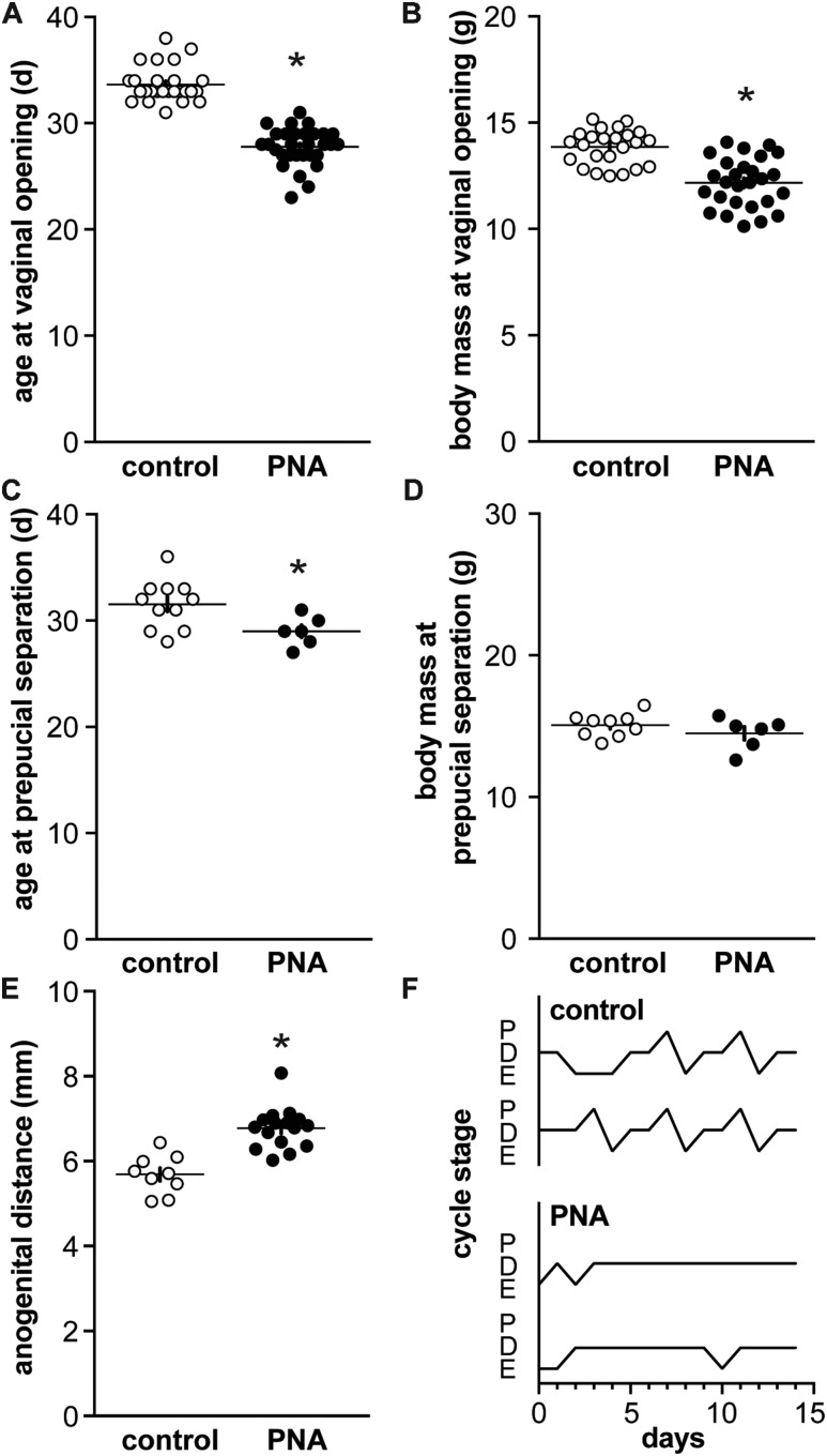
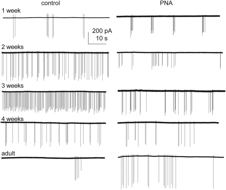
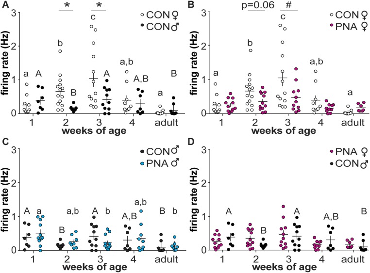
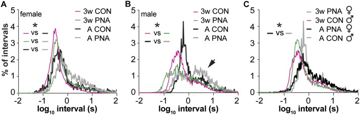
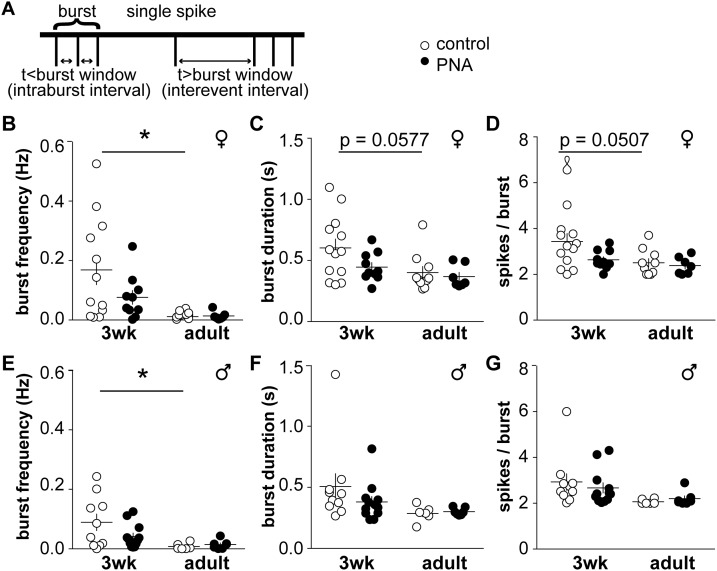
Gonadotropin-releasing hormone (GnRH) neurons regulate reproduction though pulsatile hormone release. Disruption of GnRH release as measured via luteinizing hormone (LH) pulses occurs in polycystic ovary syndrome (PCOS), and in young hyperandrogenemic girls. In adult prenatally androgenized (PNA) mice, which exhibit many aspects of PCOS, increased LH is associated with increased GnRH neuron action potential firing. How GnRH neuron activity develops over the prepubertal period and whether this is altered by sex or prenatal androgen treatment are unknown. We hypothesized GnRH neurons are active before puberty and that this activity is sexually differentiated and altered by PNA. Dams were injected with dihydrotestosterone (DHT) on days 16 to 18 post copulation to generate PNA mice. Action potential firing of GFP-identified GnRH neurons in brain slices from 1-, 2-, 3-, and 4-week-old and adult mice was monitored. GnRH neurons were active at all ages tested. In control females, activity increased with age through 3 weeks, then decreased to adult levels. In contrast, activity did not change in PNA females and was reduced at 3 weeks. Activity was higher in control females than males from 2 to 3 weeks. PNA did not affect GnRH neuron firing rate in males at any age. Short-term action potential patterns were also affected by age and PNA treatment. GnRH neurons are thus typically more active during the prepubertal period than adulthood, and PNA reduces prepubertal activity in females. Prepubertal activity may play a role in establishing sexually differentiated neuronal networks upstream of GnRH neurons; androgen-induced changes during this time may contribute to the adult PNA, and possibly PCOS, phenotype.
Copyright © 2017 Endocrine Society.
Figures
References
-
- Moenter SM, Brand RM, Midgley AR, Karsch FJ. Dynamics of gonadotropin-releasing hormone release during a pulse. Endocrinology. 1992;130(1):503–510. - PubMed
-
- Clarke IJ, Cummins JT. The temporal relationship between gonadotropin releasing hormone (GnRH) and luteinizing hormone (LH) secretion in ovariectomized ewes. Endocrinology. 1982;111(5):1737–1739. - PubMed
-
- Wildt L, Häusler A, Marshall G, Hutchison JS, Plant TM, Belchetz PE, Knobil E. Frequency and amplitude of gonadotropin-releasing hormone stimulation and gonadotropin secretion in the rhesus monkey. Endocrinology. 1981;109(2):376–385. - PubMed
-
- Haisenleder DJ, Dalkin AC, Ortolano GA, Marshall JC, Shupnik MA. A pulsatile gonadotropin-releasing hormone stimulus is required to increase transcription of the gonadotropin subunit genes: evidence for differential regulation of transcription by pulse frequency in vivo. Endocrinology. 1991;128(1):509–517. - PubMed
-
- McCartney CR, Eagleson CA, Marshall JC. Regulation of gonadotropin secretion: implications for polycystic ovary syndrome. Semin Reprod Med. 2002;20(4):317–326. - PubMed
Publication types
MeSH terms
Substances
Grants and funding
LinkOut - more resources
Full Text Sources
Other Literature Sources
