. 2017 Aug 3;8(35):57964-57980.
doi: 10.18632/oncotarget.19865. eCollection 2017 Aug 29.

CD3xPDL1 bi-specific T cell engager (BiTE) simultaneously activates T cells and NKT cells, kills PDL1+ tumor cells, and extends the survival of tumor-bearing humanized mice

Affiliations

CD3xPDL1 bi-specific T cell engager (BiTE) simultaneously activates T cells and NKT cells, kills PDL1+ tumor cells, and extends the survival of tumor-bearing humanized mice

Lucas A Horn et al. Oncotarget. .

Abstract

Bi-specific T cell engagers (BiTEs) activate T cells through CD3 and target activated T cells to tumor-expressed antigens. BiTEs have shown therapeutic efficacy in patients with liquid tumors; however, they do not benefit all patients. Anti-tumor immunity is limited by Programmed Death 1 (PD1) pathway-mediated immune suppression, and patients who do not benefit from existing BiTES may be non-responders because their T cells are anergized via the PD1 pathway. We have designed a BiTE that activates and targets both T cells and NKT cells to PDL1+ cells. In vitro studies demonstrate that the CD3xPDL1 BiTE simultaneously binds to both CD3 and PDL1, and activates healthy donor CD4+ and CD8+ T cells and NKT cells that are specifically cytotoxic for PDL1+ tumor cells. Cancer patients' PBMC are also activated and cytotoxic, despite the presence of myeloid-derived suppressor cells. The CD3xPDL1 BiTE significantly extends the survival time and maintains activated immune cell levels in humanized NSG mice reconstituted with human PBMC and carrying established human melanoma tumors. These studies suggest that the CD3xPDL1 BiTE may be efficacious for patients with PDL1+ solid tumors, in combination with other immunotherapies that do not specifically neutralize PD1 pathway-mediated immune suppression.

Keywords: T cell activation; cancer immunotherapy; checkpoint inhibitor blockade; solid tumors; tumor-induced immune suppression.


Conflict of interest statement

Conflicts of Interest The authors have no conflicts of interest.

Figures

Figure 1
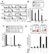
Figure 1. CHO cells transfected with the CD3xPDL1 BiTE produce a 55 kD protein
A. Supernatants from BiTE-transfected CHO cells (CHO/BiTE) cultured in serum-free HL1 medium were concentrated using 10kD spin columns, electrophoresed by SDS-PAGE, and western blotted using an anti-His mAb. B. Supernatants of CHO/BiTE transfectants were slot-blotted, probed with anti-his mAb, and BiTE content quantified by comparing to a standard his-tagged protein (CD80-Fc) using ImageJ software.
Figure 2
Figure 2. CD3xPDL1 BiTE simultaneously and specifically binds to PDL1+ human tumor cells and CD3+ human T cells
A. BiTE binds to tumor cell-expressed PDL1. PDL1+ C8161 melanoma cells and PDL1- MEL1011 cells were stained with fluorescently-tagged antibodies to PDL1 to determine their level of PDL1 expression (left panels). C8161 and MEL1011 cells were incubated with the CD3xPDL1 BiTE or an irrelevant recombinant protein (TROY-Fc) and then stained with a fluorescently-tagged anti-his mAb to detect BiTE binding (right panels). B. BiTE binds to T cell-expressed CD3. Peripheral blood mononuclear cells (PBMC) from healthy human donors were stained with fluorescently-tagged antibodies to CD4 and CD8 followed by addition of BiTE or irrelevant recombinant protein, and then fluorescently tagged antibody to his to detect BiTE binding. To ascertain that the BiTE specifically binds to CD3, aliquots of PBMC were first blocked with antibodies to CD3 to prevent BiTE binding. C. BiTE simultaneously binds CD3 and PDL1. PBMC were stained for CD4 and CD8 as in panel B, and then incubated with either BiTE or an irrelevant protein followed by soluble PDL1 (sPDL1). PDL1 binding was detected with a fluorescently-tagged antibody to PDL1. Data are representative of more than three independent experiments with at least 3 batches of BiTE.
Figure 3
Figure 3. CD3xPDL1 BiTE activates T cells that are cytotoxic for PDL1 + tumor cells
A. Healthy donor PBMC were co-cultured with C8161 tumor cells (ratio 20:1 PBMC:tumor cells) and with CD3xPDL1 BiTE (200 ng/mL) or an irrelevant protein for 48 hrs and the supernatants were analyzed for IFNγ by ELISA. Data are representative of one of two independent experiments. B. PBMC were incubated for 72 hrs with CellTrace Violet-labelled PDL1+ C8161 melanoma cells ± BiTE, and then labeled with fluorescently-coupled antibodies to CD4, CD8, and CD69 or CD25. Violet+ tumor cells were gated out and CD4+ and CD8+ T cells were gated and analyzed for the activation markers CD69 and CD25. Data are representative of 3 independent experiments. C. CellTrace Violet-stained CD3 purified PBMC were co-cultured for four days with or without BiTE or tumor, and the gated violet+ cells assessed by flow cytometry for proliferation. Data are representative of one of two independent experiments. D. C8161 cells were stained with CellTrace Violet and incubated at varying ratios with healthy donor PBMC ± BiTE. Following 72 hrs of incubation, cells were stained with the viability dye 7AAD. % dead tumor cells = [dead tumor cells (violet+7AAD+)/total tumor cells (violet+)] x 100%. Values are the average of triplicates per sample. E. C8161 and MEL1011 melanoma cells were stained with fluorescent antibodies to PDL1 (29E.2A3 mAb) or an isotype control mAb and analyzed by flow cytometry. PBMC from healthy donors were incubated ± CellTrace Violet-labeled C8161 or MEL1011 tumor cells ± BiTE and analyzed for % dead tumor cells. PBMC:tumor cell ratio is 20:1. CD8:tumor cell ratio is ~4:1. Values are the average + SE of 7 and 5 independent experiments for C8161 and MEL1011, respectively. For panels A and E, values with asterisks are significantly different from all other values.
Figure 4
Figure 4. CD3xPDL1 BiTE activates T cells and is cytotoxic for PDL1 + CML, NSCLC, and breast cancer cells
A. Tumor cells were incubated for 48 hrs with 200 units human recombinant IFNγ and stained with antibody to PDL1 (29E.2A3 mAb). B. Tumor cells were treated with IFNγ as in panel A and then labeled with CellTrace Violet and incubated ± healthy donor PBMC (20:1 ratio PBMC:tumor; 200ng BiTE/ml) ± CD3xPDL1 BiTE and analyzed 72 hrs later for percent dead tumor cells. Values are the average + SE of 5, 3, 4, and 3 independent experiments for C8161, MEL1011, MEG-01, and KU812F cells, respectively. C. H358 and MDA-MB-231 tumor cells were stained with fluorescent antibodies to PDL1 (29E.2A3 mAb) or isotype control mAb. D. Cytotoxicity assay as in B except H358 and MDA-MB-231 cells were targets. Values for H358 are the average ± SE of 7 independent experiments. Values for MDA-MD-231 are average ± SD of 3 replicates. Values with one or more asterisks are significantly different from other values for the same cell line.
Figure 5
Figure 5. Low levels of bound BiTE are sufficient for maximal killing of PDL1 + tumor cells
A. C8161 melanoma, MEL1011 melanoma, and Jurkat cells were labeled with fluorescently-tagged antibodies to PDL1 or CD3 and the corresponding isotype control mAbs. B. C8161, MEL1011, and Jurkat cells were treated with titered amounts of CD3xPDL1 BiTE followed by anti-his mAb. C. CellTrace Violet-labeled C8161 and MEL1011 cells were incubated with healthy donor PBMC plus titered amounts of BiTE as in fig. 3D. Values are from two independent experiments.
Figure 6
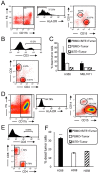
Figure 6. CD3xPDL1 BiTE activates both CD4 + and CD8+ cytotoxic T cells and NKT cells
A. PBMC from healthy human donors were either undepleted, or depleted for CD4+, CD8+, or CD4+ plus CD8+ T cells. Depleted populations were stained with fluorescently tagged antibodies to CD3, CD4, and CD8, and the CD3+ cells gated and analyzed for CD4 and CD8 expression. B. Violet-labeled PDL1+ H358 tumor cells were incubated with the depleted or not depleted PBMC ± BiTE (200ng/mL), and analyzed for % dead tumor cells. PBMC:tumor cell ratio is 20:1 for undepleted and depleted populations. For the undepleted samples the ratio of CD8 and CD4 T cells to tumor was 5.45:1 and 8.04:1, respectively. In the CD8-depleted and CD4-depleted samples the ratio of CD4:tumor was 16.08:1 and the ratio of CD8:tumor was 11.52:1. Data are the average of three independent experiments. C. The cells of panel B were stained with fluorescent antibodies to CD4, CD8, and CD107a and the CD4 and CD8 cells gated and analyzed for CD107a expression. Data are representative of one of two independent experiments. D. Violet-labeled ± C8161 tumor cells were incubated with magnetic bead purified CD3+ cells (PBMC) or purified CD3+CD56+ NKT cells ± BiTE (200ng/mL). Data are representative of three independent experiments. Values with * are significantly different from values without *.
Figure 7
Figure 7. CD3xPDL1 BiTE activates cancer patients’ PBMC that are cytotoxic for PDL1 + lung cancer cells
A. PBMC from a SCLC patient were stained for MDSC (CD11b, CD14, CD15, HLA-DR). CD11b+HLA-DR- cells were gated and analyzed by flow cytometry for CD14+CD15- M-MDSC and CD15+CD14- PMN-MDSC. Data are representative of one of three SCLC patients. B. SCLC patient's PBMC were labeled with fluorescently-tagged antibodies to CD3, CD4 and CD8, and the gated CD3+ cells were analyzed for percent CD3+CD4+ and CD3+CD8+ cells. Data are for one of three SCLC patients. C. PBMC from the SCLC patients were incubated ± CellTrace violet-labeled PDL1+ H358 or PDL1- MEL1011 tumor cells ± BiTE at a 20:1 ratio of PBMC:tumor cells. Cells were stained with 7AAD and the CellTrace violet stained tumor cells were gated and analyzed for % dead cells. The ratio of CD8+ plus CD4+ T cells to tumor cells is 2.3:1. Data are pooled from two independent experiments with PBMC from two individual patients. D. PBMC from a NSCLC patient were stained for MDSC. CD11b+HLA-DR- cells were gated and analyzed for CD14+CD15- M-MDSC and CD15+CD14- PMN-MDSC. E. NSCLC patient's PBMC were labeled with fluorescently-tagged antibodies to CD3, CD4 and CD8, and the CD3 gated cells were analyzed for percent CD3+CD4+ and CD3+CD8+ cells. F. PBMC from the NSCLC patient were incubated ± CellTrace violet-labeled H358 tumor cells ± BiTE at a 20:1 ratio of PBMC:tumor cells. Cells were stained with 7AAD and the CellTrace violet stained tumor cells were gated and analyzed for % dead cells.
Figure 8
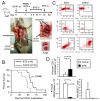
Figure 8. CD3xPDL1 BiTE significantly extends the survival time of humanized NSG mice reconstituted with human PBMC and carrying established metastatic human melanoma C8161
NSG mice were inoculated subcutaneously in the right flank with 1×10⁶ human C8161 melanoma cells on day 0. On day 7 when tumors were palpable, mice were either untreated or administered 1×10⁷ human PBMC iv in the tail vein and 0.2ng (8ng/kg) CD3xPDL1 BiTE iv in the retro-orbital sinus. On days 8, 9, 10, 11, and 32 mice were given additional iv injections of 0.2μg BiTE. A. Moribund, euthanized control mouse (tumor + PBMC, no BiTE) showing metastases in the lymph nodes and lungs on day 57. B. Kaplan-Meier plot showing survival. Data are pooled from three independent experiments. C. Representative flow cytometry profiles of splenocytes of moribund/dead BiTE-treated and untreated mice stained for human T cells (CD3, CD4, and CD8 mAbs), or mouse MDSC (Gr1 and CD11b mAbs). D. Total numbers and ratio of human T cells and mouse MDSC in the spleens of moribund/dead BiTE-treated and control mice of panel B. Data are pooled from 2-3 mice per group.

Similar articles

Cited by

References

    1. Baeuerle PA, Reinhardt C. Bispecific T-cell engaging antibodies for cancer therapy. Cancer Res. 2009;69:4941–4944. - PubMed
    1. Loffler A, Kufer P, Lutterbuse R, Zettl F, Daniel PT, Schwenkenbecher JM, Riethmuller G, Dorken B, Bargou RC. A recombinant bispecific single-chain antibody, CD19 x CD3, induces rapid and high lymphoma-directed cytotoxicity by unstimulated T lymphocytes. Blood. 2000;95:2098–2103. - PubMed
    1. Schlereth B, Kleindienst P, Fichtner I, Lorenczewski G, Brischwein K, Lippold S, da Silva A, Locher M, Kischel R, Lutterbuse R, Kufer P, Baeuerle PA. Potent inhibition of local and disseminated tumor growth in immunocompetent mouse models by a bispecific antibody construct specific for Murine CD3. Cancer Immunol Immunother. 2006;55:785–796. - PMC - PubMed
    1. Dreier T, Baeuerle PA, Fichtner I, Grun M, Schlereth B, Lorenczewski G, Kufer P, Lutterbuse R, Riethmuller G, Gjorstrup P, Bargou RC. T cell costimulus-independent and very efficacious inhibition of tumor growth in mice bearing subcutaneous or leukemic human B cell lymphoma xenografts by a CD19-/CD3- bispecific single-chain antibody construct. J Immunol. 2003;170:4397–4402. - PubMed
    1. Dreier T, Lorenczewski G, Brandl C, Hoffmann P, Syring U, Hanakam F, Kufer P, Riethmuller G, Bargou R, Baeuerle PA. Extremely potent, rapid and costimulation-independent cytotoxic T-cell response against lymphoma cells catalyzed by a single-chain bispecific antibody. Int J Cancer. 2002;100:690–697. - PubMed