PTIP promotes recurrence and metastasis of hepatocellular carcinoma by regulating epithelial-mesenchymal transition
- PMID: 28938547
- PMCID: PMC5601643
- DOI: 10.18632/oncotarget.16436
PTIP promotes recurrence and metastasis of hepatocellular carcinoma by regulating epithelial-mesenchymal transition
Abstract
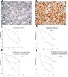
Hepatocellular carcinoma (HCC) is one of the most lethal tumors worldwide, which is mainly due to the high recurrence and metastasis rate after hepatectomy. In this study, we found that PTIP expression was dramatically upregulated in human HCC tissues and cell lines. High expression of PTIP was shown to be associated with aggressive clinicopathological features, including liver cirrhosis, vascular invasion and advanced stage. In addition, PTIP overexpression was independently associated with shorter survival and increased HCC recurrence in patients. Knockdown of the PTIP expression significantly inhibited invasion and metastasis in vitro and in vivo, whereas ectopic expression of PTIP significantly promoted invasion and metastasis. Mechanistically, PTIP promotes HCC progress by facilitating epithelial-mesenchymal transition (EMT). Notably, we also found that PTIP might increase miR-374a expression to promote EMT and metastasis in HCC. In summary, our study identified PTIP as a new potential prognostic indicator and therapeutic target for HCC.
Keywords: PAX interacting protein1 (PTIP); epithelial-mesenchymal transition (EMT); hepatocelluar carcinoma; metastasis; miRNA.
Conflict of interest statement
CONFLICTS OF INTEREST None of the authors have any conflicts of interest to declare.
Figures


Similar articles
-
MicroRNA-451: epithelial-mesenchymal transition inhibitor and prognostic biomarker of hepatocelluar carcinoma.Oncotarget. 2015 Jul 30;6(21):18613-30. doi: 10.18632/oncotarget.4317. Oncotarget. 2015. PMID: 26164082 Free PMC article.
-
FoxM1 overexpression promotes epithelial-mesenchymal transition and metastasis of hepatocellular carcinoma.World J Gastroenterol. 2015 Jan 7;21(1):196-213. doi: 10.3748/wjg.v21.i1.196. World J Gastroenterol. 2015. PMID: 25574092 Free PMC article.
-
Sorcin Predicts Poor Prognosis and Promotes Metastasis by Facilitating Epithelial-mesenchymal Transition in Hepatocellular Carcinoma.Sci Rep. 2017 Aug 30;7(1):10049. doi: 10.1038/s41598-017-10365-3. Sci Rep. 2017. Retraction in: Sci Rep. 2018 Aug 02;8(1):11857. doi: 10.1038/s41598-018-29892-8. PMID: 28855589 Free PMC article. Retracted.
-
Actin-like 6A predicts poor prognosis of hepatocellular carcinoma and promotes metastasis and epithelial-mesenchymal transition.Hepatology. 2016 Apr;63(4):1256-71. doi: 10.1002/hep.28417. Epub 2016 Jan 26. Hepatology. 2016. PMID: 26698646 Free PMC article.
-
HCRP1 downregulation promotes hepatocellular carcinoma cell migration and invasion through the induction of EGFR activation and epithelial-mesenchymal transition.Biomed Pharmacother. 2017 Apr;88:421-429. doi: 10.1016/j.biopha.2017.01.013. Epub 2017 Jan 22. Biomed Pharmacother. 2017. PMID: 28122307
Cited by
-
A set of molecular markers predicts chemosensitivity to Mitomycin-C following cytoreductive surgery and hyperthermic intraperitoneal chemotherapy for colorectal peritoneal metastasis.Sci Rep. 2019 Jul 22;9(1):10572. doi: 10.1038/s41598-019-46819-z. Sci Rep. 2019. PMID: 31332257 Free PMC article.
-
LLGL2 Increases Ca2+ Influx and Exerts Oncogenic Activities via PI3K/AKT Signaling Pathway in Hepatocellular Carcinoma.Front Oncol. 2021 Jun 10;11:683629. doi: 10.3389/fonc.2021.683629. eCollection 2021. Front Oncol. 2021. PMID: 34178676 Free PMC article.
-
Blockade of ARHGAP11A reverses malignant progress via inactivating Rac1B in hepatocellular carcinoma.Cell Commun Signal. 2018 Dec 13;16(1):99. doi: 10.1186/s12964-018-0312-4. Cell Commun Signal. 2018. PMID: 30545369 Free PMC article.
References
-
- Torre LA, Bray F, Siegel RL, Ferlay J, Lortet-Tieulent J, Jemal A. Global cancer statistics, 2012. CA Cancer J Clin. 2015;65:87–108. - PubMed
-
- Chen W, Zheng R, Baade PD, Zhang S, Zeng H, Bray F, Jemal A, Yu XQ, He J. Cancer statistics in China, 2015. CA Cancer J Clin. 2016;66:115–32. - PubMed
-
- Forner A, Llovet JM, Bruix J. Hepatocellular carcinoma. Lancet. 2012;379:1245–55. - PubMed
LinkOut - more resources
Full Text Sources
Other Literature Sources

