. 2017 May 8;8(35):58781-58789.
doi: 10.18632/oncotarget.17692. eCollection 2017 Aug 29.

Vitamin D3 induces vitamin D receptor and HDAC11 binding to relieve the promoter of the tight junction proteins

Affiliations

Vitamin D3 induces vitamin D receptor and HDAC11 binding to relieve the promoter of the tight junction proteins

Feng-Hua Liu et al. Oncotarget. .

Abstract

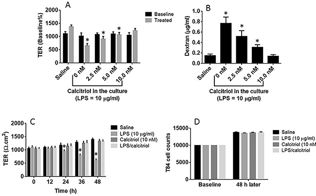
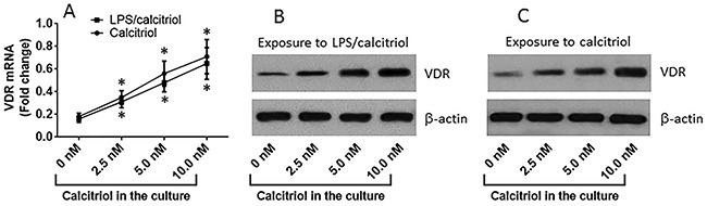
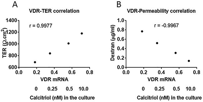
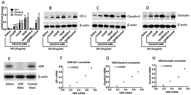
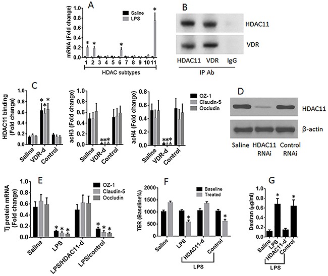
Intestinal epithelial barrier dysfunction and vitamin D (VitD)-deficiency play a critical role in a large number of diseases. The histone deacetylases (HDAC) are associated with a large number of immune diseases. This study tests a hypothesis that the interaction between VitD and HDAC is associated with the regulation of epithelial barrier functions. In this study, human intestinal epithelial cell line, T84 cells, was cultured into monolayers to be used as a model to test the epithelial barrier functions. We observed that in a VitD-deficient environment, the T84 monolayer barrier function was compromised. Exposure to calcitriol (the active form of VitD3) in the culture increased the expression of VitD receptor (VDR) in T84 cells. In a VitD-sufficient environment, VDR formed a complex with histone deacetylase-11 (HDAC11); the complex was markedly decreased in a VitD-deficient environment. We also observed that significantly more binding of HDAC11 to the promoter of the tight junction proteins inhibit the gene transcription activities of these loci in the VitD-deficient environment, which were abolished by the presence of calcitriol in the culture. In conclusion, the interaction between VDR and HDAC11 plays a crucial role in the maintenance of the epithelial barrier integrity.

Keywords: barrier function; epithelium; histone deacetylase; intestine; vitamin D.


Conflict of interest statement

CONFLICTS OF INTEREST None to declare.

Figures

Figure 1
Figure 1. VitD blocks LPS-induced epithelial barrier dysfunction
(A-B) the confluent T84 monolayers were stimulated with LPS (10 μg/ml) for 48 h with or without the presence of calcitriol at concentrations as denoted on the X axis. The bars indicate the TER levels (A) of T84 monolayers and the dextran levels in the basal chambers of Transwells (B). (C) the bars indicate the TER levels of T84 monolayers recorded at the time points denoted on the X axis after the indicated treatment. (D) T84 cell counts after culture for 48 h with the indicated treatment. The data are presented as mean ± SD. *p<0.01. The data were summarized from 3 independent experiments.
Figure 2
Figure 2. VitD up regulates VDR expression in T84 cells
(A) the curves (mean ± SD, summarized from 3 independent experiments) indicate the VDR mRNA levels in T84 cells after exposure to calcitriol or LPS (10 μg/ml) and calcitriol in the culture for 2 days. (B-C) the immune blots indicate the protein levels of VDR in T84 cells after exposure to LPS and calcitriol (B) or exposure to calcitriol alone (C). The data of B-C are from one experiment representing 3 independent experiments. *p<0.01, compared with the “0 nM” group.
Figure 3
Figure 3. Correlation between VDR mRNA and T84 monolayer barrier function
The dot plots show the correlation between VDR and TER, VDR and permeability of T84 monolayers. The T84 monolayers were exposed to LPS (10 μg/ml) and calcitriol at gradient concentrations for 48 h. The data are from one experiment representing 3 independent experiments.
Figure 4
Figure 4. VitD reverses the LPS-suppressed Tj protein expression
(A-D) the results of Tj protein expression in T84 monolayers [with or without the VDR gene knockdown (VDR-d)] after exposure to LPS (10 μg/ml) in the culture with or without the presence of calcitriol (denoted on the X axis). Control: T84 monolayers were treated with control shRNA. The data of bars (mean ± SD; *p<0.01, compared with the saline group) are summarized from 3 independent experiments. The immune blots are from one experiment representing 3 independent experiments. (E) RNAi results of VDR in T84 cells. (F-H) the dot plots show the correlation between VDR mRNA and the mRNAs of Tj proteins in T84 monolayers.
Figure 5
Figure 5. HDAC11 mediates the effects of LPS on suppression of T84 monolayer barrier function
T84 monolayers (wild type, VDR-deficient or HDAC11-deficient) were cultured in the presence or absence of LPS (10 μg/ml) for 48 h. (A) the HDAC11 mRNA levels. (B) a complex of VDR and HDAC11. (C) the levels of HDAC11, acetylated histone (acH) 3 and acH4 at the promoter loci of Tj proteins. (D) the results of HDAC11 RNAi of T84 cells. (E) the mRNA levels of Tj protein in T84 cells. (F-G) the TER (F) and permeability to dextran (G) of T84 monolayers. The data (mean ± SD) were summarized from 3 independent experiments. *p<0.01, compared with the saline group.

Similar articles

Cited by

References

    1. Halpern MD, Denning PW. The role of intestinal epithelial barrier function in the development of NEC. Tissue Barriers. 2015;3:e1000707. - PMC - PubMed
    1. Yu LC. Intestinal epithelial barrier dysfunction in food hypersensitivity. J Allergy (Cairo) 2012;2012:596081. - PMC - PubMed
    1. Landy J, Ronde E, English N, Clark SK, Hart AL, Knight SC, Ciclitira PJ, Al-Hassi HO. Tight junctions in inflammatory bowel diseases and inflammatory bowel disease associated colorectal cancer. World J Gastroenterol. 2016;22:3117–3126. - PMC - PubMed
    1. Lee SH. Intestinal permeability regulation by tight junction: implication on inflammatory bowel diseases. Intest Res. 2015;13:11–18. - PMC - PubMed
    1. Yang PC, Jury J, Soderholm JD, Sherman PM, McKay DM, Perdue MH. Chronic psychological stress in rats induces intestinal sensitization to luminal antigens. Am J Pathol. 2006;168:104–114. - PMC - PubMed

LinkOut - more resources