. 2017 Oct 1;32(10):1995-2006.
doi: 10.1093/humrep/dex269.

Complex CatSper-dependent and independent [Ca2+]i signalling in human spermatozoa induced by follicular fluid

Affiliations

Complex CatSper-dependent and independent [Ca2+]i signalling in human spermatozoa induced by follicular fluid

Sean G Brown et al. Hum Reprod. .

Abstract

Study question: Does progesterone in human follicular fluid (hFF) activate CatSper and do other components of hFF modulate this effect and/or contribute separately to hFF-induced Ca2+ signaling?

Summary answer: hFF potently stimulates CatSper and increases [Ca2+]i, primarily due to high concentrations of progesterone, however, other components of hFF also contribute to [Ca2+]i signaling, including modulation of CatSper channel activity and inhibition of [Ca2+]i oscillations.

What is known already: CatSper, the principal Ca2+ channel in spermatozoa, is progesterone-sensitive and essential for fertility. Both hFF and progesterone, which is present in hFF, influence sperm function and increase their [Ca2+]i.

Study design, size, duration: This basic medical research study used semen samples from >40 donors and hFF from >50 patients who were undergoing surgical oocyte retrieval for IVF/ICSI.

Participants/materials, setting, methods: Semen donors and patients were recruited in accordance with local ethics approval (13/ES/0091) from the East of Scotland Research Ethics Service REC1. Activities of CatSper and KSper were assessed by patch clamp electrophysiology. Sperm [Ca2+]i responses were examined in sperm populations and single cells. Computer-assisted sperm analysis (CASA) parameters and penetration into viscous media were used to assess functional effects.

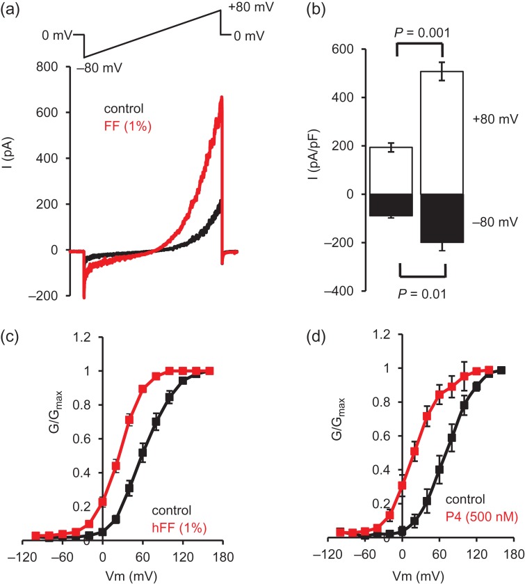
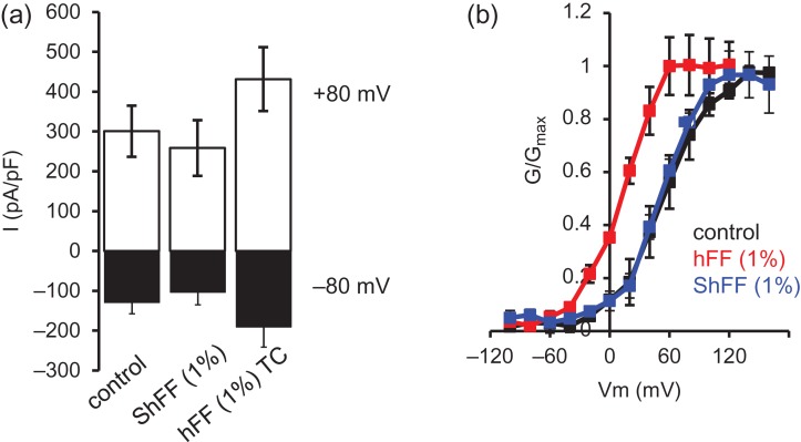
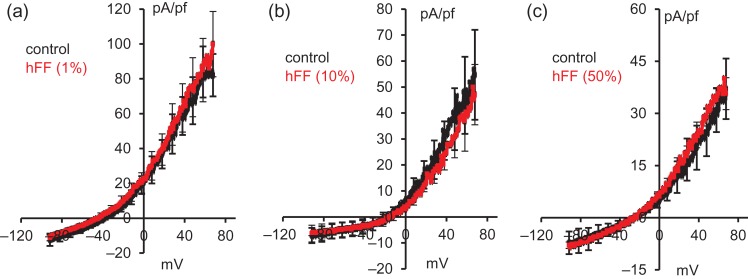
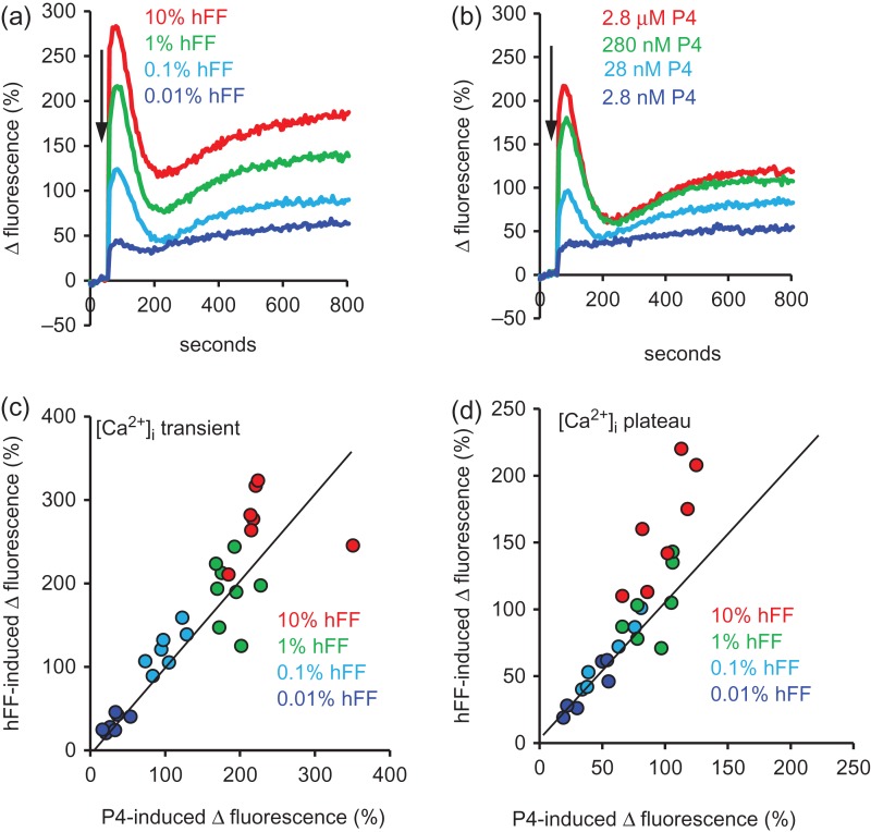
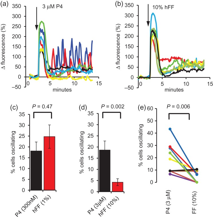
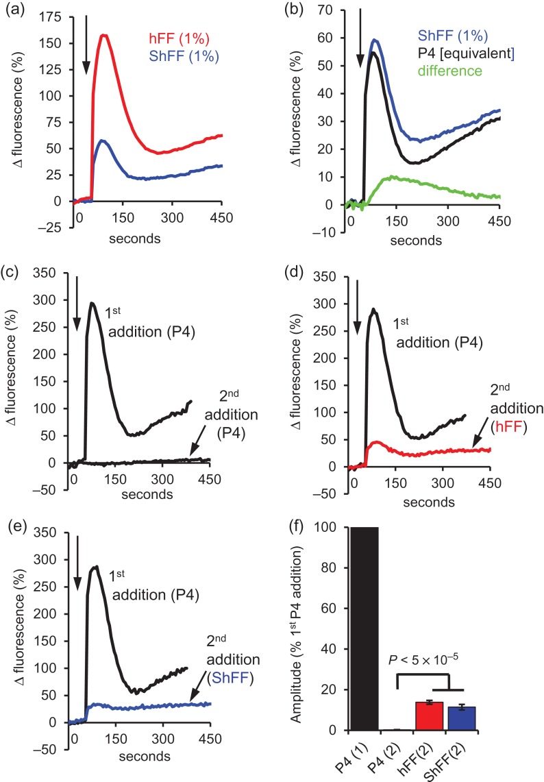
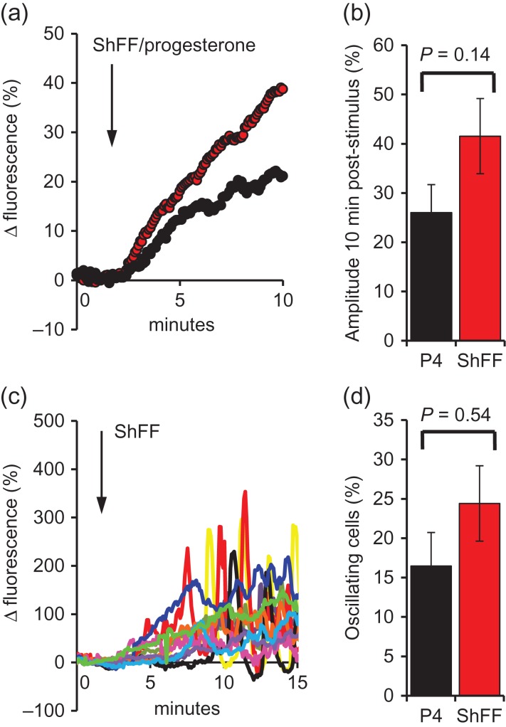
Main results and the role of chance: hFF and progesterone significantly potentiated CatSper currents. Under quasi-physiological conditions, hFF (up to 50%) failed to alter membrane K+ conductance or current reversal potential. hFF and progesterone (at an equivalent concentration) stimulated similar biphasic [Ca2+]i signals both in sperm populations and single cells. At a high hFF concentration (10%), the sustained (plateau) component of the [Ca2+]i signal was consistently greater than that induced by progesterone alone. In single cell recordings, 1% hFF-induced [Ca2+]i oscillations similarly to progesterone but with 10% hFF generation of [Ca2+]i oscillations was suppressed. After treatment to 'strip' lipid-derived mediators, hFF failed to significantly stimulate CatSper currents but induced small [Ca2+]i responses that were greater than those induced by the equivalent concentration of progesterone after stripping. Similar [Ca2+]i responses were observed when sperm pretreated with 3 μM progesterone (to desensitize progesterone responses) were stimulated with hFF or stripped hFF. hFF stimulated viscous media penetration and was more effective than the equivalent does of progesterone.

Large scale data: N/A.

Limitations, reasons for caution: This was an in vitro study. Caution must be taken when extrapolating these results in vivo.

Wider implications of the findings: This study directly demonstrates that hFF activates CatSper and establishes that the biologically important effects of hFF reflect, at least in part, action on this channel, primarily via progesterone. However, these experiments also demonstrate that other components of hFF both contribute to the [Ca2+]i signal and modulate the activation of CatSper. Simple in vitro experiments performed out of the context of the complex in vivo environment need to be interpreted with caution.

Study funding/competing interest(s): Funding was provided by MRC (MR/K013343/1, MR/012492/1) (S.G.B., S.J.P., C.L.R.B.) and University of Abertay (sabbatical for S.G.B.). Additional funding was provided by TENOVUS SCOTLAND (S.M.D.S.), Chief Scientist Office/NHS Research Scotland (S.M.D.S). C.L.R.B. is EIC of MHR and Chair of the WHO ESG on Diagnosis of Male infertility. The remaining authors have no conlicts of interest.

Keywords: CatSper; follicular fluid; patch clamp electrophysiology; potassium channel; spermatozoa.


Figures

Figure 1
Figure 1
hFF potentiates CatSper currents and shifts the voltage sensitivity to less depolarized potentials. (a) Representative Cs+-mediated CatSper current in the absence (black) and presence (red) of 1% hFF. Voltage protocol imposed is shown above. (b) Mean amplitudes (±SEM) of CatSper currents recorded in the absence (left) and presence (right) of 1% hFF (n = 8 hFF samples). Black bars show inward current (−80 mV), white bars show outward currents (80 mV; n = 13). (c and d) Show conductance-voltage (G–V) relationships for Ba2+-mediated CatSper tail currents in the absence and presence of 1% hFF (c, n = 12) and 500 nM P4 (d, n = 4). hFF, human follicular fluid.
Figure 2
Figure 2
Charcoal-stripped hFF (ShFF) does not potentiate CatSper currents. (a) Mean ± SEM inward CatSper currents at −80 mV (black) and outward currents at 80 mV (white; n = 8 cells) under control conditions, in presence of 1% stripped hFF (ShFF) and 1% time-control (hFF; 7FF samples). ShFF reduced current amplitude (P < 0.05) but subsequent application of control hFF potentiated both inward and outward currents (P < 0.01 compared to ShFF). (b) 1% stripped hFF (ShFF) failed to alter CatSper voltage sensitivity but subsequent application of control follicular fluid (hFF) caused a significant leftward shift in voltage sensitivity (V50P < 0.01 compared to control and ShFF). n = 4 cells, four hFF.
Figure 3
Figure 3
hFF does not affect K+ channel activity recorded under quasi-physiological conditions. In each panel, black trace shows mean (±SEM) control current and red trace shows mean (±SEM) of currents recorded after exposure to hFF. (a) 1% hFF; n = 6 cells, four hFF tested; (b) 10% hFF; n = 3 cells, three hFF tested; (c) 50% hFF; n = 3 cells, three hFF.
Figure 4
Figure 4
[Ca2+]i responses to hFF and progesterone are similar but not identical. (a and b) Show an example of [Ca2+]i responses induced in paired experiments using (a) four dilutions of hFF (dark blue = 0.01%, light blue = 0.1%, green = 1%, red = 10%) and (b) P4 at concentrations equivalent to those in the hFF dilutions (dark blue = 2.8 nM, light blue = 28 nM, green = 280 nM, red = 2.8 μM). (c and d) Show relative amplitudes (Δ fluorescence (%)) of the [Ca2+]i transients (c) and [Ca2+]i plateau (d, assessed 10 min post-stimulation) induced in seven sets of experiments, each using four dilutions of hFF (0.01% = dark blue, 0.1% = light blue, 1% = green, 10% = red) and P4 at concentrations equivalent to those in the hFF dilutions. Six different hFF samples were used. Line in each graph marks position of equal response amplitude. At the highest hFF concentration used (10%; red symbols), plateau responses are consistently larger than those of equivalent [P4] (P = 0.001).
Figure 5
Figure 5
Single cell [Ca2+]i responses to hFF. (a and b) Show examples of [Ca2+]i responses in a paired experiment in which cells from the same sample were exposed to 3 μM P4 (a) and 10% hFF (b). Panel c shows mean ± SEM percentage of cells in which [Ca2+]i oscillations occured after stimulation of sperm (from the same sample) with 300 nM P4 (black) or 1% hFF (red); n = 10 paired experiments. Panel d shows results from a similar series of 10 paired assessments using 3 μM P4 (black) and 10% hFF (red; P < 0.01). (e) Shows data from the 3 μM P4/10% hFF experiments summarized in panel d with paired experiments joined and shown in same colour.
Figure 6
Figure 6
Components of the hFF-induced [Ca2+]i signal are resistant to P4 desensitization and charcoal stripping. (a) Mean [Ca2+]i response from 21 experiments (5 different hFF used) in which aliquots from the same sperm sample treated with 1% hFF (red) and 1% ShFF (blue). (b) Mean [Ca2+]i response from 28 paired experiments (9 different hFF used) in which aliquots from the same sperm sample were treated with 1% ShFF (blue) or the equivalent concentration of P4 (black). Green shows the ‘non-P4’ component obtained by subtraction of traces. (ce) Examples of [Ca2+]i responses in three parallel recordings where sperm were first stimulated with 3 μM P4 (first addition-black traces) then, after an interval of 5 min, exposed to either a second 3 μM P4 stimulus (6 μM P4 total; c, second addition-black trace), 1% hFF (d, second addition-red trace) or 1% ShFF (e, second addition-blue trace). In each panel the responses to the first (3 μM P4) stimulus and to the second stimulus are overlaid (arrow at top left shows time of additions). When 3 μM P4 was followed by a second P4 stimulus the second response was negligible (desensitization). However, when either 1% hFF or 1% ShFF was added as the second stimulus there was a small transient followed by a plateau. (f) Mean amplitude (±SEM) of [Ca2+]i transients evoked by the first 3 μM P4 stimulus (P4(1) black) and by a second addition of P4 (P4(2); n = 7; black), hFF (hFF(2); n = 10; red) or stripped hFF (ShFF(2); n = 6; blue). All amplitudes are normalized to that induced by the first P4 addition in that experiment.
Figure 7
Figure 7
Single cell [Ca2+]i responses to 1% ShFF. (a) Shows mean responses to 1% ShFF (red; n = 10 experiments; 826 cells) and equivalent [P4] (black; n = 6 experiments; 447 cells), arrow marks stimulus addition. Both stimuli induced a [Ca2+]i ramp rather than the biphasic response seen in fluorimetric experiments. (b) Shows mean (±SEM) amplitude (Δ fluorescence) 9 min after stimulus application. (c) Shows responses of 12 individual cells stimulated with ShFF, arrow marks stimulus addition. Red, yellow and black cells developed oscillations 5–10 min after stimulation. (d) shows proportions of cells generating [Ca2+]i oscillations after stimulation with 1% ShFF (red; n = 10 experiments; 826 cells) or equivalent [P4] (black; n = 6 experiments; 447 cells).

References

    1. Aitken RJ, Buckingham DW, Irvine DS. The extragenomic action of progesterone on human spermatozoa: evidence for a ubiquitous response that is rapidly down-regulated. Endocrinology 1996;137:3999–4009. - PubMed
    1. Alasmari W, Barratt CL, Publicover SJ, Whalley KM, Foster E, Kay V, Martins da Silva S, Oxenham SK. The clinical significance of calcium-signalling pathways mediating human sperm hyperactivation. Hum Reprod 2013. a;28:866–876. - PMC - PubMed
    1. Alasmari W, Costello S, Correia J, Oxenham SK, Morris J, Fernandes L, Ramalho-Santos J, Kirkman-Brown J, Michelangeli F, Publicover S et al. . Ca2+ signals generated by CatSper and Ca2+ stores regulate different behaviors in human sperm. J Biol Chem 2013. b;288:6248–6258. - PMC - PubMed
    1. Baldi E, Luconi M, Bonaccorsi L, Forti G. Nongenomic effects of progesterone on spermatozoa: mechanisms of signal transduction and clinical implications. Front Biosci 1998;3:D1051–D1059. - PubMed
    1. Bedu-Addo K, Barratt CL, Kirkman-Brown JC, Publicover SJ. Patterns of [Ca2+]i mobilization and cell response in human spermatozoa exposed to progesterone. Dev Biol 2007;302:324–332. - PubMed

Publication types